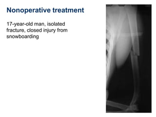
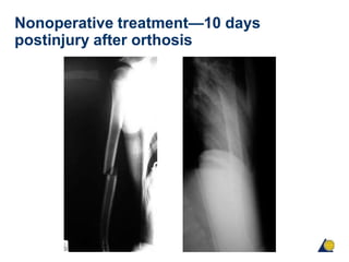
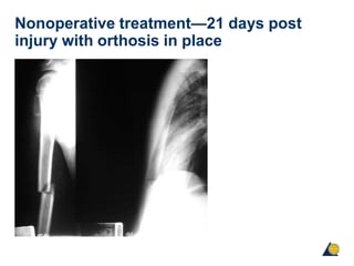
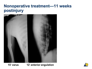
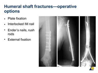
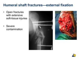
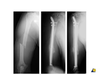
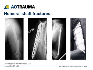
AOTrauma Principles Course
Christopher Finkemeier, US
Hank Hanff, US
Humeral shaft fractures
Objectives
• Identify several acceptable treatment strategies for
humeral diaphyseal fractures
• Learn the operative indications for humeral diaphyseal
fractures
• Understand the benefits and limitations of the various
treatment strategies
Humeral shaft fractures
• Humeral shaft fractures account for approximately 1–3%
of all fractures [Beaty 1996, Zuckerman, 1996]
• Usually the result of blunt trauma such as a fall or from
high-energy trauma including motor vehicle accidents
and gunshot injuries
• Fracture displacement is the result of muscular forces
General considerations
•
•
Energy of injury?
Associated injuries?
(isolated vs polytrauma)
Status of soft tissues?
Radial nerve function?
Patient factors?
•
•
•
-
-
Obesity
Expectations
•
•
Stable vs unstable?
Need upper extremity
weight bearing?
Open or closed?
Exploration?
Nonoperative treatment?
Do patients demand or
expect perfect x-rays?
•
•
•
•
Treatment options
Nonoperative:
•
•
•
Coaptation splint
Hanging arm cast
Functional orthosis
Operative:
•
•
•
•
•
•
•
Compression plating
Intramedullary fixation
Antegrade
Retrograde
Interlocking
Flexible
External fixation
Nonoperative treatment
Indications:
• Isolated injury
• Adequate alignment after splinting
-
-
AP bowing < 20o
lateral bowing < 300
• Preferred method
• Functional orthosis
Nonoperative treatment
Functional orthosis Collar-and-cuff
Nonoperative treatment
17-year-old man, isolated
fracture, closed injury from
snowboarding
Nonoperative treatment—10 days
postinjury after orthosis
Nonoperative treatment—21 days post
injury with orthosis in place
Nonoperative treatment—11 weeks
postinjury
15° varus 12° anterior angulation
Nonoperative treatment
•
•
15° varus
12° anterior angulation
Nonoperative treatment
Sarmiento, et al (2000) J Bone Joint Surg Am
• Followed 620 patients with humeral shaft fractures treated
with cast bracing
Non-union rate: < 2% of closed fractures
Refracture rate: 1% between 2 and 8 weeks post cast
removal
Radial Nerve Palsy: 11%
Most common was varus angulation (16%) with 10°–20° of
angulation
•
•
•
•
Humeral shaft fractures—operative
indications
•
•
•
Open fractures
Vascular injury
Radial nerve palsy after
closed reduction
Floating elbow
Failure of closed treatment
Pathologic fractures (bone
metastases)
Brachial plexus injury
•
•
•
•
Humeral shaft fractures—relative
operative indications
• Polytrauma
• Bilateral humeral
fractures
• Morbid obesity
• Segmental fractures
• Need to use crutches
Humeral shaft fractures—operative
options
•
•
•
Plate fixation
Interlocked IM nail
Ender’s nails, rush
rods
External fixation
•
Humeral shaft fractures—external fixation
• Open fractures
with extensive
soft-tissue injuries
• Severe
contamination
Humeral shaft fractures—indications for
plate fixation
• Fractures requiring
nerve or vascular
exploration
• Very distal fractures
• Very proximal
fractures
• Any diaphyseal
fracture
Humeral shaft fractures—surgical
approaches
• Anterolateral:
- Proximal and middle third
• Posterior:
- Distal and middle third
Humeral shaft fractures—anterolateral
approach
• Supine
• Good for
polytraumatized
patient
Humeral shaft fractures—anterolateral
approach
• Deltopectoral approach
proximally
• Interval between biceps
and brachialis
• Split brachialis fibers
Humeral shaft fractures—posterior
approach
• Prone or lateral
• Arm draped over padded support
Humeral shaft fractures—posterior
approach
• Interval between lateral and
long heads of triceps
• Identify radial nerve
• Split medial (deep) head
Humeral shaft fractures—posterior plate
• Tension band
• Placed beneath radial nerve
• Dictate location of nerve
Humeral shaft fractures—implants
• Broad 4.5 large fragment plate
• Small bone individuals:
-
-
Narrow large fragment plate
Small fragment plate 3.5
Absolute stability Relative stability
Humeral shaft fractures—flexible nails
• Ender nails
• Antegrade or
retrograde
• Rotational control?
• Migration problems
Intramedullary fixation—flexible nails
22-year-old with acetabular fracture and closed humeral
fracture
Intramedullary fixation—flexible nails
• Multiple, flexible,
retrograde IM nails
• Early weight bearing
allowed on upper extremity
• Callous at 4 weeks
postoperative
Nancy nails
Intramedullary fixation—flexible nails
Fracture healed at 3 months
Intramedullary fixation—flexible nails
Brumback, et al (1986) JBJS
• 63 fractures, 58 followed up
• Both antegrade and retrograde
• 94% union rate
• Retrograde insertion proximal to olecranon fossa gave
excellent results
Humeral shaft fractures—antegrade
locked IM nails
• Pathological and
osteopenic fractures
• Good rotational/length
control
• Good healing rates
• Often allows weight
bearing
Humeral shaft fractures—antegrade
locked IM nails
Concerns:
•
•
• Insertion often damages rotator
cuff tendons
Inrtamedullary canal narrows
distally
Neurovascular injury at
interlocking sites
X
Open reducation internal fixation ORIF vs
intramedullary nailing
• Two randomized prospective studies published compare ORIF to IM
nailing
• Chapman et al (2000) concluded that “neither method was shown to be
markedly superior to each other although nails were associated with a
higher incidence of shoulder discomfort.”
• McCormack et at (2000) suggested that “DCP fixation should continue to be
regarded as the best treatment for fractures of the humeral shaft which
require surgical stabilization.”
Humeral shaft fractures—locked IM nails
Design modifications to avoid
shoulder problems:
• More lateral entrance site
• Retrograde insertion
Stannard et al (2003) J Bone Joint Surg Am
• 42 consecutive patients
• 95% union rate
• All nonunions (2) occured with 7.5 mm
nail
• 10% shoulder pain
• 24% some loss of motion
• All patients with loss of motion and
shoulder pain had RETROGRADE nails
Humeral shaft fractures—retrograde IM
nailing
• Interlocked nails may also be inserted through distal site
• Care to avoid fracture at entrance site
Humeral shaft fractures—radial nerve
palsy
•
•
•
•
Incidence 1.8–24 %
Most are a neuropraxia
> 70–90% recover spontaneously
EMG (electromyography) if no evidence of recovery at
6–12 weeks
Humeral shaft fractures—radial nerve
palsy
• Associated fracture
patterns
• Transverse mid third
- Usually neuropraxia
• Spiral distal third
-
-
Holstein-Lewis fracture
Higher risk of laceration
or nerve entrapment
Humeral shaft fractures—immediate
radial nerve exploration
• Open fractures
• Distal-third spiral fractures
• Secondary palsy following closed reduction
Summary
• Most humeral shaft fractures can be treated successfully
with a functional brace
• Flexible nails are an effective treatment method
• Plates and nails have “similar” union rates, but nails
have more complications
• Plates are preferable

humerus shaft