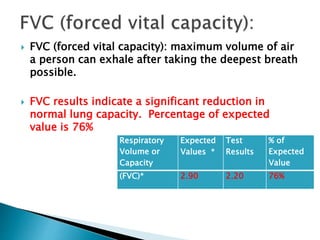
Case Study 1
 Patient 1 is a five foot two inch, 130-lb
60-year old female non-smoker who has
worked in a chemical factory for the past 34
years. She complains of shortness of breath
and a continual cough that produces sputum.
Her doctor notes the patient is breathing with
pursed lips and her skin that has a slight
bluish color.
 Environmental factors (occupational
exposure to chemicals) and other symptoms
noted, including: shortness of breath,
productive cough, pursed lips breathing, and
cyanosis, are all possible indicators for
COPD (emphysema).
 A stethoscope can
be used to listen
to the lungs.
 However,
sometimes the
lungs sound
normal even when
COPD is present.
 X-rays and CT scans
can also be useful.
 But sometimes they
look normal even
when a person has
COPD.
 A blood test (called
arterial blood gas) can
also be done to measure
the amounts of oxygen
and carbon dioxide in
the blood.
 The carbon dioxide
measure gives some idea
of lung function.
 Check for reversibility by
administrating
Bronchodilators.
 If reversibility levels are
low then => COPD.
 Body Plethysmography -
results would show
Emphysema (>120%
predicted).
 The spirometry results would indicate the
possibility of obstructive lung disease, which
could include a number of different things:
emphysema, chronic obstructive pulmonary
disease (COPD), and asthma.
Respiratory Volume or
Capacity
Expected
Values *
Test Results % of
Expected
Value
Forced Vital Capacity
(FVC)*
2.90 2.20 76%
Forced Expiratory Volume
after 1 second (FEV1)*
2.30. 1.35 59%
FEV1/FVC*, ** 81.7% 61.4%
** Normal lung function usually
produces values > 80%.
 FVC (forced vital capacity): maximum volume of air
a person can exhale after taking the deepest breath
possible.
 FVC results indicate a significant reduction in
normal lung capacity. Percentage of expected
value is 76%
Respiratory
Volume or
Capacity
Expected
Values *
Test
Results
% of
Expected
Value
(FVC)* 2.90 2.20 76%
 FEV1 (forced expired volume in 1 second): volume expired in the
first second of maximal expiration after a maximal inspiration. This
is a measure of how quickly the lungs can be emptied in 1 second.
 FEV1 results indicate a significant reduction in lung emptying.
Percentage of expected value is 59%
Respiratory
Volume or
Capacity
Expected
Values *
Test
Results
% of
Expected
Value
(FEV1)* 2.30. 1.35 59%
 FEV1/FVC: FEV1 expressed as a percentage
of the FVC, gives a clinically useful index of
airflow limitation.
 FEV1/FVC results indicate a reduction.
FEV1/FVC is the ability of air to move through
airways, which would be expected to
accompany obstructive lung diseases.
Respiratory
Volume or
Capacity
Expected
Values *
Test
Results
FEV1/FVC*, ** 81.7% 61.4%
 By looking at the symptoms it
looks like this patient has COPD
Emphysema.
 Emphysema is a lung condition
where the alveoli overinflates with
air and causes shortness of breath
(dyspnea)
 The patient is also showing
symptoms of hypoxia-induced
cyanosis or the appearance of a
blue-to-purplish discoloration of
the skin. It is due to increased
levels of deoxyhaemoglobin in the
blood.
 Emphysema could be due to exposure to air pollution
as the patient has worked in a chemical factory for 34
years.
 Other factors which could cause emphysema includes;
Deficiency of an enzyme alpha-1-antitrypsin and/or
other chemicals and toxins.
Smoking tobacco and/or from second-hand smoke.

Human Physiology Case Study 1 complete.pptx

  • 1.
  • 2.
     Patient 1is a five foot two inch, 130-lb 60-year old female non-smoker who has worked in a chemical factory for the past 34 years. She complains of shortness of breath and a continual cough that produces sputum. Her doctor notes the patient is breathing with pursed lips and her skin that has a slight bluish color.
  • 3.
     Environmental factors(occupational exposure to chemicals) and other symptoms noted, including: shortness of breath, productive cough, pursed lips breathing, and cyanosis, are all possible indicators for COPD (emphysema).
  • 4.
     A stethoscopecan be used to listen to the lungs.  However, sometimes the lungs sound normal even when COPD is present.
  • 5.
     X-rays andCT scans can also be useful.  But sometimes they look normal even when a person has COPD.
  • 6.
     A bloodtest (called arterial blood gas) can also be done to measure the amounts of oxygen and carbon dioxide in the blood.  The carbon dioxide measure gives some idea of lung function.
  • 7.
     Check forreversibility by administrating Bronchodilators.  If reversibility levels are low then => COPD.  Body Plethysmography - results would show Emphysema (>120% predicted).
  • 8.
     The spirometryresults would indicate the possibility of obstructive lung disease, which could include a number of different things: emphysema, chronic obstructive pulmonary disease (COPD), and asthma.
  • 9.
    Respiratory Volume or Capacity Expected Values* Test Results % of Expected Value Forced Vital Capacity (FVC)* 2.90 2.20 76% Forced Expiratory Volume after 1 second (FEV1)* 2.30. 1.35 59% FEV1/FVC*, ** 81.7% 61.4% ** Normal lung function usually produces values > 80%.
  • 10.
     FVC (forcedvital capacity): maximum volume of air a person can exhale after taking the deepest breath possible.  FVC results indicate a significant reduction in normal lung capacity. Percentage of expected value is 76% Respiratory Volume or Capacity Expected Values * Test Results % of Expected Value (FVC)* 2.90 2.20 76%
  • 11.
     FEV1 (forcedexpired volume in 1 second): volume expired in the first second of maximal expiration after a maximal inspiration. This is a measure of how quickly the lungs can be emptied in 1 second.  FEV1 results indicate a significant reduction in lung emptying. Percentage of expected value is 59% Respiratory Volume or Capacity Expected Values * Test Results % of Expected Value (FEV1)* 2.30. 1.35 59%
  • 12.
     FEV1/FVC: FEV1expressed as a percentage of the FVC, gives a clinically useful index of airflow limitation.  FEV1/FVC results indicate a reduction. FEV1/FVC is the ability of air to move through airways, which would be expected to accompany obstructive lung diseases. Respiratory Volume or Capacity Expected Values * Test Results FEV1/FVC*, ** 81.7% 61.4%
  • 13.
     By lookingat the symptoms it looks like this patient has COPD Emphysema.  Emphysema is a lung condition where the alveoli overinflates with air and causes shortness of breath (dyspnea)  The patient is also showing symptoms of hypoxia-induced cyanosis or the appearance of a blue-to-purplish discoloration of the skin. It is due to increased levels of deoxyhaemoglobin in the blood.
  • 14.
     Emphysema couldbe due to exposure to air pollution as the patient has worked in a chemical factory for 34 years.  Other factors which could cause emphysema includes; Deficiency of an enzyme alpha-1-antitrypsin and/or other chemicals and toxins. Smoking tobacco and/or from second-hand smoke.

Editor's Notes

  • #14 From looking at the results they indicate that the patient has COPD now copd comes in different types such as asthma it could be chronic bronchitis – reduced levels of oxygen in haemoglobin.