
Recommended
More Related Content
Similar to Human Foot Measurement-Foot Measurement.pptx
Similar to Human Foot Measurement-Foot Measurement.pptx (20)
Recently uploaded
Recently uploaded (20)
Human Foot Measurement-Foot Measurement.pptx
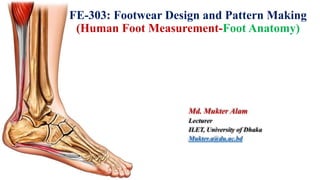
- 1. FE-303: Footwear Design and Pattern Making (Human Foot Measurement-Foot Anatomy) Md. Mukter Alam Lecturer ILET, University of Dhaka Mukter.a@du.ac.bd
- 2. The anatomy of the human foot 2
- 3. The surface anatomy of the human foot Medial malleolus First metatarsal Calcaneus Tubercle of the Tendon of navicular tibialis posterior Tarsal sinus Cuboid Lateral malleolus Tubercle of thefifth metatarsal Calcaneus Tendons of peroneus longusand brevis Tendon of tibialis anterior Tendon of extensor hallucislongus Tendons ofextensor digitorum longus (a ) (b ) Archilles tendon Medial malleolus Lateral malleolus Foot dorsum Posterior aspect Medial aspect Lateral aspect 3
- 4. Arches of the human foot 4
- 5. 5 Anthropometric measurements such as lengths, widths, heights, and girths are directly acquired by- Foot anthropometric measurements Figure: The Brannock device for measuring the foot • Ruler • Caliper • Cloth tape Other special devices: • Brannock device • Indirectly measured from footprints • Foot laser scans (generally in the form of 3D points cloud)
- 6. 6 There are eighteen basic foot measurements obtained from three methods: (i) Traditional manual measures (MM) using simple devices such as rulers, calipers, and cloth tapes (ii) Commercial software-generated measures (CP) from foot laser scans (iii) Simulated measures (SM) obtained from their coded algorithms. Foot anthropometric measurements
- 7. 7 5 lengths 1. Foot length: The distance along the Brannock axis (X- direction) from pternion to the tip of the longest toe. 2. Arch length: The distance along the Brannock axis from pternion to the most medially prominent point on the 1st metatarsal head. 3. Heel to medial malleolus: Length from pternion to the most medially protruding point of the Medial Malleolus, measured along the Brannock axis. 4. Heel to lateral malleolus: Length from pternion to the most laterally protruding point of the Lateral Malleolus measured along the Brannock axis. 5. Heel to 5th toe: The distance along the Brannock axis from pternion to the anterior 5th toe tip. Foot anthropometric measurements
- 8. 8 4 widths 6. Foot width: Maximum horizontal breadth (Y-direction), across the foot perpendicular to the Brannock axis in the region in front of the most laterally prominent point on the 5th metatarsal head. 7. Heel width: Breadth of the heel 40 mm forward of the pternion. 8. Bimalleolar width: Distance between the most medially protruding point on the medial malleolus and the most laterally protruding point on the lateral malleolus measured along a line perpendicular to the Brannock axis. 9. Mid-foot width: Maximum horizontal breadth, across the foot perpendicular to the Brannock axis at 50% of foot length from the pternion. Foot anthropometric measurements
- 9. 9 3 heights 10. Medial malleolus height: Vertical (Z-direction) distance from the floor to the most prominent point on the medial malleolus. 11. Lateral malleolus height: Vertical (Z-direction) distance from the floor to the most prominent point on the lateral malleolus. 12. Height at 50%-foot length: Maximum height of the vertical cross-section at 50% of foot length from the pternion. Foot anthropometric measurements
- 10. 10 6 girths 13. Ball girth: Circumference of foot, measured with a tape touching the medial margin of the head of the 1st metatarsal bone, top of the 1st metatarsal bone and the lateral margin of the head of the 5th metatarsal bone. 14. Instep girth: Smallest girth over middle cuneiform prominence. 15. Long heel girth: Girth from instep point around back heel point. 16. Short heel girth: Minimum girth around back heel point and dorsal foot surface. 17. Ankle girth: Horizontal girth at the foot and leg intersection 18. Waist girth: Circumference at the approximate center of the metatarsal, measured in a vertical plane, perpendicular to the Brannock axis. Foot anthropometric measurements
- 11. 11 Subject factors such as • Ethnicity • Gender • Growth environment • Load-bearing condition • Foot side Variations in foot anthropometric measurements The average length growth of South African schoolboys’ feet is 0.32 inches per year between the ages of approximately 6 and 16, but after 16, the growth rate fell off dramatically. Feet stops growing in length in 75% of girls after 14 years, even if their statures are still increasing. A survey of girls’ feet in New Zealand confirmed earlier observations on foot length, at the same time; it was found that joint girths continue to increase for a further 18 months after cessation of foot length growth. • Age
- 12. 12 Different load-bearing conditions can significantly change the foot shape. Based on nine-foot measurements of thirty Hong Kong Chinese under three different load-bearing conditions, it was found that the foot becomes significantly longer, wider, and reduced in height while rotating to the medial side (everting) with increased loading on the foot. Load-bearing Foot side It has been reported that 15% of the population have differences in foot length of more than 5mm, other considerable foot-side differences in foot width, ball girth, medial malleolus height, and lateral malleolus height are also found. Hence, it is always good practice to check the fit of both shoes before purchasing footwear. When the full bodyweight acts on each foot, the foot stretches by more than 3mm relative to its ‘unloaded’ condition. However, the dimensional changes (in percentage) are relatively small and within 3.0% for all investigated measurements except two (the midfoot height and midfoot width) in the midfoot.
- 13. 13 • In general, the feet of the Scotch are large, flat, and bony, while the feet of the Irish are short and chubby. • The English have broad feet at the instep and joints. • The French have long and high-arched feet. • The German have chubby and arched feet. • Compared with Caucasoid and Australoid populations, Mongoloid populations, including Japanese, have wider feet for a given foot length. • East Asian populations have a smaller foot length for height compared to southeast Asian and Africans. Ethnicity Based on an unsubstantiated observation, feet can be grouped into three main groups: : The Negroid foot is broad at the forefoot and narrow at the heel. : The Oriental foot is short and broad in both the forefoot and the heel. : The Caucasoid foot is broad with straight toes. 1. Negroid feet 2. Oriental feet 3. Caucasoid feet
- 14. 14 Gender Even though the average female foot is shorter and more slender compared with the male foot (its length is approximately 91% of that of the male and its volume is around 81% of that of the male), the female foot is not just a scaled-down version of the male foot. Different shape characteristics are found; for example, it has been reported that the female foot has larger calf height, plantar arch height, ankle circumference, and calf circumference than the male foot after being normalized by foot length. Additionally, using the radiography technique, it is found that the African female group has a significantly higher calcaneal angle than that of the male group. All this evidence indicates that, if possible, the shoe last should be designed separately for each gender group. Growth environment While the extreme deforming and growth-inhibiting effects of ancient practices such as Chinese foot binding, other environmental factors such as nutritional status, heat, and moisture can affect the size and shape of the foot. Even within the same day, due to different thermal conditions at the end of the day when compared to early morning, foot volume can differ by 5%.
- 15. 15 15 Anthropometry-based foot type classification Directly based on some dimensions of the foot (simple ratio (R) of foot width to foot length) the foot shapes can be classified into three types- Based on the cluster analysis technique feet can be classified into three types- • Slender type • Standard type • Broad type • Voluminous feet Very small ball width Very small heel width Short medial ball length Small ball angle • Flat and pointed feet • Slender feet Low instep length Average ball width Narrow heel Long medial ball length Large ball angle
- 16. 16 16 The length, shape and alignment of your toes largely dictates what type of feet you have. Research suggests there are at least 10 different foot types. 2. Greek 4. Celtic 5. Germanic 7. Square 8. Stretched 9. Simian 10. Wide-set toes shape Anthropometry-based foot type classification 1. Egyptian 3. Roman 6. Peasant
- 17. 17 17 Egyptian feet: This is where the big toe is the longest toe and the lesser toes slant downward at a 45º angle. Egyptian feet are often longer and narrower. A common condition of the Egyptian foot is bunion. With this type of foot, the big toe will deviate towards the other toes. Greek feet: This foot type is characterized by a 2nd toe that is larger than the others and has been known to be defined by Morton’s toe that juts out past the big toe for a more pointed formation. Greek feet are also known as the Flame Foot or Fire Foot. With a Greek foot, the second toe will determine the size of the shoe.
- 18. 18 18 Roman feet: Roman feet can be prone to painful hammer toes. Roman feet are defined by the largest three toes being the same height and the smaller two descending in length and all of lesser toes may curve slightly inward and descending. Roman feet tend to have high arches. Peasant feet: Peasant feet are similar to Roman feet in that the three biggest toes are equally long or short with the lesser 2 toes descending. Peasant feet often belong to people with all-around smaller feet. Structurally, peasant feet are flat, which can lead to bad posture and back pain.
- 19. 19 19 Germanic feet/Warrior Toes: With this type of foot shape the big toe is longer than all the other toes forming a straight line that steps down from the big toe, with the pinky often much smaller. Celtic Feet: This foot type is a combination of Germanic toes, one big toe and all lesser toes of the same length and a pronounced second digit like the Greeks, with descending toe size from the third toe onwards.
- 20. 20 20 Square feet: In this case all five toes are about the same length, giving the foot a boxier appearance. Sometimes, peasant feet and square feet are used interchangeably, but there are subtle differences. Square feet often have a wide ball and narrow heel, which can lead to painful inflammation of the balls known as metatarsalgia. Stretched feet: This is when an individual who has stretched toes has a foot shape in which the big toe stays far away from the rest of the toes, with a considerable gap in between all lesser toes. If the toes are noticeably spaced out or fan-shaped, then this is stretched or splayed feet. Widely spaced metatarsal bones are the main factor in stretched feet.
- 21. 21 21 Simian feet – This is a foot type where the big toes look as if they are veering toward your little toes. Simian feet can also resemble an ape’s digits or foot type. Bunions are an enemy of simian feet and are particularly problematic for individuals who wear narrow or pointy shoes. Wide-set toes shape: The wide-set toe pattern which is also referred to as the traveler’s foot has in between the toes a lot of gap. The toes are stretched quite far apart. Individuals with this toe shape are naturally hard workers, who hate waiting around. They are constantly on the move, love travelling and exciting adventures. They are happiest when they go out of their environment.
- 22. 22 22 Motion in the ankle joint Basically, a joint, as you already know, is a connection of two bones with each other through a system of ligaments. The ankle joint is a complex structure consisting of many bones and soft tissues. The ankle joint is divided into: Plantarflexion Ankles mortise Ankles tarsus Normal Dorsiflexion
- 23. 23 23 Posterior ankle joint Ankles tarsus Anterior ankle joint Inversion Eversion
- 24. 24 24 Neutral position Adduction Abduction Anterior ankle joint
- 25. 25 25 Foot shape modeling 4D 2D 3D Two-dimensional modeling
- 26. 26 26 Three-dimensional modeling In general, the key techniques for laser-scanning-based 3D foot shape measurement systems include laser stripe extraction, laser stripe coordinate transformation from a camera image coordinates system to a laser plane coordinates system, laser stripe assembly of cameras, and elimination of image noise and disturbance. Three-dimensional foot surface shapes can be captured by laser-scanning-based measurement systems quickly and accurately.
- 27. 27 27 For example, the YETITM laser foot scanner by Vorum Research Corporation can obtain completed surface data of the human foot within 10 seconds, with an error of less than 1mm. When a YETI laser foot scanner is used, four lasers shine a line of light on the foot surface; eight cameras then capture images of the reflected laser light as the light advances along the scanned surface. The camera information is then used to create the 3D coordinates of 360 points in each section, spaced at a certain interval (normally 1.0mm), along the length of the foot. Considering the relatively high cost of laser-scanning-based 3D foot shape measurement systems, a parameterized approach to generate an individual 3D foot shape from a standard/average foot, based on four-foot dimensions (foot length, foot width, foot height, and foot curvature). The experimental results showed that this approach can achieve a mean absolute modeling error of less than 2.5 mm for both left and right feet. Three-dimensional modeling
- 28. 28 28 Four-dimensional modeling A 3D foot FE model is capable of generating 3D foot shape changes over time to create a four-dimensional foot model. Due to foot FE modeling having the capability of modeling complex foot structures and simulating complicated boundary and loading conditions between the foot and footwear, it is efficient for evaluating the effects of footwear design modifications without the prerequisite of fabricated footwear and replicating experimental trials. To build the FE model, the geometry of a human foot, including both the surface shape and the inner structure, should be obtained first through magnetic resonance imaging (MRI) or computed tomography (CT) scans. The 3D model of bones can be reconstructed afterward by combining a series of computed 2D outlines of MRI/CT images. Then, the 3D model of bones and the whole foot shape are converted into small elements, usually 3D-tetrahedral or hexahedral elements. MPa +2.30e-01 +1.50e-01 +1.38e-01 +1.27e-01 +1.15e-01 +1.04e-01 +9.23e-02 +8.07e-02 +6.92e-02 +5.76e-02 +4.61e-02 +3.45e-02 +2.30e-02 +1.14e-02 +0.00e+00 Peak 0.23 MPa Adding the biomechanical properties of each tissue and bone, such as Young’s modulus and Poisson’s ratio, a foot FE model can be built. Once the foot FE model has been successfully built and its loading and boundary conditions have been clearly specified, it can be used to conduct many biomechanical investigations.
- 29. 29 29 Limitations of Foot FE models Firstly, they lack external validity since most FE models are built based on MRI or CT scans of one participant’s foot. Additionally, the parameters used to characterize the biomechanical properties of each tissue and bone of the foot are usually estimated from the existing literature and these may not be similar to the foot being investigated. Secondly, foot FE models generally involve certain simplifications and assumptions such as simplified foot shape/structure, assumptions of linear material properties, no friction and slip boundary conditions, etc. Thirdly, even though pressure measurements have been widely used to validate the developed foot FE models, slight adjustments in the biomechanical parameters or boundary conditions of the FE model may generate similar pressure patterns.
- 30. 30 30 Bespoke Footwear Measuring Time: Evening
- 33. 33 33