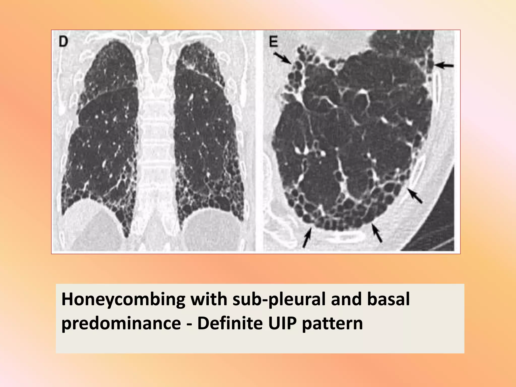
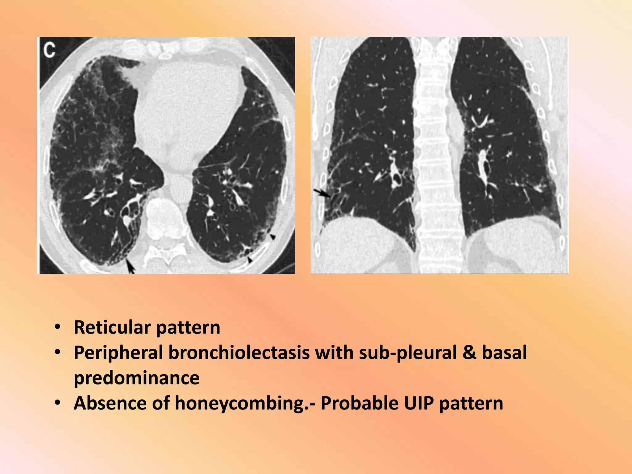
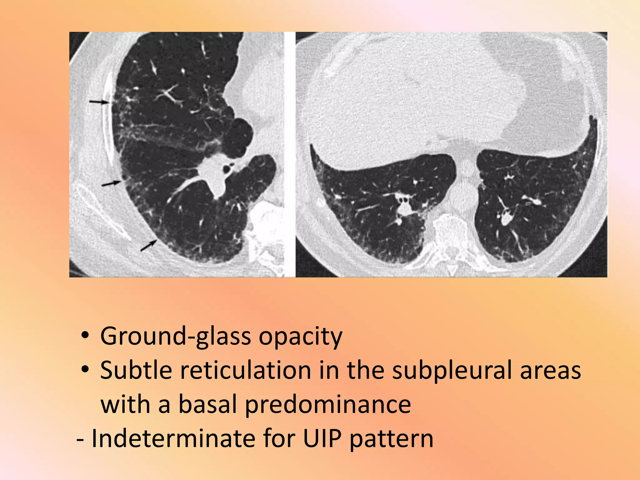
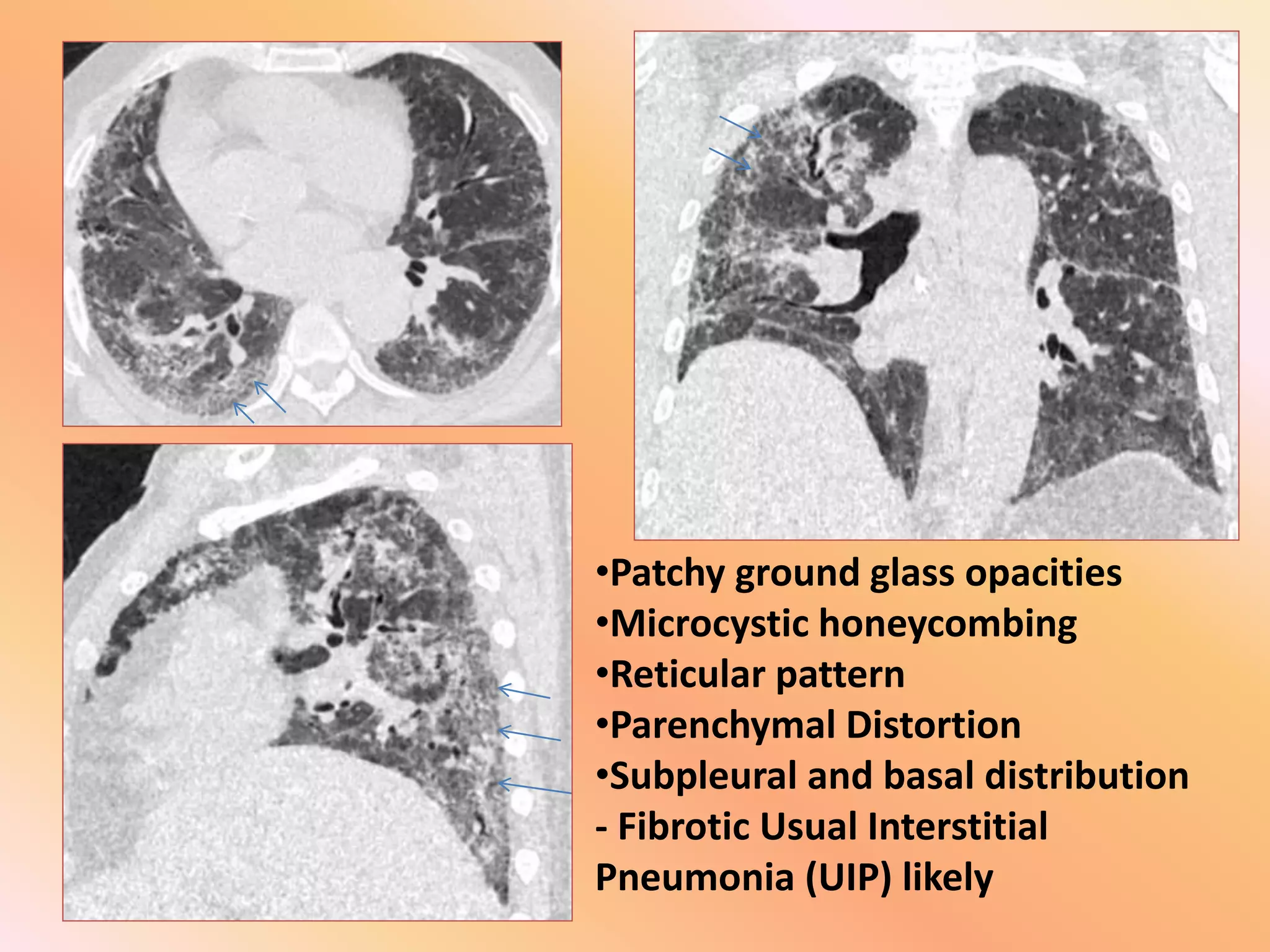
HRCT Basics of
Usual Interstitial Pneumonia
E-Poster presentation by -
Dr. Vaibhavkumar Vekaria (Resident
doctor)
Under guidance of –
Dr. Nalin G. Patel
Professor & H.O.D. of Radiology dept.
Dr. M.K. Shah Medical college.
(A) UIP pattern
(Given in 2018 update of ATS/ERS classification)
• 1. Honeycombing with or without traction
bronchiectasis (hallmark).
• 2. Reticular abnormality
• 3. The typical sub-pleural and basal
predominance
Honeycombing with sub-pleural and basal
predominance - Definite UIP pattern
Probable UIP pattern
• Absent honeycombing
• Reticular abnormalities
• Peripheral tractional bronchiectasis or
bronchiolectasis
• Typical distribution: Sub-pleural and basal
predominance
• Reticular pattern
• Peripheral bronchiolectasis with sub-pleural & basal
predominance
• Absence of honeycombing.- Probable UIP pattern
Indeterminate for UIP pattern
• Subtle reticular abnormalities with/without
ground glass opacification superimposed on a
fine reticular pattern.
• Non specific distribution
• Does not explicitly suggest an "alternative
diagnosis”.
• Ground-glass opacity
• Subtle reticulation in the subpleural areas
with a basal predominance
- Indeterminate for UIP pattern
Alternative diagnosis for UIP
• Upper or mid lung predominance
• Peri-broncho-vascular or Peri-lymphatic
distribution
• Marked mosaic attenuation
• Predominant ground-glass opacification
• Profuse micro-nodules
• Pleural Effusion
• Pleural Thickening or plaques
Case:
64Y /Male patient
• C/O shortness of breath
• On ambulatory O2 therapy
• H /O chronic bidi smoking
• On lab. Reports – elevated D-DIMER
•Patchy ground glass opacities
•Microcystic honeycombing
•Reticular pattern
•Parenchymal Distortion
•Subpleural and basal distribution
- Fibrotic Usual Interstitial
Pneumonia (UIP) likely
THANK YOU

HRCT basics of UIP POSTER.pptx

  • 1.
    HRCT Basics of UsualInterstitial Pneumonia E-Poster presentation by - Dr. Vaibhavkumar Vekaria (Resident doctor) Under guidance of – Dr. Nalin G. Patel Professor & H.O.D. of Radiology dept. Dr. M.K. Shah Medical college.
  • 2.
    (A) UIP pattern (Givenin 2018 update of ATS/ERS classification) • 1. Honeycombing with or without traction bronchiectasis (hallmark). • 2. Reticular abnormality • 3. The typical sub-pleural and basal predominance
  • 3.
    Honeycombing with sub-pleuraland basal predominance - Definite UIP pattern
  • 4.
    Probable UIP pattern •Absent honeycombing • Reticular abnormalities • Peripheral tractional bronchiectasis or bronchiolectasis • Typical distribution: Sub-pleural and basal predominance
  • 5.
    • Reticular pattern •Peripheral bronchiolectasis with sub-pleural & basal predominance • Absence of honeycombing.- Probable UIP pattern
  • 6.
    Indeterminate for UIPpattern • Subtle reticular abnormalities with/without ground glass opacification superimposed on a fine reticular pattern. • Non specific distribution • Does not explicitly suggest an "alternative diagnosis”.
  • 7.
    • Ground-glass opacity •Subtle reticulation in the subpleural areas with a basal predominance - Indeterminate for UIP pattern
  • 8.
    Alternative diagnosis forUIP • Upper or mid lung predominance • Peri-broncho-vascular or Peri-lymphatic distribution • Marked mosaic attenuation • Predominant ground-glass opacification • Profuse micro-nodules • Pleural Effusion • Pleural Thickening or plaques
  • 9.
    Case: 64Y /Male patient •C/O shortness of breath • On ambulatory O2 therapy • H /O chronic bidi smoking • On lab. Reports – elevated D-DIMER
  • 10.
    •Patchy ground glassopacities •Microcystic honeycombing •Reticular pattern •Parenchymal Distortion •Subpleural and basal distribution - Fibrotic Usual Interstitial Pneumonia (UIP) likely
  • 11.