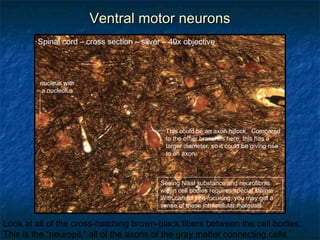
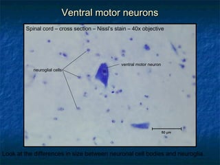
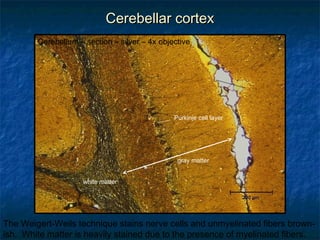
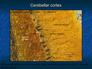
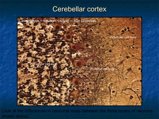
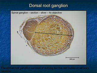
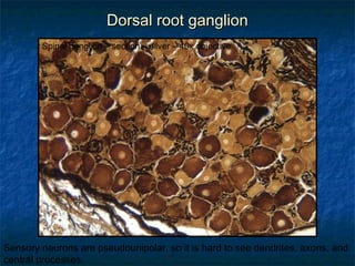
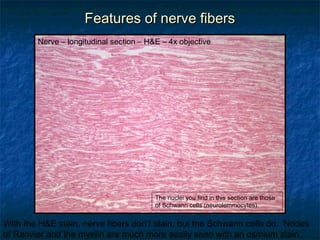
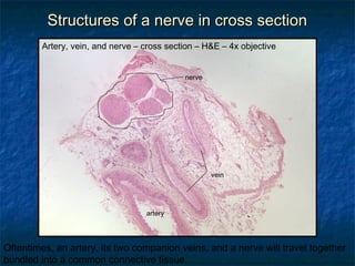
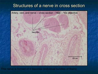
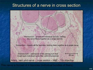
HISTOLOGY REVIEW 
Nervous Tissue 
Dr. Tim Ballard 
Department of Biology and Marine Biology
OOrriieennttaattiioonn ttoo tthhee ssppiinnaall ccoorrdd 
Spinal cord – cross section – silver – 4x objective 
dorsal horns of gray matter 
dorsal 
ventral 
gray 
commissure 
central 
canal 
ventral horns of gray matter 
Silver stains nerve cells and unmyelinated fibers brownish-black. The white 
matter is lightly stained because there is so much myelin around the fibers.
VVeennttrraall mmoottoorr nneeuurroonnss 
motor 
neurons 
Spinal cord – cross section – silver – 10x objective 
In the ventral horn there are very large motor neurons. The brown fibers you 
see leaving the ventral horn are axons from these neurons.
VVeennttrraall mmoottoorr nneeuurroonnss 
Spinal cord – cross section – silver – 40x objective 
These much smaller cells are 
likely to be neuroglial cells. 
neuronal cell body 
neuronal cell body 
nucleus with 
a nucleolus 
dendrites 
Think in three dimensions. Where would the dendritic zone or the axon be 
related to this two-dimensional section?
VVeennttrraall mmoottoorr nneeuurroonnss 
Spinal cord – cross section – silver – 40x objective 
This could be an axon hillock. Compared 
to the other branches here, this has a 
larger diameter, so it could be giving rise 
to an axon. 
nucleus with 
a nucleolus 
Seeing Nissl substance and neurofibrils 
within cell bodies requires special stains. 
With careful fine-focusing, you may get a 
sense of these intracellular materials. 
Look at all of the cross-hatching brown-black fibers between the cell bodies. 
This is the “neuropil,” all of the axons of the gray matter connecting cells.
VVeennttrraall mmoottoorr nneeuurroonnss 
Spinal cord – cross section – Nissl’s stain – 40x objective 
neuroglial cells 
ventral motor neuron 
Look at the differences in size between neuronal cell bodies and neuroglia.
CCeerreebbeellllaarr ccoorrtteexx 
Cerebellum – section – silver – 4x objective 
white matter 
Purkinje cell layer 
gray matter 
The Weigert-Weils technique stains nerve cells and unmyelinated fibers brown-ish. 
White matter is heavily stained due to the presence of myelinated fibers.
CCeerreebbeellllaarr ccoorrtteexx 
Cerebellum – section – silver – 10x objective 
granular 
cell layer 
molecular cell layer 
Purkinje cell layer 
Look at the differences in neuronal sizes between the three layers of neurons 
shown above.
CCeerreebbeellllaarr ccoorrtteexx 
Cerebellum – section – silver – 40x objective 
granular cell layer 
molecular cell layer 
Purkinje cell layer 
Look at the differences in neuronal sizes between the three layers of neurons 
shown above.
DDoorrssaall rroooott ggaanngglliioonn 
Spinal ganglion – section – silver – 4x objective 
The dorsal root ganglion consists of nothing but the cell bodies of sensory 
neurons.
DDoorrssaall rroooott ggaanngglliioonn 
Spinal ganglion – section – silver – 40x objective 
Sensory neurons are pseudounipolar, so it is hard to see dendrites, axons, and 
central processes.
FFeeaattuurreess ooff nneerrvvee ffiibbeerrss 
Nerve – longitudinal section – H&E – 4x objective 
The nuclei you find in this section are those 
of Schwann cells (neurolemmocytes). 
With the H&E stain, nerve fibers don’t stain, but the Schwann cells do. Nodes 
of Ranvier and the myelin are much more easily seen with an osmium stain.
FFeeaattuurreess ooff nneerrvvee ffiibbeerrss 
Nerve – teased fibers – osmium – 20x objective 
node of Ranvier 
Osmium stains the lipid of myelin gray-black. The segments you see are 
individual pieces of myelin. Between each would reside nodes of Ranvier.
SSttrruuccttuurreess ooff aa nneerrvvee iinn ccrroossss sseeccttiioonn 
Artery, vein, and nerve – cross section – H&E – 4x objective 
nerve 
artery 
vein 
Oftentimes, an artery, its two companion veins, and a nerve will travel together 
bundled into a common connective tissue.
SSttrruuccttuurreess ooff aa nneerrvvee iinn ccrroossss sseeccttiioonn 
Artery, vein, and nerve – cross section – H&E – 10x objective 
fascicles 
This nerve is formed of 4 fascicles (individual bundles of fibers)
SSttrruuccttuurreess ooff aa nneerrvvee iinn ccrroossss sseeccttiioonn 
Perineurium – invests an individual fascicle, holding 
the nerve fibers together as a single fascicle 
Epineurium – invests all the fascicles, holding them together as a single nerve 
Endoneurium – extensions of the perineurium that 
invest each individual nerve fiber within the fascicle 
Artery, vein, and nerve – cross section – H&E – 10x objective 
This nerve is formed of 4 fascicles (individual bundles of fibers) 
end

Histology: Nerves

  • 1.
    HISTOLOGY REVIEW NervousTissue Dr. Tim Ballard Department of Biology and Marine Biology
  • 2.
    OOrriieennttaattiioonn ttoo tthheessppiinnaall ccoorrdd Spinal cord – cross section – silver – 4x objective dorsal horns of gray matter dorsal ventral gray commissure central canal ventral horns of gray matter Silver stains nerve cells and unmyelinated fibers brownish-black. The white matter is lightly stained because there is so much myelin around the fibers.
  • 3.
    VVeennttrraall mmoottoorr nneeuurroonnss motor neurons Spinal cord – cross section – silver – 10x objective In the ventral horn there are very large motor neurons. The brown fibers you see leaving the ventral horn are axons from these neurons.
  • 4.
    VVeennttrraall mmoottoorr nneeuurroonnss Spinal cord – cross section – silver – 40x objective These much smaller cells are likely to be neuroglial cells. neuronal cell body neuronal cell body nucleus with a nucleolus dendrites Think in three dimensions. Where would the dendritic zone or the axon be related to this two-dimensional section?
  • 5.
    VVeennttrraall mmoottoorr nneeuurroonnss Spinal cord – cross section – silver – 40x objective This could be an axon hillock. Compared to the other branches here, this has a larger diameter, so it could be giving rise to an axon. nucleus with a nucleolus Seeing Nissl substance and neurofibrils within cell bodies requires special stains. With careful fine-focusing, you may get a sense of these intracellular materials. Look at all of the cross-hatching brown-black fibers between the cell bodies. This is the “neuropil,” all of the axons of the gray matter connecting cells.
  • 6.
    VVeennttrraall mmoottoorr nneeuurroonnss Spinal cord – cross section – Nissl’s stain – 40x objective neuroglial cells ventral motor neuron Look at the differences in size between neuronal cell bodies and neuroglia.
  • 7.
    CCeerreebbeellllaarr ccoorrtteexx Cerebellum– section – silver – 4x objective white matter Purkinje cell layer gray matter The Weigert-Weils technique stains nerve cells and unmyelinated fibers brown-ish. White matter is heavily stained due to the presence of myelinated fibers.
  • 8.
    CCeerreebbeellllaarr ccoorrtteexx Cerebellum– section – silver – 10x objective granular cell layer molecular cell layer Purkinje cell layer Look at the differences in neuronal sizes between the three layers of neurons shown above.
  • 9.
    CCeerreebbeellllaarr ccoorrtteexx Cerebellum– section – silver – 40x objective granular cell layer molecular cell layer Purkinje cell layer Look at the differences in neuronal sizes between the three layers of neurons shown above.
  • 10.
    DDoorrssaall rroooott ggaanngglliioonn Spinal ganglion – section – silver – 4x objective The dorsal root ganglion consists of nothing but the cell bodies of sensory neurons.
  • 11.
    DDoorrssaall rroooott ggaanngglliioonn Spinal ganglion – section – silver – 40x objective Sensory neurons are pseudounipolar, so it is hard to see dendrites, axons, and central processes.
  • 12.
    FFeeaattuurreess ooff nneerrvveeffiibbeerrss Nerve – longitudinal section – H&E – 4x objective The nuclei you find in this section are those of Schwann cells (neurolemmocytes). With the H&E stain, nerve fibers don’t stain, but the Schwann cells do. Nodes of Ranvier and the myelin are much more easily seen with an osmium stain.
  • 13.
    FFeeaattuurreess ooff nneerrvveeffiibbeerrss Nerve – teased fibers – osmium – 20x objective node of Ranvier Osmium stains the lipid of myelin gray-black. The segments you see are individual pieces of myelin. Between each would reside nodes of Ranvier.
  • 14.
    SSttrruuccttuurreess ooff aanneerrvvee iinn ccrroossss sseeccttiioonn Artery, vein, and nerve – cross section – H&E – 4x objective nerve artery vein Oftentimes, an artery, its two companion veins, and a nerve will travel together bundled into a common connective tissue.
  • 15.
    SSttrruuccttuurreess ooff aanneerrvvee iinn ccrroossss sseeccttiioonn Artery, vein, and nerve – cross section – H&E – 10x objective fascicles This nerve is formed of 4 fascicles (individual bundles of fibers)
  • 16.
    SSttrruuccttuurreess ooff aanneerrvvee iinn ccrroossss sseeccttiioonn Perineurium – invests an individual fascicle, holding the nerve fibers together as a single fascicle Epineurium – invests all the fascicles, holding them together as a single nerve Endoneurium – extensions of the perineurium that invest each individual nerve fiber within the fascicle Artery, vein, and nerve – cross section – H&E – 10x objective This nerve is formed of 4 fascicles (individual bundles of fibers) end