This document provides information about different types of biopsies. It discusses:
- Biopsy is the removal of tissue from the body for examination to make a diagnosis. The main types are excisional, incisional, and aspiration biopsies.
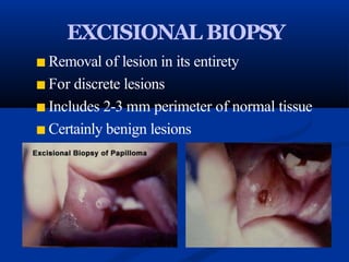
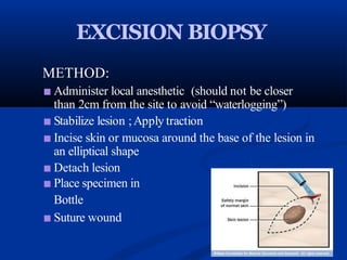
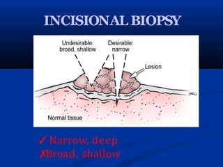
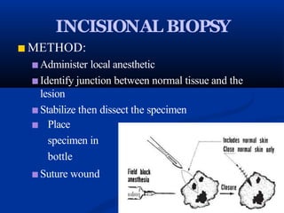
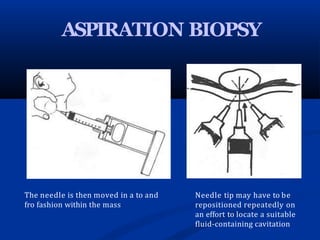
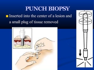
- Excisional biopsy removes the entire lesion. Incisional biopsy removes only a portion. Aspiration biopsy uses a needle to remove fluid.
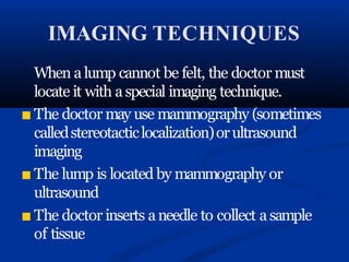
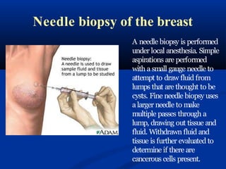
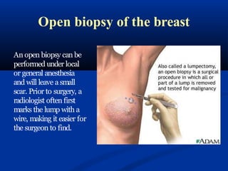
- Breast biopsies include needle biopsies like fine needle and core needle, and open biopsies like incisional and excisional. Imaging helps locate non-palpable lumps.
- New techniques like ABBI and Mammotone aim to obtain


![BIOPSY IN SURGERY 1 [Auto-savedd] 3.pptx](https://cdn.slidesharecdn.com/ss_thumbnails/biopsyinsurgery1auto-saved3-251018145913-707215d9-thumbnail.jpg?width=640&height=640&fit=bounds)


