Science – Grade8
Alternative Delivery Mode
Quarter 4 – Module 2: Cellular Reproduction
First Edition, 2020
Republic Act 8293, section 176 states that: No copyright shall subsist in any work of
the Government of the Philippines. However, prior approval of the government agency or office
wherein the work is created shall be necessary for exploitation of such work for profit. Such
agency or office may, among other things, impose as a condition the payment of royalties.
Borrowed materials (i.e., songs, stories, poems, pictures, photos, brand names,
trademarks, etc.) included in this module are owned by their respective copyright holders.
Every effort has been exerted to locate and seek permission to use these materials from their
respective copyright owners. The publisher and authors do not represent nor claim ownership
over them.
Published by the Department of Education
Secretary: Leonor Magtolis Briones
Undersecretary: Diosdado M. San Antonio
Printed in the Philippines by ________________________
Department of Education – CARAGA
Office Address: Teacher Development Center
J. P. Rosales Avenue, Butuan City, Philippines 8600
Telefax: (085) 342-8207/ (085) 342-5969
E-mail Address: caraga@deped.gov.ph
Development Team of the Module
Writer: Marilou C. Salazar
Editor: Daisy Ruby S. Sinday
Reviewers: Bernabe L. Linog, Corazon P. Roa, Juvy B. Luna, Arlene L. Abala,
Kathyleen S. Torculas and Nonita C. Patalinghug
Illustrators: Marilou C. Salazar and Rosa Mia L. Pontillo
Layout Artist: Christopher David G. Oliva
Layout Evaluators: Celeste Faith R. Almanon and Jay S. Ayap
Management Team: Francis Cesar B. Bringas
Isidro M. Biol, Jr.
Maripaz F. Magno
Josephine Chonie M. Obseñares
Gregoria T. Su
Marvilyn C. Francia
Jay S. Ayap
Nonita C. Patalinghug
Introductory Message
This Self-LearningModule (SLM) is prepared so that you, our dear learners,
can continue your studies and learn while at home. Activities, questions,
directions, exercises, and discussions are carefully stated for you to
understand each lesson.
Each SLM is composed of different parts. Each part shall guide you step-by-
step as you discover and understand the lesson prepared for you.
Pre-tests are provided to measure your prior knowledge on lessons in each
SLM. This will tell you if you need to proceed on completing this module or if
you need to ask your facilitator or your teacher’s assistance for better
understanding of the lesson. At the end of each module, you need to answer
the post-test to self-check your learning. Answer keys are provided for each
activity and test. We trust that you will be honest in using these.
In addition to the material in the main text, Notes to the Teacher are also
provided to our facilitators and parents for strategies and reminders on how
they can best help you on your home-based learning.
Please use this module with care. Do not put unnecessary marks on any part
of this SLM. Use a separate sheet of paper in answering the exercises and
tests. And read the instructions carefully before performing each task.
If you have any questions in using this SLM or any difficulty in answering the
tasks in this module, do not hesitate to consult your teacher or facilitator.
Thank you.
5.
1 CO_Q4_Science 8_Module 2
What I Need to Know
This module was designed and written with you in mind. It is here to help you
master cellular reproduction. The scope of this module permits it to be used in many
different learning situations. The language used recognizes the diverse vocabulary
level of students. The lessons are arranged to follow the standard sequence of the
course. But the order in which you read them can be changed to correspond with
the learner’s material you are now using.
This module contains:
Lesson 1 – Cellular Reproduction
After going through this module, you are expected to:
1. Understand cell reproduction at the molecular level, giving
significance to the roles of DNA and chromosomes;
2. Describe the cell cycle;
3. Explain mitosis and meiosis; and
4. Compare mitosis and meiosis, and their role in the cell division cycle.
(MELC Week 2 S8LT-IVd-16)
6.
2 CO_Q4_Science 8_Module 2
What I Know
Directions: Choose the letter of the correct answer. Write your answers on a
separate sheet of paper.
1. Which statement about mitosis is INCORRECT?
A. The first phase is prophase.
B. It forms four daughter cells.
C. It makes diploid or haploid nuclei.
D. Separation of sister chromatids occurs.
2. What is the form of reproduction whose benefit is variability of the offspring?
A. asexual
B. binary fission
C. mitosis
D. sexual
3. The diploid (2N) chromosome number in an organism is 42. What is the
normal chromosome number of its sex cells?
A. 21
B. 42
C. 63
D. 84
4. These activities of the cell occur in both mitosis and meiosis EXCEPT:
A. cytokinesis
B. DNA replication
C. karyokinesis
D. synapsis
For items 5 -10, answer each statement using the given choices.
Write the letter only.
A. Meiosis I B. Meiosis II
5. Occurrence of synapsis between tetrads.
6. Separation of homologous chromosomes.
7. Results in four sperm cells in human males.
8. Daughter cells have chromosomes with two sister chromatids.
9. Daughter nuclei have chromosomes with single chromatids.
10. Occurrence of crossing-over of homologous chromosome.
7.
3 CO_Q4_Science 8_Module 2
For items 11-15, match the cell phase to its description. Write the letter only.
A. DNA replication occurs
B. last phase of nuclear division
C. condensed chromosomes become visible
D.chromatids separate at the centromere
E. chromosomes line up at the equatorial plane
11.Metaphase
12.Telophase
13.Anaphase
14.Prophase
15.Interphase
Lesson
1 Cellular Reproduction
What’s In
Directions: Recall the previous lesson about digestive system. Rearrange the parts
of digestive system in correct sequence during digestion process. Write
the letter of the correct answer on a separate sheet of paper.
Have you ever watched a tadpole turn into an adult frog? If so, you are perhaps
familiar with the idea of a life cycle. Frogs go through some interesting life cycle
transitions: from egg to larva (tadpole), then finally, to an adult frog. Other
organisms, such as humans, plants, and bacteria, also have life cycles, a series of
Digestion Process
A. anus 1. ________
B. esophagus 2. ________
C. large intestine 3. ________
D. mouth 4. ________
E. rectum 5. ________
F. small intestine 6. ________
G. stomach 7. ________
8.
4 CO_Q4_Science 8_Module 2
Note to the Teacher
Provide extra copies of the puzzle for students’ use.
developmental steps that an individual goes through from birth until the time it
reproduces.
The same thing happens with the living cells. The cell cycle can be compared
to as the life cycle of a cell, a series of growth and developmental steps a cell
undergoes between its “birth” and reproduction.
Every living thing undergoes reproduction. The nutrients taken by an
individual will provide energy for metabolic processes, for growth and development
as well as for reproduction.
In this module you will learn the importance of cell division for growth, repair,
and reproduction of eukaryotic organisms. During mitosis, the resulting two new
daughter cells have the same type and number of genes as the original parent cell,
thereby preserving and maintaining the stability of genetic material of a particular
population. But in more complex organisms, meiotic division produces gametes that
possess half of the genetic information. During fertilization, these gametes unite
allowing genes from each parent to combine which results to differences in the DNA
composition or genotypes resulting to genetic variability of offspring.
What’s New?
Activity 1 – Puzzle Solving
Directions: Locate ten (10) words that are associated to cellular reproduction in the
puzzle. They can be read horizontally, vertically, or diagonally. Write
your answers on a separate sheet of paper.
E L C Y C E F I L N
A C O W B U C I E O
T G A M E T E S F I
R E F I A G H F I S
E L O T M U S G N I
P P A O R P I R E V
R S I S R T S O O I
O U V I A W O W X D
D Y N S A Z I T E L
U G E N E S E H B L
C A C E D O M U F E
E E L C Y C L L E C
9.
5 CO_Q4_Science 8_Module 2
What is It
The Chromosome
All living things contain a self-replicating genetic material that directs the
activities and functions of the cells. Deoxyribonucleic acid or DNA is the genetic
material located inside a chromosome in the nucleus of the cell. The DNA from the
parents is transmitted to the offspring to ensure the continuity of life. The DNA is a
helical structure consisting of two strands as shown in Figure 1. Figure 1 also shows
the organization or packaging of DNA molecules by proteins or histones to form
different levels of chromosome packaging. This is necessary so that the long and
numerous DNA molecules can be organized and be accommodated inside the
nucleus of a eukaryotic cell. The DNA helix illustration in Figure 1 shows a structure
called nucleosomes which is composed of globular structures known as histones
where the DNA strands are attached, and coiled looking like beads attached on a
string in a form of chromatin measuring up to 11 nm. The next level of organization
is a series of chromatin molecules forming a 30-nanometer chromatin fiber of packed
coiled nucleosomes called solenoid. This solenoid level of packaging becomes
supercoiled forming loops that are visible and are usually called chromatin loops
which further leads to condensation of the chromosomes up to 700 nm. When the
DNA molecules is replicated and undergoes also packaging and coiling, it would form
the entire mitotic chromosome or metaphase chromosome which measures up to
1,400 nm which is illustrated at the bottom of the diagram in Figure 1.
Figure 1. The Organization/Packaging of the DNA into Chromosomes
Illustrated by: Rosa Mia L. Pontillo
10.
6 CO_Q4_Science 8_Module 2
Figure 2. Parts of the Chromosome
Parts of the Chromosome
1. Chromatids – two identical halves of a replicated chromosome after the
Synthesis phase or the S phase of the cell cycle.
2. Centromere – the attachment points of the two chromatids of a
chromosome. It is also described as the constriction point which divides
the chromosome into two sections, or “arms.”
3. Short arm – or p arm - upper arms of the chromosome which is usually
shorter.
4. Q arm - lower arms of the chromosome which is usually longer.
The number of chromosomes in a cell is a characteristic of the species to which
it belongs. For example, fruit flies have 8 chromosomes while sunflowers have 34.
Table 1 summarizes the chromosome numbers of some organisms.
Table 1. Chromosome Number of Selected Organisms
Organism Chromosome number
Drosophila melanogaster
(fruit fly)
8
Canis familiaris
(dog)
78
Homo sapiens
(man)
46
Oryza sativa
(rice)
24
Zea mays
(corn)
20
Illustrated by: Rosa Mia L. Pontillo
11.
7 CO_Q4_Science 8_Module 2
The Cell Cycle
The chromosomes of a cell change their form as they undergo cell transitions
from one stage to another in a typical cell cycle as shown in Figure 3. The cell cycle
may be divided into two stages: the interphase where the chromosomes are long,
and extended, and the cell division or mitotic phase where the chromosomes
become condensed or thickened.
Interphase is the interval between two cell divisions. During this stage, the
cell is not dividing; it merely grows. The chromosome doubles or replicates itself
because the DNA molecule contained in the chromosome produces a precise copy of
itself.
Figure 3. The Cell Cycle
Interphase is the interval
between two cell divisions. During this
stage, the cell is not dividing; it obtains
nutrients and metabolizes, grows,
replicates its DNA in preparation for
mitosis.
The interphase is divided into
three sub-stages, namely:
Figure 4. The Cell in the Interphase Period
1. First gap period or G1 where
cell grows initially
synthesis of protein and ribonucleic acid or RNA occurs
mitochondria increase in number
2. Synthesis stage or S phase where
DNA are synthesized thus replicating the chromosomes in
preparation for the next cell division.
3. Second gap period or G2 where
cell grows rapidly
cell prepares for the actual cell division
Illustrated by: Rosa Mia L. Pontillo
Illustrated by: Rosa Mia L. Pontillo
12.
8 CO_Q4_Science 8_Module 2
Cell Division
Cell division phase occurs every after interphase. In eukaryotic cells, these
types of cell division occur: mitosis and meiosis.
1. Mitosis
Each time a child goes to the doctor, a nurse measures his height and mass.
A child’s height and mass increases because the number of cells in his body increases
as he develops. Our body cells increase its number through the process known as
mitosis.
Mitosis is a cellular process wherein two nuclei and two cells are produced
due to the division of the original nucleus, each of which contains the same
chromosome number as the parent cell. Mitosis is divided into four stages namely:
prophase, metaphase, anaphase, and telophase. Figure 5 shows the different stages
of mitosis.
Figure 5. The Stages of Mitosis
Prophase Stage
The repeated coiling of chromosomes occurs resulted to its thicker and
shorter structure. These are made up of two sister chromatids that are
identical to each because of the replication of DNA during the S phase.
The two chromatids are still attached at the centromere.
The nuclear membrane breaks down.
Metaphase Stage
Chromosomes align at the equatorial plane.
Each spindle fiber from both centrosomes connects to each chromosome
through its kinetochore.
Anaphase Stage
Spindle fibers begin to contract and become shorter. Continued
contraction causes the separation of the genetically identical sister
chromatids.
Centromeres divide.
The single chromatids move towards the opposite poles.
Illustrated by: Rosa Mia L. Pontillo
13.
9 CO_Q4_Science 8_Module 2
Telophase Stage
The chromosomes are now at the opposing poles of the spindle.
The microtubules disappear.
Two sets of chromosomes are surrounded by new nuclear membranes,
completing the nuclear division process known as karyokinesis.
Cytoplasmic division called cytokinesis occurs concurrently, splitting the
cell into two.
What happens after telophase?
Two new nuclear membranes are formed, and two new nuclei are seen. There
are two new daughter cells that are produced from one dividing parent cell. Thus,
mitosis has come to an end.
Figure 7 shows the formation of a cell plate in plant cells. During telophase,
membrane-enclosed vesicles from the Golgi complex of the cell move towards the
center where the metaphase plate of the cell is located and become part of the cell
plate.
In telophase, the cell plate continues to grow and gets attached with the cell
membrane. This results to the formation of two daughter cells. Each cell is bounded
with cell membrane. New cell walls form between the two cell membranes at the area
where the cell plate was formed earlier.
In animal cells, two grooves or indentations known as cleavage furrows
form at both ends of the metaphase plate during telophase. These grooves
deepen and lengthen and meet, to separate and form the two new daughter
cells. This event is shown in the illustration below in Figure 6.
Figure 6: Formation of two new daughter cells in animal cells
Plant cells do not form cell furrows. Instead, a new cell wall known as
cell plate forms between the two new nuclei.
Illustrated by: Rosa Mia L. Pontillo
14.
10 CO_Q4_Science 8_Module 2
Figure 7. Cell Plate Formation
2. Meiosis
There are two main types of cells possessed by multicellular eukaryotic
organisms: somatic, or body cells and gametes, or sex cells. Majority of the cells are
called somatic or body cells. These consist of two complete sets of chromosomes,
making them diploid in number (2N).
Multicellular eukaryotic organisms that undergo sexual reproduction use
gametes, or sex cells, to produce offspring. Gametes are haploid cells, union of which
result to creation of a new organism with diploid number of chromosomes in all its
somatic or body cells hence, to create new individuals for the species, two parents
are very necessary to provide the gametes.
How are sex cells, the sperms and eggs, formed? Another form of cell division
known as meiosis produces sex cells. In meiosis, the nucleus will undergo two
divisions: Meiosis I and Meiosis II. The cell divides twice in these processes.
All human somatic cells have forty-six chromosomes. When human cells
reproduce through mitosis, each new cell will also have forty-six chromosomes. But
in reproductive cells or gametes, each of these cells produced has only twenty-three
chromosomes. Thus, meiosis decreases the chromosome number by half.
Why must meiosis take place to produce sperm and egg?
When a sperm unites with an egg, each of them contributes only one half of
the total number of chromosomes (twenty-three chromosomes) to the new zygote
which is diploid in number of chromosomes (forty-six chromosomes). Meiosis creates
cells that are destined to become gametes (or reproductive cells), this reduction in
chromosome number is critical — without it, the union of two gametes during
fertilization would result in offspring with twice the normal number of chromosomes.
Thus, in humans, a new life originates with the normal diploid number (2N)
of forty-six chromosomes. Then the zygote undergoes the process of mitosis,
producing cells with forty-six chromosomes each.
Illustrated by: Rosa Mia L. Pontillo
15.
11 CO_Q4_Science 8_Module 2
Meiosis involves two divisions, Meiosis I and Meiosis II. Each follows similar
stages as mitosis (prophase, metaphase, anaphase, and telophase). Before meiosis,
the reproductive cell is in the interphase stage whereby DNA replicates to produce
chromosomes having two sister chromatids. Then, the cell will undergo second
growth phase called interkinesis. This stage happens between Meiosis I and II,
however, DNA does not replicate in this stage.
Meiosis I.
The first meiotic division, also known as Meiosis I, is a reduction division
phase (diploid - haploid). There are two daughter cells produced after Meiosis 1, each
daughter cell is carrying haploid number of chromosomes. This consists of four
stages, namely, prophase I, metaphase I, anaphase I, and telophase I.
Prophase I Stage
Meiosis starts with this stage and includes the following substages: leptotene,
zygotene, pachytene, diplotene, and diakinesis. Figure 8 shows the different
substages of prophase I.
Figure 8. Substages of Prophase I
Substage 1: Leptotene
Each chromosome is made up of sister chromatids. These are
long threadlike structures which result from the replication of DNA
during the Synthesis or S phase of the cell cycle.
Substage 2: Zygotene
The homologous chromosomes start to pair off through the
process known as synapsis. Pairs of chromosomes that are similar in
size and shape are called homologous chromosomes or tetrads.
Substage 3: Pachytene
The repeated coiling of chromosomes occurs resulting to its
contraction and thickening making the homologous pair of
chromosomes to be very close to each other. At this stage, the process
called crossing over happens. Here, the exchange of segments between
Illustrated by: Rosa Mia L. Pontillo
16.
12 CO_Q4_Science 8_Module 2
the sister chromatids of the homologous chromosomes occurs. The
exchanging process form a cross-linkage called a chiasma. After
crossing over, the sister chromatids of each chromosome may not be
identical with each other based on the genetic material they contain.
Crossing-over is a complicated process that results to genetic
variability.
Figure 9 shows the pairing of two homo-logous chromosomes during synapsis.
The paired chromosome then exchanges DNA segments during crossing over which
results to exchange of genetic material. Chiasma shows the place where the two sister
chromatids of the paired homo-logous chromosomes touched each other.
Figure 9. Crossing Over of Homologous Chromosomes during Meiosis
Substage 4: Diplotene
The two homologous chromosomes forming a tetrad begin to
repel one another and move apart. They are held only by the chiasma.
Substage 5: Diakinesis
This is the last stage of meiosis prophase 1. Diakinesis stage is
characterized by chiasmata terminalization. After diakinesis, the dividing
cell enters metaphase.
At this stage, bivalents or homologous pair chromosomes
distribute them evenly in the nucleus. The nuclear membrane breaks
down and the nucleolus disappears. Chiasma moves towards the end,
which is called terminalization. Chromatids remain attached only at the
terminal chiasmata and enter the metaphase stage.
Metaphase I Stage
Spindle fibers from the centrosomes of each pole connect to bivalents or
tetrads through the kinetochores. Homologous chromosomes line up at the
equatorial plane. There is double alignment of the chromosomes.
Anaphase I Stage
Spindle fibers begin to contract and separate the bivalent or tetrads.
Homologous chromosomes separate and migrate to each pole of the cell.
Telophase I Stage
Chromosomes decondense.
Nuclear membranes reform.
Cytokinesis or the cytoplasmic division occurs and two haploid (N) daughter
cells with chromosomes with two sister chromatids are formed.
Illustrated by: Rosa Mia L. Pontillo
17.
13 CO_Q4_Science 8_Module 2
Meiosis II.
The second meiotic division forms four daughter cells, each carrying haploid
number of chromosomes. This consists of the following stages, namely, prophase II,
metaphase II, Anaphase II, and telophase II.
Prophase II Stage
Chromosomes (chromatids) condense to form metaphase chromosomes.
Nuclear membrane dissolves and nucleolus disappears.
Centrosomes move towards each pole of the cell.
Metaphase II Stage
Spindle fibers attach to chromatids at the kinetochores.
Chromosomes line up at the equatorial plane. (Single alignment of
chromosomes).
Anaphase II Stage
Spindle fibers shortened and separated the sister chromatids.
Chromatids that are now called chromosomes move towards each pole of the
cell.
Telophase II Stage
Single-stranded chromosomes decondense.
Nuclear membrane and nucleolus reforms.
Cytoplasm divides (cytokinesis).
Four haploid (n) daughter cells are formed.
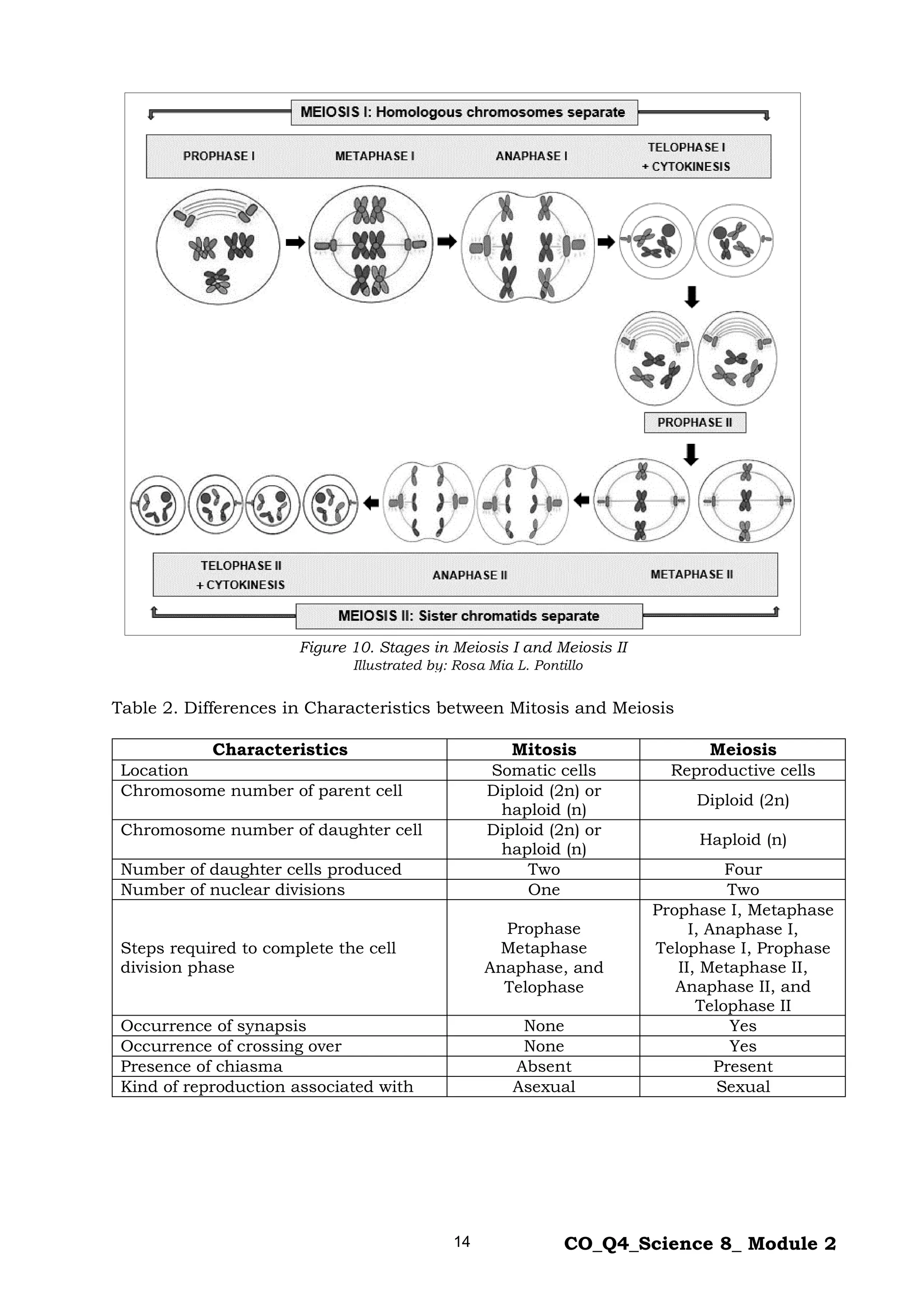
Figure 10 shows the stages in Meiosis I and Meiosis II. In meiosis I, the two
homologous chromosomes separate which results to two haploid (n) daughter cells
with chromosomes with two chromatids each. In meiosis II, four haploid (n) daughter
cells are formed. Each cell is carrying haploid number of chromosomes.
18.
14 CO_Q4_Science 8_Module 2
Figure 10. Stages in Meiosis I and Meiosis II
Table 2. Differences in Characteristics between Mitosis and Meiosis
Characteristics Mitosis Meiosis
Location Somatic cells Reproductive cells
Chromosome number of parent cell Diploid (2n) or
haploid (n)
Diploid (2n)
Chromosome number of daughter cell Diploid (2n) or
haploid (n)
Haploid (n)
Number of daughter cells produced Two Four
Number of nuclear divisions One Two
Steps required to complete the cell
division phase
Prophase
Metaphase
Anaphase, and
Telophase
Prophase I, Metaphase
I, Anaphase I,
Telophase I, Prophase
II, Metaphase II,
Anaphase II, and
Telophase II
Occurrence of synapsis None Yes
Occurrence of crossing over None Yes
Presence of chiasma Absent Present
Kind of reproduction associated with Asexual Sexual
Illustrated by: Rosa Mia L. Pontillo
19.
15 CO_Q4_Science 8_Module 2
Table 3. Roles of Mitosis and Meiosis in the Cell Division
Mitosis Meiosis
1. For somatic or body cell
production
- The repeated cell division through
mitosis increases the number of
somatic cells which is important for
the growth of organisms.
1. For gametes or sex cell production
- The diploid parent sex cells divide twice
resulting to four genetically different haploid (N)
daughter cells.
2. For asexual reproduction
- Unicellular (single-celled) orga-
nisms reproduce fast and easily by
mitosis that will result to the produc-
tion of genetically identical offspring.
Among plants, reproduction is also
possible through cloning, grafting and
marcotting, all of which do not involve
gametes.
2. For sexual reproduction
- Most multicellular organisms start as a single
cell – a fertilized egg known as zygote. This zygote
is the result of fertilization or the union of a female
gamete, an egg, with a male gamete, a sperm that
are produced through meiotic cell division.
3. For genetic stability
- During mitosis, the resulting two
daughter cells have the same type and
number of genes as the original parent
cell, thereby preserving and
maintaining the genetic composition of
a particular population.
3. For genetic diversity
- Complex or multicellular organisms produce
gametes that contain only one half of the
information carried by the parent gamete. During
fertilization, these gametes unite allowing genes
from each parent to combine which results to
differences in the DNA sequences of offspring.
4. For the repair of damaged cells/
tissues
- Mitosis helps in the repair of
worn-out body cells and replaces
damaged cells and tissues through
repeated cell division.
4. Aids in the repair of genetic
defects
- Meiotic recombination is also used in DNA
repair, whereby pieces of DNA are broken and
recombined to produce new combination of alleles
(form of gene). Recombination replaces defective
gene with the healthy allele giving way to healthy
offspring.
20.
16 CO_Q4_Science 8_Module 2
What’s More
Activity 2 – Cellular Activities in Mitosis
Directions: From the choices given below, complete the table by writing the letter
corresponding to the cellular activity that describes the phase indicated.
Write your answers on a separate sheet of paper.
A. DNA replicates.
B. Nuclear membrane reforms.
C. Cytoplasm completely divides.
D. Protein and RNA synthesis occur.
E. Cell prepares for the actual cell division.
F. Chromosomes align at the equatorial plane.
G. Chromosomes condense and become visible.
H. Sister chromatids separate and migrate to the opposite pole.
Stages Phases Cellular Activities
Interphase
G₁ phase 1.
S phase 2.
G₂ phase 3.
Mitosis
Prophase 4.
Metaphase 5.
Anaphase 6.
Telophase 7.
Cytokinesis 8.
21.
17 CO_Q4_Science 8_Module 2
Activity 3 – Cellular Activities in Meiosis
Directions: Match the cellular activities with their appropriate phases in meiosis.
The stages can be used more than once or not at all. Write your answers
on a separate sheet of paper.
1. Synapsis occurs.
2. Crossing over occurs.
3. DNA replication occurs.
4. Homologous pairs separate.
5. Two daughter cells are created.
6. Chromatids align along equator.
7. Chromatids move to opposite poles.
8. Daughter cells divide forming four haploid cells.
9. Homologous chromosomes or tetrads align at the equator.
10. Spindle fibers attach to tetrads through their kinetochores.
Activity 4. Comparing Mitosis and Meiosis
Directions: Put a check mark (√) in the appropriate column to tell whether the
characteristics and roles describe mitosis or meiosis or both. Write your
answers on a separate sheet of paper.
Characteristics and Roles Mitosis Meiosis Both
1. Produces body cells
2. Ensures genetic stability
3. Divides the parent cell once
4. Divides the parent cell twice
5. Produces four daughter cells
6. Gives way to genetic diversity
7. Produces gametes or sex cells
8. Produces daughter cells with same number of
chromosomes like the mother cell
9. Aids in the repair of genetic defects
10. Associated with sexual reproduction
11. Associated with asexual reproduction
12. Produces two identical daughter cells
13. Occurs in the gonads (testes and ovaries)
14. Produces diploid or haploid daughter cells
15. Helps in the repair of damaged cells/tissues
A. Prophase I D. Telophase I G. Anaphase II
B. Metaphase I E. Prophase II H. Telophase II
C. Anaphase I F. Metaphase II I. Interphase
22.
18 CO_Q4_Science 8_Module 2
What I Have Learned
Activity 5. Fill in the Blank
Directions: Complete the statement by writing the appropriate word or phrase on the
blank. Write your answers on a separate sheet of paper.
1. Genes consist of ________.
2. Chromosomes are structures found in the cell __________ that contain a person’s
genes.
3. Every normal human somatic cell contains 23 pairs of chromosomes, or a total of
__________ chromosomes.
4. The cell undergoes a cycle that may be divided into two stages, the __________ and
the cell division or mitotic phase.
5. The interphase is divided into three phases namely: the gap one phase (G1), the
__________ phase, and the gap two phase (G2).
6. There are four distinct stages of mitosis namely: prophase, __________, anaphase,
and telophase.
7. During mitosis, two things occur. These are the nuclear division and the
cytoplasmic division called ____________.
8. Each spindle fiber from both ___________ connects to the kinetochore of each
chromosome.
9. In plant cells, _________ forms and becomes a new cell wall dividing the cytoplasm
into two parts.
10.The outcome of meiosis is the production of four _________ (N) daughter cells.
23.
19 CO_Q4_Science 8_Module 2
What I Can Do
Activity 6. Concept Mapping
Directions: Select from the word bank the appropriate word/s that will complete the
concept map below. Each word should be used only once. You may not copy
the boxes. Write your answers on a separate sheet of paper.
Anaphase Interphase Synthesis phase
Cell cycle Metaphase Telophase
G₁ phase Mitotic phase
G₂ phase Prophase
1.
2. 3.
7. 8. 9. 10.
4. 5. 6.
into two stages
is divided
is divided into three phases (in order) is divided into four stages (in order)
24.
20 CO_Q4_Science 8_Module 2
Assessment
Directions: Choose the letter of the correct answer. Write your answers on a separate
sheet of paper.
1. Which factor controls hereditary traits?
A. cells C. genes
B. chromosomes D. parents
2. Which phase of the cell cycle does DNA replication occur?
A. G1 phase C. S phase
B. G2 phase D. M phase
3. Which stage in the life of a cell is spent most?
A. cytokinesis phase C. mitotic phase
B. interphase D. synthesis phase
4. Which statement describes what happens during karyokinesis?
A. DNA replication C. doubling of cell size
B. division of nucleus D. synthesizing enzymes for mitosis
5. Humans have diploid chromosome number (2N) which is equal to 46
chromosomes. What is the chromosome number of each daughter cell produced
during meiosis?
A. 1 C. 46
B. 23 D. 92
For items 6 – 7, refer to the statement below.
Solanum tuberosum or potato has a chromosome number of 24 (2N).
6. How many chromosomes are there during metaphase?
A. 12 C. 36
B. 24 D. 48
7. How many daughter cells are there by the end of telophase?
A. 1 C. 12
B. 2 D. 24
8. Which stage of mitosis where the chromatids of chromosomes separate and begin
to move away from each other?
A. anaphase C. prophase
B. metaphase D. telophase
9. There are 64 chromosomes of a fern plant. After mitosis, each daughter cell
formed will have how many chromosomes?
A. 2 C. 32
B. 4 D. 64
25.
21 CO_Q4_Science 8_Module 2
10. Which diagram correctly represents the process of meiosis?
A.
B.
D.
C.
11. The following statements are true about meiosis EXCEPT:
A. It occurs in reproductive cells.
B. It results in four haploid (N) daughter cells.
C. Exchanging of genetic material does not occur.
D. Pulling apart of homologous pairs of chromosomes occurs.
For item 12, refer to the table below.
Basis of Comparison Mitosis Meiosis
Number of daughter cells 2
Chromosome number 2N or N
12. What information is provided to complete column 3 under meiosis?
A. 2 – 2N or diploid C. 4 – 2N or diploid
B. 2 – 2N or haploid D. 4 – N or haploid
13. What process is shown in the illustration of chromosomes below?
A. synapsis only
B. crossing over only
C. synapsis and crossing over
D. pulling apart of chromosomes
14. What is the substage of prophase I where the pairing of chromosomes begins?
A. diplotene C. pachytene
B. leptotene D. zygotene
15. Which stage of your development as a human being when you were just one cell?
A. baby C. infant
B. fetus D. zygote
2N N
2N 2N
2N
2N
2N
2N
N
N
N
N
Illustrated by: Rosa Mia L. Pontillo
26.
22 CO_Q4_Science 8_Module 2
Additional Activities
Activity 7. Learn More!
Directions: Use the correct word from the word bank to tell the correct stages of
cell division shown below. Each word should be used only once. Write
the letter of your answers on a separate sheet of paper. (Hint: Notice the
traces of synapsis and crossing over in the chromosomes during meio-
sis.)
1. 7.
2. 8.
3. 9.
4. 10.
A. Anaphase D. Metaphase G. Prophase J. Telophase
B. Anaphase I E. Metaphase I H. Prophase I K. Telophase I
C. Anaphase II F. Metaphase II I. Prophase II L. Telophase II
27.
23 CO_Q4_Science 8_Module 2
5. 11.
6. 12.
Illustrated by: Rosa Mia L. Pontillo
28.
25 CO_Q4_Science 8_Module 2
References
Books:
Pia C. Campo, May R. Chavez, Maria Helen D.H. Catalan, Ph.D., Leticia V. Catris,
Ph.D.,et al., Science Learner’s Module Philippines: Vibal Publishing House,
Inc., 2013.
Zonia M. Gerona, Rebecca C. Nueva Espaňa, Lolita M. Salmorin, and Aurora M.
Villamil. Science and Technology SEDP Quezon City: Abiva Publishing
House, Inc., 1997.
Rodolfo S. Treyes, Ph.D., Ma. Dulcelina O. Sebastian, Maria Helen D.H. Catalan,
Ph.D., Pedro Jesus Orbita, et al., Lesson Plans in Science II Philippines:
Bureau of Secondary Education. Department of Education, 2003.
29.
For inquiries orfeedback, please write or call:
Department of Education - Bureau of Learning Resources (DepEd-BLR)
Ground Floor, Bonifacio Bldg., DepEd Complex
Meralco Avenue, Pasig City, Philippines 1600
Telefax: (632) 8634-1072; 8634-1054; 8631-4985
Email Address: blr.lrqad@deped.gov.ph * blr.lrpd@deped.gov.ph