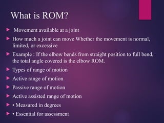
What is ROM?
Movement available at a joint
How much a joint can move Whether the movement is normal,
limited, or excessive
Example : If the elbow bends from straight position to full bend,
the total angle covered is the elbow ROM.
Types of range of motion
Active range of motion
Passive range of motion
Active assisted range of motion
• Measured in degrees
• Essential for assessment
4.
Why ROM IsImportant in Physiotherapy
ROM assessment helps to:
Identify joint stiffness or restriction
Detect muscle tightness or contracture
Plan appropriate treatment
Monitor improvement or deterioration
Document objective findings
Compare affected and unaffected sides
5.
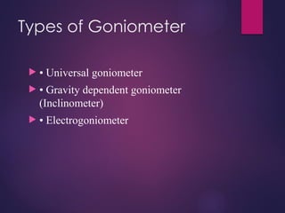
Definition of Goniometery
GONIOMETRY is the most widely used method for measuring
joint range of motion
In Greek gonio = angle and metron = measurement
Both AROM and PROM can be measured
It is a technique used to measure and document amount of
available range of motion of joints
Goniometer is the instrument which is used to measure joint
range of motion
6.
Why IsGoniometry Important ?
Converts visual estimation into numerical values
Helps identify joint stiffness or hypermobility
Assists in clinical decision-making
Allows comparison: Side to side Pre-treatment vs
post-treatment
Essential for legal and documentation purposes
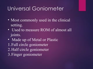
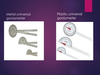
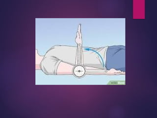
Universal Goniometer
• Mostcommonly used in the clinical
setting.
• Used to measure ROM of almost all
joints.
• Made up of Metal or Plastic
1.Full circle goniometer
2.Half circle goniometer
3.Finger goniometer
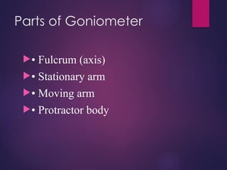
Parts :
Body :- The body resembles of a protractor, and may form full
circle or half circle.
- Fulcrum of the goniometer is present at centre of the body,
and is placed at axis of measuring joint.
Stationary arm : - The stationary arm is a structural part of the
body of the goniometer and cannot be moved independently
from the body.
- Aligned with proximal segment of the joint.
Movable arm : - The moving arm is attached to the center of
the body of goniometer, that permits the arm to move freely on
the body.
- Aligned with distal segment of the joint.
13.
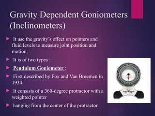
Gravity Dependent Goniometers
(Inclinometers)
It use the gravity’s effect on pointers and
fluid levels to measure joint position and
motion.
It is of two types :
Pendulum Goniometer :
First described by Fox and Van Breemen in
1934.
It consists of a 360-degree protractor with a
weighted pointer
hanging from the center of the protractor
15.
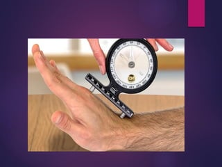
Fluid (Bubble)Goniometer :
Developed by Schenkar in 1956.
It has a fluid-filled circular chamber
containing an air bubble, and has a 360-
degree scale motion.
16.
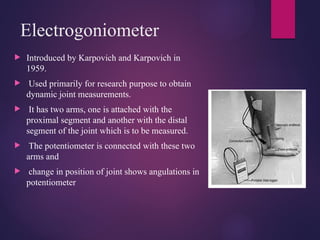
Electrogoniometer
Introduced byKarpovich and Karpovich in
1959.
Used primarily for research purpose to obtain
dynamic joint measurements.
It has two arms, one is attached with the
proximal segment and another with the distal
segment of the joint which is to be measured.
The potentiometer is connected with these two
arms and
change in position of joint shows angulations in
potentiometer
17.
Advantages
Simple andnon-invasive
Inexpensive
Portable
Widely accepted tool
Easy to learn for beginners
18.
Procedure
The examinermust have the skill to perform the following for each
joint and motion.
• Position & stabilize correctly
• Move a body part through appropriate ROM.
• Determine end range of motion (end-feel).
• Palpate the appropriate bony landmarks.
• Align the measuring instrument with landmarks.
• Read the measuring instrument.
• Record measurements correctly.
19.
Principles of Goniometer
1. POSITIONING:
Positioning is an important part of goniometer. Testing positions refers to the
selection of appropriate starting position for obtaining goniometric measurements.
The series of testing positions are designed to;
• Place the joint in a starting position of 0 degree.
• Permit a complete ROM.
• Provide stabilization of the proximal joint segment.
Testing position involve a variety of positions such as supine, prone, sitting &
standing.
When an examiner has to test several joints & motion during one testing session,
the goniometric examination should be planned to avoid moving the subjects
unnecessarily. For e.g. if the subject is in prone all possible measurements in this
position should be taken before the subject moved to another position.
20.
2. STABILISTION:
The testing position helps to stabilize the subjects body &
proximal joint segment so that the motion can be isolated to
the joint being examined.
Stabilization may be given manually by the examiner.
The amount of manual stabilization applied by an examiner
must be sufficient to keep the proximal joint segment fixed
during movement of distal joint segment.
21.
3. ALIGNMENT:
Goniometer alignment refers to the alignment of the arms
of the goniometer with the proximal & distal segments of
the joint being evaluated.
Instead of depending on the soft tissue contour the
examiner should use bony anatomical landmarks to more
accurately visualize the joint segments.
The fulcrum of the goniometer may be placed over the
approximate location of the axis of the motion of the joint
being measured.
22.
4. RECORDING:
The following points are recommended to be included in
the recording;
• Subject’s name, age & gender.
• Examiner’s name
• Date & time of measurement.
• Side of the body, joints & motion being measured.
• ROM that is measured.
• Type of motion being measured that is passive or active.
• Any subjective information such as discomfort or pain
that is reported by the subject during the testing.
23.
Contraindication
Joint dislocation
Unhealed fracture
Post surgery
Severe pain aggravated by movement
Infection or inflammation around a joint
Open wound
24.
Summary
What isROM
What is goniometry
Why is goniometer important
Types of goniometer
Its advantages
Principles of goniometer