
Recommended
More Related Content
What's hot
What's hot (20)
Similar to GI System by Rupam Bhowmik.pptx help full for NORCET EXAM
Similar to GI System by Rupam Bhowmik.pptx help full for NORCET EXAM (20)
More from Rupam Bhowmik
More from Rupam Bhowmik (8)
Recently uploaded
Recently uploaded (20)
GI System by Rupam Bhowmik.pptx help full for NORCET EXAM
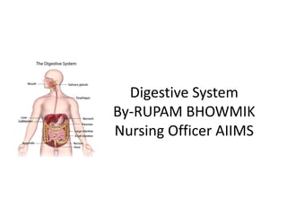
- 1. Digestive System By-RUPAM BHOWMIK Nursing Officer AIIMS
- 3. The Digestive system consists of two groups of organ 1. Gastrointestinal Tract (GIT) or Alimentary Tract or Primary Digestive Organ : • The organ where actual digestion takes place called primary digestive organ. • GIT is a 5-7 meter long continuous tube from mouth to anus.
- 4. The length of GIT in a cadaver (a dead human body preserved for anatomical study) is about 7- 9 meter (23-30 feet), but in living person it is much shorter (5-7 meter) because of muscles are in contracted state 2. Accessory Digestive Organ : • It help primary digestive organ in the process of digestion, examples are teeth, tongue, salivary gland, pancreas, liver, gall bladder etc.
- 5. FUNCTION OF DIGESTIVE SYSTEM The digestive system perform following 6 basic process – Eating or Ingestion - Taking food and liquid into mouth. Secretion – The cells of GIT or accessory organs secrete about 7 liter of water, acid, buffer and enzymes. Mixing & Propulsion - GIT mix food & secretion and then propel toward the anus.
- 6. Digestion -Break down of ingested food into small and simple chemical molecules by mechanical and chemical process called digestion. A. Mechanical digestion-Cutting & churning of foods. B. Chemical digestion-Breaking up of larger molecules into smaller molecules by digestive enzymes.
- 7. Absorption – Taking up small digested molecules (glucose amino-acid, fatty acid etc.) into blood through epithelial cells of GIT called absorption. Defecation- Wastes, undigested substance, bacteria and cells sloughed from lining of GIT leave the body through the anus called defecation.
- 9. Layers of GIT • INNER To OUTER : Is mucosa, submucosa, muscularis and serosa/adventitia. • Mucosa:- inner most layer, contain goblet cell which secrete mucus. • Submucosa :- contain nerves, blood vessels. • Muscularis :- helps in peristalsis. • Serosa/adventitia:- prevent any frictional damage from the intestine rubbing against other tissue.
- 10. MUCOSA • Mucosa is consisting of stratified squamous epithelium (mouth, pharynx, upper part of esophagus and anal canal) and columnar epithelium (lower part of esophagus, stomach and intestine). • Within every 5-7 days, epithelial cells of GIT are replaced by new cells.
- 11. SUB-MUCOSA • Extensive networks of neurons are also located in submucosa which called submucosal plexus (Meissner's plexus). • Vagus nerve also supplies most of part of GI tract but last half of the large intestine supplied by nerve of sacral spinal cord.
- 12. MUSCLE LAYER • Muscles of mouth, pharynx, upper part of esophagus and external anal sphincter contain skeleton muscles (voluntary in nature) and rest of tract contains smooth muscles (involuntary in nature). • A Muscle layer help in mixing and propulsion of food by peristalsis. • Between layers of muscles, nerve tissue present which called myenteric plexus (Auerbach's plexus).
- 13. SAROSA/ADVENTITIA • Prevent any frictional damage from the intestine rubbing against other tissue.
- 14. Mouth or Oral Cavity or Buccal Cavity • The roof of month formed by palates. • Anterior part of palate called hard palate (formed by maxilla & palatine bone) and posterior part called soft palate (formed by muscles).
- 15. • Posterior free hanging border of soft palate called uvula, which during swallowing close naso-pharynx to prevent entry of food and liquid into nasal cavity.
- 16. • Pain upon swallowing- called odynophagia. • Inflammation of the mouth (including the lips, tongue and mucous membranes) called stomatitis. • Normal flora or commonsale microbes of oral cavity mainly contain lactobacillus acidophilus, bacteria that produces lactic acid by fermenting the sugars present in milk
- 17. TEETH 1. Primary Teeth or Temporary Teeth or Deciduous Teeth or Baby Teeth or Milk Teeth: • Total numbers are 20 (10 in each jaw). • It starts to erupt at 6 month of age (lower central incisor) and completed at 24 month (second molar). • It lost between 6 year to 12 year of life.
- 19. • Formula - 2102/2102 (sequence from anterior to posterior : 2 incisor, 1 canine, 0 premolar, 2 molar). • These teeth called diphyodont (having two sets of teeth) because erupt two times in the life.
- 20. 2. Secondary Teeth or Permanent Tecth: It starts to erupt in 6 year of age (first molar) and complete 32 teeth in 24 years (3rd molar). Formula - 2123/2123 (sequence from anterior to posterior : 2 incisor, 1 canine, 2 premolar, 3 molar).
- 21. Incisor and canine are called- cutting teeth and premolar and molar are called- grinding, crushing or chewing teeth. 3rd molar teeth called wisdom teeth. 3rd molar and premolar teeth called monophyodont (having a single permanent set of teeth) because erupt one times in the life.
- 22. Structure of Teeth • A tooth has three major region 1. Crown- Visible portion of teeth. 2. Root - Embedded portion in sockets. 3. Neck - Constricted junction of crown and root near the gum.
- 24. • Teeth are composed of dentin (calcified connective tissue composed of 70% calcium salt). • The dentin encloses a cavity within crown called pulp cavity, which contain blood vessels and nerves.
- 25. • The dentin in crown is covered by a hardest substance of body (composed of about 95% of calcium salt) called enamel. • Inflammation of the gums or gingiva characterized by redness, swelling and tendency to bleed called Gingivitis.
- 26. Salivary Gland • 3 pairs of the glands secrete saliva called salivary gland : 1. PAROTID GLAND (25% SECRETION OF SALIVA) • Largest salivary gland, weight 20-30gm • Secretion of these glands comes in to oral cavity by parotid duct or Stensen's duct (40 mm long) and it open at upper second molar teeth.
- 27. • Ptyalin secreted by only parotid gland. • A Inflammation and enlargement of parotid gland by paramyxovirus called mumps or epidemic parotitis or parotiditis.
- 28. 2. SUBMANDIBULAR (70% secretion of saliva) : • Situated beneath the base of tongue. • Weight 8-10gm. Opens by submandibular duct or Wharton's duct (40 mm long) at side of frenulum of tongue.
- 30. 3. SUBLINGUAL GLAND (5% secretion of saliva) : • Smallest salivary gland, Weight 2-3gm. • Open their secretion by 5-15 ducts called minor sublingual duct (duct of Rivinus) or a large sublingual duct (Bartholin's ducts).
- 31. • Volume of Saliva: 1000 -1500 ml/day or 1 ml/minute . • Excessive secretion of saliva called ptyalism or sialorrhea. • Xerostomia (mouth dryness) means decreased production or lack of saliva.
- 32. • Composition of Saliva : • 99.5%- H2O • 0.5% Solutes like Na+ , K+, Cl-, Phosphate,HCO3, • Immunoglobulin A, lysozyme (bactericidal) & enzymes. • pH of saliva - 6.35 to 6.85 • Specific gravity-1.002 to 1.012.
- 33. • Food may remain in the fundus of stomach about an hour without mixing with gastric juice, during this time, digestion by salivary amylase is continues. A
- 34. • Tongue Tongue is a voluritary muscular organ. • It attached to floor of mouth or hyoid bone by frenulum (a thin fold of mucosa in middle of floor of the mouth). • If a person's lingual frenulum is abnormally short or rigid which called Ankyloglossia (tongue tied) result into speech impairment.
- 37. • A Inflammation of tongue called glossitis. • Superior surface of tongue consist of stratified squamous epithelium with numerous papillae (little projections).
- 38. • Papillae containing taste buds (nerve ending for sense of taste, sensory branch of glossopharyngeal nerve). • Types of papillae There are three types of papillae based on their shape -
- 39. • 1) Vallate (circum-vallate) papillae: • It arranged in inverted V shape at the base of tongue. • These are larger papillae, can most ensily seen on tongue. • 2) Filiform papillae (thread-like) : Smallest type and situated on anterior 2/3 of tongue. • 3) Fungiform papillae(mushroom-like): Situated mainly at tip & edges of tongue.
- 40. TASTE BUDS • Sweet and salty taste buds situated mainly on tip. • Sour taste buds situated at the edge & side of tongue. • Bitter taste buds situated at the back of tongue.
- 41. QUESTIONS • Al of the following accessory organs help in digestion except: (a) Liver (b)Gall bladder (c) Pancreas (d)Duodenum
- 42. • The process of taking food into the digestive system is (a) Ingestion (b)Propulsion (c) Digestion (d)Elimination
- 43. • Inflammation of mucus membrane of the teeth is called: (a) Stomatitis (b)Halitosis (c) Gingivitis (d)Cheilosis
- 44. • Halitosis means offensive odor of the breath. • Cheilosis means a condition in which lips become reddened and develop fissures at the angles, mainly due to vitamin B complex deficiencies, especially riboflavin.
- 45. • Commonest commonsale microbes present into oral cavity (a) Helicobacter pylori (b)E. Coli (c) Bacillus acidophilus (d)All of above
- 46. • Lactobacillus acidophilus is a species of bacteria that produces lactic acid by fermenting the sugars present in milk. • It is found in milk, feces of bottle-fed infants, adults whose diets include high milk content. • It is also part of oral and vaginal commonsale mierobes (flora). • Helicobacter pylori mainly present in stomach and E. coli into colon,
- 47. • Which acid convert milk into curd (a) Citric acid (b)Mallic acid (c) Lactic acid (d)Tartaric acid
- 48. • Which of the following taste is one most limited to the tip of the tongue? (a) Bitter (b)Sweet (c) Salty (d)Sour
- 49. • Taste buds for sensing bitterness are located on which part of the tongue? (a) Anterior part of the tongue (b)Posterior part of the tongue (c) Lateral part of the tongue (d)Under surface of the tongue
- 50. • Which one of the following enzymes is found/ secreted in the Saliva? (a) Rennin (b)Tenin (c) Ptyalin (d)Resin milk. .
- 51. • Saliva contains an enzyme, ptyalin (salivary amylase), which start to digest carbohydrates like starch and glycogen to maltose and glucose into mouth. • The optimum pH for ptyalin activity is 6.8. • Saliva also contains an enzyme, lingual lipase, which digest pre-emulsified fat present into milk. • Rennin or chymosin is an enzyme present in the gastric juice of young ruminants that curdles milk.
- 52. • Which part of food first start to digest into mouth? (a) Carbohydrate (b)Fat (c) Protein (d)Vitamin
- 53. • Digestion of food in human starts from (a) Small intestine (b)Mouth (c) Duodenum (d)Large intestine
- 54. • The salivary gland secretes saliva which contains the enzyme (a) Pepsin (b)Renin (c) Lipase (d)Ptyalin
- 55. • Amount of saliva produced in one day is: (a) 0.5 liter (b)1.5 liter (c) 1 liters (d)2 liter
- 56. • Deficient salivation is called (a) Xerophthalmia (b)Xerostomia (c) Ptyalism (d)Halitosis
- 57. ESOPHAGUS • Esophagus is a 25cm long collapsible muscular tube that lies posterior to the trachea. • Laryngo-pharynx continues to esophagus at 2nd thoracic vertebrae.
- 59. • Through esophageal hiatus (an opening for esophagus), esophagus cross the diaphragm and ends in the superior portion of the stomach.
- 61. • Sometimes part of the stomach protrudes above the diaphragm through esophageal hiatus called hiatal hernia.
- 62. • A sphincter between upper part of esophagus and laryngopharynx called upper esophageal sphincter or cricopharyngeal sphincter.
- 63. • A sphincter between the lower end of esophagus and stomach called lower esophageal sphincter (LES) or cardiac sphincter.
- 64. • If LES or cardiac sphincter fails to close adequately after entering the food into stomach, the stomach content can reflux or back up into esophagus; this condition is known as gastroesophageal reflex disease (GERD).
- 67. • Pharmacological Management of GERD: - • H2 histamine receptor antagonists (decrease acidity) . • Reduce gastric acidity: Ranitidine. • Proton pump inhibitors (decrease acidity and increase lower esophageal sphincter tone):Pantoprazole and Omeprazole. • Gastric emptying: Prokinetic agents: Reglan.
- 68. • Management of GERD : • Dietary modification: Encourage low fat diet; avoid caffeine, tobacco, beer, carbonated beverages and milk. • Avoid eating and drinking 2 hours before the bed time Maintain normal body weight . • Avoid tight fitting clothes. • Elevate head end of the bed and elevate upper part of the body over a pillow
- 69. • Hcl (hydrochloric acid) from stomach content irritate the esophageal wall, causes burning sensation which is termed heart burn or pyrosis. • Drinking alcohol & smoking cause cardiac sphincter to relax, which result into heart burn.
- 70. • Pharynx continues to esophagus at : (a) 2nd cervical vertebra (b)3rd cervical vertebra (c) 2nd thoracic vertebra (d)3rd thorneic vertebra
- 71. • These tissues are found in stomach: (a) Longitudinal fibres (b)Circular fibers (c) All of these Nurse (d)Oblique fibers
- 72. • The muscular layer of stomach contains three. layers of smooth muscle fibers; an outer layer contain longitudinal fibers, a medial layer circular and an inner layer oblique fiber of muscles. • These three layer of smooth muscles of stomach are responsible for mechanical digestion of food.
- 73. • Mechanical Digestion in the stomach is accomplished by which of the following structures? (a) Mucosa (b)Smooth muscle layer (c) Striated muscle layer (d)Gastric glands
- 74. • Chyme is called: (a) Food in the mouth (b)Food in stomach (c) Food reaches in duodenum (d)Food reaches in the rectum
- 75. • The mixture of partially digested food digestive secretions in the stomach, or partially digested food which is entering into first part of small intestine (duodenum) called chyme.
- 77. • Food reservoir is known as: (a) Oesophagus (b)Small intestine (c) Stomach (d)Large intestine
- 78. • Stomach is a "J" shaped distensible muscular sac like part of the alimentary tube, serve as reservoir of food. • Fatty food remains longest time, protein less than fat and carbohydrate least into stomach.
- 79. • Identify the part of Human Stomach which connects/joins with Oesophagus (a) Fundus (b)Body (c) Cardia (d)Pylorus
- 81. • Human body needs a constant supply of proteins to survive. The first part of digestive system to begin digesting proteins is (a) Mouth (b)Small intestine (c) Stomach (d)Large intestine
- 82. • The process of digestion of protein first started into stomach by proteolytic enzyme pepsin (an enzyme that catalyzes the conversion of proteins into peptides and amino acids). • Pepsin secreted by chief cells of gastric glands in inactive form of pepsinogen
- 83. STOMACH • Stomach is a J- shaped distensible enlarged part of GIT. • It is situated just inferior to diaphragm in the epigastric, umbilical & left hypochondriac region. • Stomach serves as a mixing chamber (make chyme) or food reservoir (maximum capacity 1.5 liter).
- 84. • Cardia – Region around the opening of lower esophageal sphincter or cardiac sphincter. • Fundus - A dome shaped part above the cardiac sphincter. • Body - Central largest part (80%) of stomach. • Pylorus – Lower part of stomach which connects the body of stomach to the duodenum.
- 85. • It has two parts- Pyloric antrum, which connect to the body of stomach. • Pyloric canal, which open into duodenum.
- 86. Region of stomach Stomach has divided in four main region-
- 87. • The sphincter between stomach & duodenum called pyloric sphincter.
- 88. • Major clinical manifestation : • Non-bilious projectile vomiting . • Dehydration and electrolyte imbalance • Olive like mass on palpation of right upper quadrant of the abdomen - Left to right peristaltic movement across the abdomen
- 89. • Management - Pyloromyotomy known as Ramstedt's procedure: Dividing the muscle of the pylorus to open up the gastric outlet.
- 90. • Narrowing of pyloric sphincter called pyloric stenosis. • Projectile vomiting is a hallmark symptom of pyloric stenosis.
- 91. • Surface mucosa of stomach is renewed about every 3 day, but steroids decrease cell renewal. • Prostaglandins are protective for gastric mucosa, which synthesis inhibit when patient taking NSAID.
- 92. • Gastric glands contains 3 types of exocrine giand cells that secrete different content of gastric juice, like- 1. Chief cells – it secrete pepsinogen (digest protein )to peptone and gastric lipase (digest fat).
- 93. 2. Parietal cells - It produce intrinsic factor and hydrochloric acid (HCI). • Intrinsic factor is needed for absoption of vitamin B12 & HCl convert pepsinogen (inactive) to pepsin (active form).
- 94. 3. Mucus neck cells - It secrete mucus. • Mucus protects the wall from HCl and pepsinogen.
- 95. About 2-3 liter of gastric juice secreted per day by exocrine cells of gastric glands. The pH of gastric juice is 1.5 to 3.5 (strongly acidic), which destroys pathogens.
- 96. Gastric gland also contains enteroendocrine cells (c.g. G cells) which secrete gastrin hormone directly into blood stream. Rennin (chymosinin, an enzyme that curdles milk) in human present only in newborn, in adult curdling of milk occurs by Hcl.
- 97. Small Intestine • Small intestine has three parts: 1. Duodenum (25 cm),shortest part. 2. Jejunum (8 feet) 3. Ileum (12 feet), Absorption of vitamin B12 take place in ileum.
- 101. HEPATIC BILIARY SYSTEM Liver is the largest gland in the body, located in the right upper quadrant. It weighs about 1500 gm and divided into 4 Lobes Lobes are further divided into lobules. Kupffer cells are the phagocytic cells present in the liver. Smallest bile duct called canaliculi are located between the lobules of liver.
- 102. Canaliculi carry bile secreted by hepatocytes to larger bile duct, which eventually become hepatic duct. Hepatic duct from liver and cystic duct from gallbladder join to form common bile duct which empties bile into the duodenum of small intestine.
- 104. Functions of Liver • Glucose metabolism: After digestion, glucose is converted to glycogen and whenever the body requires, glycogen is converted to glucose. • Glycogenesis: Glucose is converted and stored as glycogen. • Glycolysis: Glycogen is converted back to glucose. • Gluconeogenesis: Glucose is synthesized from non-carbohydrate substances such as protein and amino acids.
- 105. • Ammonia conversion: Protein and amino acid metabolism leads to ammonia generation. Liver converts this highly toxic ammonia to urea for excretion. • Protein metabolism: Liver synthesizes all plasma protein (except gamma globulin) such as albumin, alpha and beta globulins.
- 106. • Fat metabolism: When glucose is not available fatty acids are broken down for the production of energy. • Vitamin and iron storage: Liver store large amount of vit A, D, E, K and vit. B12 and it also store iron and copper. • Drug metabolism: Liver is a major site for drug metabolism • Bile formation: Hepatocytes synthesizes bile and store in gallbladder. Bile salts are formed from cholesterol.
- 108. • Between small intestine and large intestine is the caecum which is located in the right lower quadrant. Ileocecal valve located here. • Ileocecal valve control the entry of content into the large intestine as well as it prevent the reflex of bacteria from large to small intestine.
- 110. •Vermiform appendix is located near to ileocecal junction in the right lower quadrant. Deficiency of vitamin B12 results in pernicious anaemia.
- 111. Large Intestine • 5 feet (1.5 metre) long. • Absorbs water and eliminate waster. • Intestinal bacteria play a vital role in the synthesis of some vitamins and vitamin K . • The large intestine is divided into three parts caecum, colon and rectum.
- 113. • Colon is further divided as : 1. Ascending: shortest person of colon 2. Transverse 3. Descending 4. Sigmoid • Rectum continuous in an external opening called anus.
- 115. Waste Product of Digestion • The brown colour of the fecal matter is due to breakdown of bile by intestinal bacteria. • Chemical formed by intestinal bacteria such as indole and skatole are responsible for fecal odor. • Internal anal sphincter is under automatic nervous system control. • External anal sphincter is under voluntary control.
- 116. Stomatitis: inflammation of oral/buccal mucosa.
- 118. • Surgery: Neissan fundoplication; Wrapping of the gastric fundus around the sphincter area of the esophagus.
- 119. Hiatal hernia • Eesophagus enter into the stomach through an opening in the diaphragm known HIATUS. • In Hiatal hernia this opening becomes large and upper portion of the stomach moves into lower thorax. • A condition in which part of the stomach pushes up through the diaphragm muscle
- 122. Peptic Ulcer Disease • It is the erosion of the mucosal wall of stomach, duodenum or in esophagus. • Based upon the location, it is also called as esophageal ulcer, gastric ulcer or duodenal ulcer. • Peptic ulcer is more commonly seen in duodenum.
- 124. Etiology 1. Hyper-secretion of gastric juice, by- • Increase stimulation of vagus nerve. • Increase number or capacity of parietal cells to secrete acids. 2. Impair mucosal barrier protection.
- 125. Risk factor • Use of alcohol, smoking and ulcerogenic drugs like aspirin, NSAID & steroids (so it never gives empty stomach). • Infection by helicobacter pylori bacteria (gram- negative bacteria that causes 90% of peptic ulcers. • Family history of peptic ulcer & blood group O. • Zollinger- Ellison syndrome (ZES) - Abnormal secretion of gastrin from tumor of pancreatic islets, which cause excessive secretion of gastric juice.
- 126. • Stress especially cause duodenal ulcer (due to vasoconstriction). • Curling's ulcer (ulcer caused by severe burn). • Cushing's ulcer (ulcer caused by increased secretion of gastric acid due to vagus nerve stimulation in increased ICP during head injury cause).
- 127. GASTRIC ULCER DUODENAL ULCER Age- 50 or more Male : Female – 1:1 Clinical manifestation : Normal or hyposecretion of HCL. Wt loss. Pain occur 30-60 min after a meal. Pain Relieved by Vomiting. Ingestion of food may increase the pain. Vomiting and Hematemesis seen. Age- 30 -60 Male : Female – 2-3:1 Clinical manifestation : Hyper secretion of HCL. May Wt gain Pain occur 2-3 hours after a meal. Wakes up in the night due to pain. Pain Relieved by Vomiting. Ingestion of food may decrease the pain. Vomiting is uncommon. Malena is coomon.
- 128. • Client education : 1. Avoid consuming alcohol and substances that contain caffeine or chocolate. 2. Avoid smoking. 3. Avoid aspirin or NSAIDS. 4. Obtain adequate rest and reduce stress.
- 129. • Management - Histamine blocker such as ranitidine or cimetidine is used to decrease gastric acid production. • Proton pump inhibitor such as omeprazole, lansoprazole, pantoprazole is used to suppress acid production. • Avoid smoking, alcohol use and caffeinated beverages and coffee.
- 130. Surgical management • Total gastrectomy: removal of entire stomach and some portion of esophagus attached to it and anastomosis of esophagus to jejunum or duodenum. • So it is also called as esophagojejunostomy or esophagoduodenostomy.
- 131. • Vagotomy: Removal of the vagus nerve supply to stomach and there by decreases the stimuli for gastric acid secretion Gastric resection: Removal of the lower portion of the stomach usually along with vagotomy. It is also called as antrectomy.
- 132. • Billroth I: Partial gastrectomy and anastomosis of remaining stomach to duodenum. It is also called as gastroduodenostomy.
- 133. • Billroth II: Reconstruction after a partial gastrectomy, the duodenal stump is closed and a gastrojejunostomy is created.
- 135. • Pyloroplasty: Enlargement of pylorus to prevent or decrease pyloric obstruction and thereby enhancing gastric emptying.
- 136. • Postoperative interventions: • Monitor vital signs. • Place in a Fowler's position for comfort and to promote drainage. • Administer fluids and electrolyte replacements intravenously as prescribed; monitor intake and output. • Assess bowel sounds. • Monitor NG suction as prescribed. • Maintain NPO status as prescribed for 1 to 3 days until peristalsis returns. • Progress the diet from NPO to sips of clear water to 6 small bland meals a day, as prescribed when bowel sounds return. • Monitor for postoperative complications of hemorrhage, dumping syndrome, diarrhea, hypoglycemia, and vitamin B12 deficiency.
- 137. Dumping Syndrome • Also called rapid gastric emptying. Occurs when food moves from stomach to intestine too quickly. • It is one of the major complications of patients who underwent partial or complete resection of stomach.
- 138. • Manifestations: • Sensation of fullness, weakness, faint, dizziness, palpitation, diaphoresis, cramping pain, diarrhea and borborygmi (loud gurgles indicating hyperperistalsis).
- 139. • Patient education to prevent dumping syndrome : • Avoid sugar, salt and milk. • Eat high protein, high fat low carbohydrate food items. • Eat small meals and avoid consuming fluids along with meal. • Lie down after eating.
- 140. • The part of the large intestine that is joined to the rectum is called a) Ascending colon b) Descending colon c) Transverse colon d) Sigmoid colon
- 141. • The basic general layout of the gastrointestinal tract wall from inside to outside follows this sequence: a.Submucosa → mucosa → muscular → serosa b. Muscular → serosa → mucosa → submucosa c. Mucosa → submucosa → muscular → serosa d. Serosa → muscular → submucosa → mucosa
- 142. • In the liver, bacteria that found their way into portal circulation is destroyed by : (ESIC 2019) a) Hydrochloric acid b) Kupffer cells c) Cilia d) Leukocytosis
- 143. APPENDICITIS • Appendix is a small finger like projection attached to cecum below ileo-cecal valve. • Inflammation of this projection is knows as appendicitis. • Fecalith is the most common cause of appendicitis. • Most commonly occur in adolescents
- 144. Clinical Manifestation • Low grade fever, nausea and vomiting • Rebounding tenderness on palpation at Mc Burney's point. (it is a point 2/3rd of the way umbilicus to Anterior Superior Iliac Spine). • Rovsing sign: Palpation of left lower quadrant paradoxically elicit pain in the right lower quadrant.
- 145. Diagnosis • Blood test: Elevation of WBCS • Abdominal X-ray, USG, CT • Complication : Perforation of appendix which leads to peritonitis
- 147. • KEY POINTS 1. Enema, laxatives and hot applications are contraindicated in case of appendicitis. It may lead to rupture of appendix and peritonitis. 2. Fowler's position to minimize pain. 3. After surgery, place the client ina semi fowler's position. This will help to reduce tension in suture and abdominal organ.
- 148. PERITONITIS • Inflammation of peritoneum, the serous membrane covering the abdominal cavity. • Content of abdominal organ leaks into the abdominal cavity as a reşult of trauma, perforation, infection, or inflammation • Immediate response of intestinal tract in peritonitis is hyper motility followed by paralytic ileus.
- 149. Manifestations • Diffused pain in the abdomen which becomes constant and localized. • Movement may aggravate the pain. • Rebound tenderness and paralytic ileus may be present. • Elevated leukocyte count and electrolyte abnormalities.
- 150. Diagnosis • X-ray shows air and fluid levels and distended bowel loops. • Culture and sensitivity of peritoneal fluid to identify the cause.
- 151. Management Manifestations • Fluid and electrolyte replacement is the major focus of management. • Lung expansion is compromised due to the compression of intra-abdominal fluid over the lung. So oxygen therapy or intubation is often necessary for airway management. • Massive antibiotic therapy.
- 152. • Surgical management is needed if peritonitis occurred as a result of appendicitis, perforation etc. • Peritonitis patients placed on side with knee flexed. This position will reduce the tension on the abdominal organs.
- 153. Pancreatitis • Inflammation of the pancreas due to the any etiological factor which causes injury to pancreatic cells or activation of pancreatic enzyme into pancreas rather than intestine that may result in auto digestion of pancreas by its own enzymes. • Types -There are two types of pancreatitis.
- 154. 1. ACUTE PANCREATITIS : • Sudden and single attack of inflammation of pancreas. • Attack may be recurrent with resolution also called acute pancreatitis. Etiology : • Alcohol abuse (may be increase secretion of pancreatic enzyme) and obstructions of the pancreatic duct by gallstones are the most common causes. • Biliary tract discase (most common in female) such as gall bladder stone and cystic fibrosis. • Viral infection e.g. HIV, mumps and coxsackievirus. • Some drugs like corticosteroids, NSAID and oral contraceptive.
- 155. diagnosis • Elevated serum level of WBC, bilirubin, lipase (diagnostic marker) & amylase.
- 156. C/M • Sudden abdominal pain in left upper quadrant then radiates to back. • Pain increase by intake of alcohol and fatty foods. • Abdominal tenderness & guarding. • Cullen Sign and Turner's Sign
- 157. • Cullen's sign is superficial edema and bruisingin the subcutaneous fatty tissue around the umbilicus. • It is a sign hemorrhage. • It is also a sign of rupture of fallopian tube or ectopic Pregnancy.
- 158. • Grey Turner's sign refers to bruising of the flanks, the part of the body between the last rib and the top of the hip. • It is a sign of retroperitoneal hemorrhage, or bleeding behind the peritoneum. • Gray turner's sign (flank ecchymosis) may also be rupture of aneurysm.
- 159. 2. CHRONIC PANCREATITIS : • After repeated attack of acute pancreatitis, the inflammatory process results in scarring and calcification of pancreatic tissue. • The damage is irreversible & affects both endocrine & exocrine function of pancreas.
- 160. • Etiology – Excessive and chronic intake of alcohol. • Biliary tract disease like cholelithiasis, which cause inflammation of sphincter of oddi (most common cause). • C/M – Sign symptoms of diabetes mellitus. Jaundice, weight loss. • Steatorrhea (fats in stool) which cause foul smelling stool. • Abdominal pain & tenderness which radiate to back.
- 161. MANAGEMENT • The patient should receives nothing by mouth until pain, nausea and vomiting resolved. • Total parenteral nutrition, fluid & electrolytes for patients with severe pancreatitis. • Administer somatostatin for inhibition of gastric motility and gastric acid secretion, blockage of the exocrine and endocrine function of the pancreas in the patient with acute pancreatitis. • Administer pancreatin (a mixture of enzymes, chiefly amylase, lipase and protease; used in patients with chronic pancreatitis, who do not secrete adequate amounts of their own pancreatic enzymes). • Meperidine (pethidine) is a kind of opioid analgesic which is believed to induce less biliary spasm (spasm of pancreatic duct and sphincter of oddi) than morphine and codeine, so meperidine is preferred analgesic in to cholecystitis, pancreatitis and biliary colic.
- 162. • Morphine causes spasm of biliary duct, so it is contraindicated in pancreatitis as an analgesic. • A nasogastric tube may be inserted and placed on low, intermittent suctioning to reduce hydrochloric acid levels or relieve gastric distention, because increase level of gastric juice or gastric distension may cause pancreatic stimulation. • Advice to avoid alcohol consumption and limited intake of fats & protein.
- 163. Surgical Interventions: 1. Pancreaticojejunostomy: Anastomosis of pancreatic duct to jejunum. 2. Whipple resection (Pancreaticoduodenectomy) The head of the pancreas. the first part of the small intestine (duodenum), the gallbladder and the bile duct will be removed. The remaining organs are reattached to allow digestion of food normally after surgery.
- 165. PANCREAS • It is a pale grey, retroperitoneal gland. • It situated into epigastric and left hypochondriac region. • Its weight is about 60gm. • The pancreas consists of a head, a body and a tail. • The head is expanded portion which lies into curve of duodenum.
- 166. Structure of Pancreas • 99% part of pancreas is exocrine and remaining 1% part is endocrine (pancreatic islets or islets of Langerhans). • "Islets of Langerhans" are concentrated in to tail portion of pancreas. • The acinar cells of Pancreas secrete "pancreatic juice".
- 167. • Just before entering the duodenum the pancreatic duct join the common bile duct to form Hepato-Pancreatic ampulla (Vater's ampulla or papilla of Vater). • Vater's ampulla opens into duodenum by hepato-pancreatic sphincter or Sphincter of Oddi. • pH- alkaline (8 to 8.9) due to high concentration of HCO3, (110-115mEq/liter).
- 168. Exocrine gland : 1. Secretes sodium bicarbonate to neutralize the acidity of the stomach contents that enter the duodenum. 2. Pancreatic juices contain enzymes for digesting carbohydrates, fats, and proteins.
- 169. Endocrine gland : 1. "Secretes glucagon to raise blood glucose levels and secretes somatostatin to exert hypoglycemic effect . 2. The islets of Langerhans secrete insulin. 3. Insulin is secreted into the bloodstream and is important for carbohydrate metabolism.
- 170. Pancreatic Enzyme in Pancreatic Juice • Protein - digesting enzyme : • Trypsinogen (converted to active trypsin ) and chymotrypsinogen (convert to active chymotrypsin). • Both trypsin and chymotrypsin digest protein and forms amino-acids.
- 171. • Fat - digesting (lipolytic) enzyme : • Pancreatic lipase (powerful lipolytic enzyme) digests triglyceride in to fatty acid & glycerol.
- 172. • Carbohydrate digesting enzyme : • Pancreatic amylase digests starch into oligosaccharide, disaccharides & monosaccharide. • Note - Protein digesting enzymes such as trypsin or chymotrypsin are produced in inactive form in pancreas as trypsinogen and chymotrypsinogen, so do not digest cells of pancreas.
- 173. Portal Circulation or entero-hepatic circulation • In the portal circulation venous blood from capillaries of digestive system, spleen & pancreas to passes through a Secondary capillaries bed (hepatic sinusoids) in the liver before entering into the IVC. • It is essential because high concentration of digested nutrients absorbed by digestive system must first göes to liver. •
- 174. • Portal Veins - Portal vein is formed by union of following 5 veins • 1) Splenic vein - Drain blood from spleen and pancreas. • 2) Inferior mesenteric vein - Drain blood from a part of transverse colon, descending colon & rectum. • 3) Superior mesenteric vein - Drain blood from small intestine, ascending colon and part of transverse colon. • 4) Gastric vein - Drain blood from stomach. • 5) Cystic vein - Drain blood from gall bladder.
- 175. • Secretion of bile Volume 500 to 1000 ml/day. COMPOSITION OF BILE : • Water, mineral salt, mucus, bile pigment (bilirubin is most common bile pigment) bile salts (derived from bile acid). • Bile acids are the complex acid that occurs as salts in bile. • It reabsorbed from the intestine to be used again by the liver, so the circulation of bile acids is called enterohepatic circulation. • Bile does not contain any digestive enzyme so it called pseudodigestive juice. • Bile salts into small intestine helping in fat digestion.
- 176. Diagnostic Procedure Upper GI tract study (barium swallow) 1. Description: Examination of the upper GI tract under fluoroscopy after the client drinks barium sulfate 2. Preprocedure: Withhold foods and fluids for 8 hours prior to the test. 3. Postprocedure: • A laxative may be prescribed. • Instruct the client to increase oral fluid intake to help pass the barium. • Monitor stools for the passage of barium stools will appear chalky white for 24 to 72 hours postprocedure) because barium can cause a bowel obstruction.
- 177. Fiberoptic colonoscopy • Fiberoptic endoscopy study in which the lining of the large intestine is. visually examined; biopsies can be performed. • Cardiac and respiratory function is moni- tored continuously during the test. • Colonoscopy is performed with the client lying on the left side with the knees drawn up to the chest; position may be changed during the test to facilitate passing of the scope.
- 178. • Preprocedure : • A clear liquid diet is started on the day before the test. Red, orange, and purple (grape) liquids are to be avoided. • The client receiving oral liquid bowel cleansing prep- arations or enemas is at risk for fluid and electrolyte imbalances.
- 179. Signs of Bowel Perforation and Peritonitis • Guarding of the abdomen • Increased temperature and chills • Pallor • Progressive abdominal distention and abdominal pain • Restlessness • Tachycardia and tachypnea
- 180. Liver biopsy • A needle is inserted through the abdominal wall to the liver to obtain a tissue sample for biopsy and microscopic examination. Preprocedure : • Assess results of coagulation tests (prothromtime, partial thromboplastin time, platelet count). • Adminbin ister a sedative as prescribed • The client is placed in the supine or left lateral position during the proce- dure to expose the right side of the upper abdomen.
- 181. Post procedure : • Assess vital signs • Assess biopsy site for bleeding. • Monitor for peritonitis • Maintain bed rest for several hours as prescribed. • Place the client on the right side with a pillow under the costal margin for 2 hours to decrease the risk of bleeding, and instruct the client to avoid coughing and straining • Instruct the client to avoid heavy lifting and strenuous exercise for 1 week.
- 182. UREA BREATH TEST • The urea breath test detects the presence of Helicobacter pylori, the bacteria that cause peptic ulcer disease. • The client consumes a capsule of carbon-labeled urea and provides a breath sample 10 to 20 minutes later. • Certain medications may need to be avoided before testing. These may include antibiotics for 1 month before the test; 15 days sucralfate and omeprazole for 1 week before the test; and ranitidine, and nizatidine for 24 hours before breath testing. • H. pylori can also be detected by assessing serum antibody levels. and
- 183. Liver and pancreas laboratory studies • Liver enzyme levels (alkaline phosphatase [ALP], aspartate aminotransferase [AST], and alanine aminotransferase [ALT]) are elevated with liver damage or bilary obstruction. • Normal reference intervals: ALP, 0.5 to 2.0 mckat/L (35 to 120 U/L); AST, O to 35 U/L (0 to 35 U/L); ALT, 4 to 36 U/L(4 to oibo 36 U/L).
- 184. • Prothrombin time is prolonged with liver dam- age. • Normal reference interval: 11 to 12.5 seconds. • An increase in cholesterol level indicates pancreatitis or biliary obstruction. • Normal reference interval: <200 mg/dL (<5.0 mmol/L).
- 185. • An increase in bilirubin level indicates liver damage or biliary obstruction. • Normal reference intervals: Total, 0.3 to 1.0 mg/dL, indirect 0.2 to 0.8 mg/dL, direct 0.1 to 0.3 mg/dL • Increased values for amylase and lipase levels indicate pancreatitis
- 186. CHOLECYSTITIS Inflammation of the gallbladder that may occur as an acute or chronic process Acute inflammation is associated with gallstones (cholelithiasis). A Chronic cholecystitis results when inefficient bile emptying and gallbladder muscle wall disease cause a fibrotic and contracted gallbladder. Acalculous cholecystitis occurs in the absence of gallstones and is caused by bacterial invasion via the lymphatic or vascular system.
- 187. • Assessment : Nausea and vomiting Indigestion Flatulence Epigastric pain that radiates to the right shoulder or scapula Pain localized in right upper quadrant and trig- gered by high-fat or high-volume meal Guarding, rigidity, and rebound tenderness Mass palpated in the right upper quadrant
- 188. Murphy's sign (cannot take a deep breath when the examiner's fingers are passed below the hepatic margin because of pain) Elevated temperature Tachycardia Signs of dehydration
- 189. • In case of Biliary obstruction : Jaundice Dark orange and foamy urine Steatorrhea (Oily, smelly stools) and clay- colored feces Pruritis
- 190. Interventions : Maintain NPO status during nausea and vomiting episodes. Maintain NG decompression as prescribed for severe vomiting. Administer antiemetics as prescribed for nausea and vomiting. Administer analgesics as prescribed to relieve pain and reduce spasm.
- 191. Administer antispasmodics (anticholinergics) as prescribed to relax smooth muscle. Instruct the client with chronic cholecystitis to eat small, low-fat meals. Instruct the client to avoid gas-forming foods. Prepare the client for nonsurgical and surgical procedures as prescribed.
- 192. Surgical interventions • Cholecystectomy is the removal of the gallbladder. • Choledocholithotomy requires incision into the common bile duct to remove the stone. • Surgical procedures may be performed by laparoscopy.
- 193. • Postoperative interventions : 1. Monitor for respiratory complications caused by pain at the incisional site. 2. Encourage coughing and deep breathing. 3. Encourage early ambulation. 4. Instruct the client about splinting the abdomen to prevent discomfort during coughing. 5. Administer antiemetics as prescribed for nausea and vomiting.
- 194. • Administer analgesics as prescribed for pain relief. • Maintain NPO status and NG tube suction as prescribed. • Advance diet from clear liquids to solids when prescribed and as tolerated by the client. • Maintain and monitor drainage from the T- tube, if present.
- 195. THANK YOU