SlideShare a Scribd company logo
FULL MOUTH
REHABILITATION
CASE REPORT
GUIDED BY:
DR NARENDER PADIYAR U.
DR PRAGATI KAURANIDR
DR SUDHIR MEENA
DR DEVENRA PAL SINGH
DR AJAY GUPTA
DR HEMANT SHARMA
DR PRAJEKTA
PRESENTED BY:
DR ISHA SETHI
PG FINAL YEAR
DEPARTMENT OF PROSTHODONTICS, CROWNS AND BRIDGES
All ceramic fixed partial denture with a precision attachment
1
CONTENTS
2
Introduction01
Objectives of full mouth rehabilitation02
Reasons for full mouth rehabilitation03
Indications and contraindications04
CONTENTS
Classifications of patients requiring FMR05
Etiology of worn dentition06
Diagnosis and treatment planning07
Vertical relation consideration08
Mandibular deprogramming
09
PART
1
3
INTRODUCTION
The personality of an individual
is often judged by his looks.
A beautiful smile always gives
pleasure. However, the
personality may be falsely
interpreted by ugly and impaired
teeth.
4
INTRODUCTION
“The time should be over where we are the dentists
of the tooth or may be of two or three teeth at a time.
Let us be the doctors of the mouth”
McCollum
5
INTRODUCTION
Peter E. Dawson stated, ”Patient lose their teeth in two
ways: either the teeth break down, other supporting
structures break down”
6
DEFINITION
The term occlusal rehabilitation has been defined as the restoration of the functional integrity
of the dental arches by use of inlays, crowns, bridges and partial dentures.
DAWSON PE EVALUATION, DIAGNOSIS AND TREATMENT OF OCCLUSAL
PROBLEMS, ST. LOUIS CV MOSBY, 2nd Edition, 164-68
Mouth Rehabilitation: Restoration of the form and function of the masticatory apparatus to as near normal
as possible
(GPT-9) Definition FULL MOUTH REHABILITATION The Glossary of Prosthodontic
Terms, 9th Edition J Prosthet Dent 2017;117(5s) .
7
CHERYL COLE
8
Her teeth were straightened and the fangs were
reshaped to give her a perfect smile. Teeth Whitening
was also carried out on her stained teeth and she now
sports beautiful porcelain veneers to give her that
perfect celebrity smile . (May 23, 2014)
Hollywood Stars Before & After Cosmetic Dentistry www.ewanbramley.com
› Cosmetic Dentistry
9
Introduction
The word rehabilitation implies “To restore to the good condition or to restore to former privilege”
The term ‘full mouth rehabilitation’ is used to indicate extensive and intensive restorative procedures in
which the occlusal plane is modified in many aspects in order to accomplish “EQUILIBRATION”.
Complete mouth rehabilitation is a dynamic functional endeavor and it embodies the correlation and
integration of all component parts into one functioning unit.
10
Introduction
Planning and executing the restorative
rehabilitation of a decimated occlusion is
probably one of the most intellectually and
technically demanding tasks facing a
PROSTHODONTIST. The stakes are high and
failure is costly.
AHUJA.P., OCCLUSAL REHABILITATION. A CASE REPORT. JIPS 1999, 25-
11
Objective Of Full Mouth Rehabilitation
Our objective is to minimize these stresses so that they are not destructive.
In order to prevent this stress from being destructive, the best thing to do is to
distribute it evenly or as great area as possible, over as many teeth and as much
tissue as possible, with the teeth providing a means by which the forces are
distributed.
All patients requiring full mouth rehabilitation have one problem in common:
stress and strain.
Usually the stress is due to malfunction or to poorly related parts of the oral
mechanism.
Irving Goldman: The goal of full mouth rehabilitation, J PROSTHET DENT 1951, vol 2, 246-251
12
Reasons For Full Mouth Rehabilitation
The most common reason for doing full mouth rehabilitation is to obtain and maintain the health of
periodontal tissues.
Temperomandibular joint disturbance is another reason. (Dawson, Lindhe & Nyman)
Need for extensive dentistry as in case of missing teeth, worn down teeth and old fillings that need
replacement.
Esthetics, as in case of multiple anterior worn down teeth and missing teeth.
Lucia W. O. : Modern Gnathological Concepts St.Louis: C.V.Mosby co.1961
13
Indications Of Occlusal Rehabilitation
Restore impaired occlusal function
Preserve longevity of remaining teeth
Maintain healthy periodontium
Improve objectionable esthetics
Eliminate pain and discomfort of teeth and
surrounding structures.
Lucia W. O. : Modern Gnathological Concepts St.Louis: C.V.Mosby co.1961
14
INDICATIONS
15
Contraindications Of Full Mouth Rehabilitation
Malfunctioning mouths that do not need extensive dentistry and have no joint
symptoms should be best left alone. Prescribing a full mouth rehabilitation
should not be taken as a preventive measure unless there is a definite evidence
of tissue breakdown.
In short, it can be concluded that : No pathology- No treatment.
Lucia W. O. : Modern Gnathological Concepts St.Louis: C.V.Mosby co.1961
16
Classifications of patients requiring FMR
The patients were classified into three categories –
Category 1 - Excessive wear with loss of vertical
dimension.
Category 2 - Excessive wear without loss of vertical
dimension of occlusion but with space available.
Category 3 - Excessive wear without loss of vertical
dimension of occlusion but with limited space available
Kenneth Turner & Donald Missirlian:Restoration of the extremely worn dentition, J PROSTHET DENT 1984, vol 52, 467-474
Classification by Turner
and Missirlain (1984)
17
CATEGORY 1
A typical patient in this category has few
posterior teeth and unstable posterior occlusion.
There is excessive wear of anterior teeth.
Closest speaking space of 3 mm and
interocclusal distance of 6 mm. There is some
loss of facial contour that results in drooping of
the corners of mouth.
Kenneth Turner & Donald Missirlian: Restoration of the extremely worn dentition, J PROSTHET DENT 1984, vol 52, 467-474
18
Category 1
Patients with dentinogenesis imperfecta
with excessive occlusal attrition, around
35 years of age and appearing prognathic
in centric occlusion also belongs to this
category.
Closest speaking space of 5 mm and inter occlusal distance of 9 mm indicates there is loss of occlusal
vertical dimension with concomitant occlusal wear.
Goud, Anil, and Saee Deshpande. "Prosthodontic rehabilitation of dentinogenesis imperfecta." Contemporary clinical dentistry 2.2 (2011): 138. (pictures)
19
Category 2
-Patient has adequate posterior support and history of gradual wear.
Closest speaking space of 1 mm and inter occlusal distance of 2-3
mm.
-Continuous eruption has maintained occlusal vertical dimension
leaving insufficient inter occlusal space for restorative material.
-Manipulation of mandible into centric relation will often reveal
significant anterior slide from centric relation to maximum
intercuspation.
Kenneth Turner & Donald Missirlian: Restoration of the extremely worn dentition, J PROSTHET DENT 1984, vol 52, 467-474
20
Category 3
Posterior teeth exhibit minimal wear but anterior teeth show excessive
gradual wear over a period of 20-25 years.
Centric relation and centric occlusion are coincidental with closest
speaking space 1mm and interocclusal distance 2- 3mm.
It is most difficult to treat because vertical space must be obtained for
restorative material.
Kenneth Turner & Donald Missirlian: Restoration of the extremely worn dentition, J PROSTHET DENT 1984, vol 52,
21
22
Classification by Breaker
Group I
Class I – Patients with collapse of vertical dimension of occlusion because of shifting of existing teeth caused by failure
to replace missing teeth.
Class II – Patients with collapse of vertical dimension of occlusion because of loss of all posterior teeth in one or both
jaws with remaining teeth in unsatisfactory occlusal relationship.
Class III – Patients with collapse of vertical dimension of occlusion because of excessive attritional wear of occlusal
surfaces.
Breaker S.C, Clinical procedures in occlusion Rehabilitation,W. B. Saunders, Philadelphia 1958
23
Group II
Class I – Patients with all or sufficient natural teeth present, with satisfactory occlusal relationship.
Class II – Patients with limited teeth present but in satisfactory occlusal relationship requiring aid in the form
of occlusal rims.
Group III
Patients requiring maxillofacial surgery of orthodontic treatment as an aid in restoring the lost vertical
dimension.
Group IV
Patients in whom sectional treatment is required over extended periods of time because of status of health of
the patient, age or economic factor.
Breaker S.C, Clinical procedures in occlusion Rehabilitation,W. B. Saunders, Philadelphia 1958
24
Etiology Of Extremely Worn Dentition
Occlusal wear is most often attributed to attrition.
Attrition is defined as ‘ the wearing away of one tooth surface by another tooth surface’.
The causes for worn dentition are
1. Congenital abnormalities:
• Amelogenesis imperfecta
• Dentinogenesis imperfecta
Bernard smith :Tooth wear : Etiology and diagnosis Gerodontology Text Book 1994
25
Amelogenesis Impertecta
Khodaeian, Niloufar, Mahmoud Sabouhi, and Ebrahim Ataei. "An Interdisciplinary Approach for Rehabilitating a Patient
with Amelogenesis Imperfecta: A Case Report." Case reports in dentistry 2012 (2012).
26
Goud, Anil, and Saee Deshpande. "Prosthodontic rehabilitation of dentinogenesis imperfecta." Contemporary clinical
dentistry 2.2 (2011): 138.
Dentogenesis Impertecta
27
The causes for worn dentition are
2. Parafunctional occlusal habit
3. Abrasion
4. Erosion
5. Loss of posterior support:
Posterior collapse that results from missing,
tipped, rotated , broken down teeth,
malposition and occlussal interference exerts
undue force on anterior teeth resulting in teeth
mobility and excessive wear of clinical crown.
Bernard smith :Tooth wear : Etiology and diagnosis Gerodontology Text Book 1994, 88-102
28
DIAGNOSIS AND TREATMENT PLANNING
29
Complete mouth periapical
radiographs and orthopentamograph
Radiographs
Dental history
Behaviour evaluation
Medical history
The following aids should be used
Computer imaging
CBCT
John Bowley, John Stockstill : A preliminary diagnostic and treatment protocol, D. Clin. North America1992, vol 36, 551-597
Photographs
Clinical examination
Diagnostic wax-up
30
Diagnostic wax-up
Before diagnostic wax-up, the occlusal discrepancies in centric and eccentric occlusion should be eliminated.
Diagnostic preparation of gypsum stone teeth that will require prospective crowns is carried out. This will reveal any
resistance or retention form problems caused by short axial walls.
Thus planning of subgingival margins or surgical crown lengthening required can be done.
Then wax is used to appropriately shape all crowns and final prosthesis is planned.
This diagnostic wax-up can be used to prepare an elastomeric putty mould and used for temporization or sectioned
through long axis of tooth to act as reduction guide intra-orally.
John Bowley, John Stockstill : A preliminary
diagnostic and treatment protocol, D. Clin. North
America1992, vol 36, 551-597
31
Treatment plan
Pre- prosthetic phase
Maintenance phase
Prosthetic phase
John Bowley, John Stockstill : A preliminary diagnostic and treatment protocol, D. Clin. North America1992, vol 36, 551-597)
Comprehensive treatment plan must be established prior to start of the treatment .
Communication and patient education are essential in order to match the dentist’s and patient’s
definition of success.
Treatment plan is divided into:-
32
1) Pre- prosthetic phase
To develop proficiency in diagnosing the need of
occlusal rehabilitation, all specialties (POEOP) be
integrated in establishing an environment conducive
to oral health.
Harry Shrunik : Treatment Planning For Occlusal Rehabilitation, J PROSTHET DENT 1959, vol 9, 988-100
Periodontist
Orthodontist
Endodontist
Prosthodontist
Oral Surgeon
33
2) Prosthetic phase
Prosthetic full mouth rehabilitation is divided into - Immediate treatment and Definitive treatment
Harry Shrunik : Treatment Planning For Occlusal
Rehabilitation, J PROSTHET DENT 1959, vol 9,
988-100
Immediate treatment
In some cases like amelogenesis imperfecta in a child, postponing treatment until adulthood may cause adverse
psychological effect and impair correct relationship between maxillary and mandibular teeth.
Preformed nickel-chromium crowns are placed on first permanent molars and second deciduous molars to stabilize
occlusion and halt attrition.
Vertical dimension is not altered. As anterior teeth and premolars erupt, polycarbonate resin crowns are given.
Second molar is fitted with nickel crown to preserve vitality. After all permanent teeth are erupted, these restorations
serve as transitional treatment until adulthood.
34
Definitive treatment
Once all teeth have erupted and adulthood is reached, the size of
pulp horns decreases compared to newly erupted teeth.
A definitive treatment can then be planned.
Harry Shrunik : Treatment Planning For Occlusal Rehabilitation, J PROSTHET DENT 1959, vol 9, 988-100
35
Maintenance Phase
Stimulate
meticulous
plaque control
habits
To monitor the
dental health
Identify
incipient
disease
After placement and cementation of a prosthesis the patient treatment continues with carefully structured sequence of
follow-up appointments.
Recall
schedule
After maintaining adequate
oral hygiene, patient is
recalled at 1 month, 3
months, 6 and 12 months.
After 1 year patient is
recalled annually for check-
up and prophylaxis.
Follow ups
Introduce any
corrective
measure if
required
Shetty et al, PHILOSOPHIES IN FULL MOUTH REHABILITATION – A SYSTEMATIC REVIEW , Int J Dent Case Reports,Nov-Dec 2013, Vol.3, ,No. 3
Adequate scaling is done
periodically to maintain gingival
health.
Margins of restoration must be
evaluated to detect secondary
caries.
Oral hygiene aids prescribed are
tooth brushes, oral floss, interdental
brush, oral irrigation devices and
oral rinses.
36
Prosthetic phase
Diagnostic
impression
Facebow
transfer
Articulation
01
03 02
Encyclopedia of Biomedical Engineering, 2019
Articulator
From Wikipedia, the free encyclopedia
37
Determining the occlusal plane
Simplified Occlusal Plane
Analyzer
Main Contents
The average plane established by the incisal and occlusal surfaces of the teeth. Generally,
it is not a plane but represents the planar mean of the curvature of these surfaces.
Broadrick’s Occlusal
Plane Analyzer (BOPA)
Custom Made Occlusal
Plane Analyzer
The Glossary of Prosthodontic Terms, 9th Edition J Prosthet Dent 2017;117:05
Definition
Various Occlusal Plane Analyser
Dr. Lawson K Broadrick
Availbility :-
Broadrick flag
Teledyne Water Pik
Fort Collins
Colo
It is used for analyzing the Curve of
Spee & developing an acceptable curve
of Occlusion
This simplified method reduces the
time required for occlusal plane
analysis because the analysis point for
surveying the occlusal plane is already
related to the condylar axis.
Availability:
Denar® Simplified Occlusal Plane
Analyzer Whip Mix Corporation –
West, CO 80525 38
Preparation of
the Analyzer
Selection of
anterior
survey point
Occlusal
Plane Survey
Line
Determination
of posterior
survey point
Broadrick’s Occlusal
Plane Analyzer
1Step 2Step
3Step4Step
Steps
Gupta R, Luthra RP, Sheth H.H. Broadrick’s occlusal plane analyzer: A review. International Journal of Applied Dental Sciences 2019; 5(1): 95-98 39
VERTICAL DIMENSION CONSIDERATIONS
40
Can Vertical Dimension Be Altered?
Out of the experience gained in occlusion of natural teeth has come an
awareness that there are certain underlying treatment principles.
These principles are so important that they cannot be overemphasized.
Sicher (1949) and Silverman(1952). They concluded that as the teeth
wear or become abraded, the teeth and alveolar bone elongate through
growth to maintain the original vertical dimension with the maintenance of
the same closest speaking space. However, occlusal wear may occur
more rapidly than continuous eruption depending upon the etiology of the
wear.
Meyer Silverman : Vertical dimension must not be increased, J PROSTHET DENT 1952, v0l 2, pg 756-779
41
Harry Kazis and Albert Kazis stated that treatment of reduced vertical
dimension is not designed to increase the vertical dimension beyond
the normal, but is intended to restore the amount of vertical
dimension that has been lost.
A young person will tolerate a greater correction of vertical dimension
and become adjusted more easily to a reduction in the interocclusal
distance as necessitated by the changes.
Harry Kazis : Complete mouth rehabilitation through Fixed denture prosthodontics, J
PROSTHET DENT 1969, vol 10, pg 296-303
42
Meyer Silverman : Vertical dimension must not be increased, J PROSTHET DENT
1952, v0l 2, pg 756-779
Silverman (1956) said that closest speaking space can
range from 0 to 10mm in different patients and that there
is no average closest speaking space.
But it is constant in an individual. Vertical dimension
must not be increased beyond the normal for each
patient.
Increasing the vertical dimension only 1mm will cause
discomfort to the patient .
43
44
Joseph Landa : The freeway space and its significance in the rehabilitation of the
masticatory apparatus,J PROSTHET DENT 1952, vol 2, pg 756-779
Landa (1955) stated that increasing the vertical dimension places the muscles of mastication and
temperomandibular joint under strain.
The crown to root ratio is also affected and hence ‘bite raising’ is contraindicated.
The state of health of the temporomandibular joint structures, the neuromuscular reflexes of the
masticatory mechanism, and the habitual postural position of the head and mandible should be taken into
consideration in the determination of the vertical dimension of an individual case under treatment.
45
Dawson PE Functional Occlusion. From TMJ to Smile Design, 1st edition (2009)
Dawson (1974) stated that even when the teeth have grown
down to the gum line the vertical dimension is not lost
because of the eruption of the teeth along with the alveolar
bone.
Increase in vertical dimension interferes with the optimum
length of the resting muscles which serve as a stimulus to
produce hypertonicity.
46
When it is not practical to restore severely worn dentition without restoring the vertical dimension, to obtain space
for the restorative material, the dimension can be increased to 1- 1.5 mm.
The potential problems of restoring the vertical dimension are clenching, muscle fatigue, soreness of teeth,
muscles and joints, headache, intrusion of teeth, fracture of porcelain , occlusal instability due to shifting of
restored teeth and continual wear.
In such cases, checking and periodic occlusal adjustment must be done up to a year before normal stability
returns.
Dawson PE Functional Occlusion. From TMJ to Smile Design, 1st edition (2009)
47
Carlsson et al : Effect of increasing vertical dimension on the masticatory system in
subjects with natural teeth,J PROSTHET DENT 1979, vol 41, pg 284- 289
Carlsson et al (1979) increased the vertical dimension in natural
dentition by cementing acrylic resin splints in lower canines, premolars
and molars for 7 days.
He found that subjects experienced moderate symptoms of discomfort
initially but symptoms decreased later and no clinically demonstrable
symptoms were found.
A moderate increase in the vertical dimension of occlusion does not
seem to be a hazardous procedure, provided that occlusal stability
is established.
48
Increasing occlusal vertical dimension — Why, When & How •
VD is unrelated to temporomandibular disease (TMD) and there is no evidence to suggest that by changing VD one can
treat TMD. However, VD can be increased or decreased for the best functional and aesthetic anterior contact in centric
relation.
Carlsson G E, Ingervall B, Kocak G. The effect of increasing vertical dimension on the masticatory system in subjects with natural teeth. J Prosthet Dent 1979; 41: 284-289.
The vertical dimension of occlusion (VDO) is determined by the repetitive contracted length of the closing muscles,
hence increase in VDO cannot be maintained as the jaw to jaw relationship will always return to the original dimension
ie the MUSCLES always WIN.
Kohno S ,Bando E. Die funktionelle anpassung der Kaumuskulatur Bei Starker Bissagbung (functional adaptation of masticatory muscles as a result of large increases in vertical dimension). Dtsch
Zahnarztl ZI1983; 38: 759-764.
49
Wear does not result in loss of VD, as the alveolar process lengthens to
make up for this.
But the position of the condyles does affect muscle length and hence the
VDO.
When looking at changes in VD it is paramount to mount the study casts
in centric relation (CR).
Dawson P E. Evaluation, diagnosis and treatment of occlusal problems. pp 280- 285. St Louis, MO: CV Mosby, 1989.
50
Treatment options
Reposition
Restore
Surgical
osteotomy
Orthogna
thic
surgery
Equilibrate
D. R. Bloom and J. N. Padayachy, Increasing occlusal vertical dimension — Why, When & How, BRITISH DENTAL JOURNAL VOLUME 200, NO.5 MARCH 2006 C 51
Restore the lost
Vertical
dimension
Grind
opposing
teeth
Possible esthetic and
pulpal problems
Methods of Obtaining Space To Restore Worn Anterior Teeth
May be required to increase axial
wall height to aid in crown retention
Crown
Lengthening
Rarely indicated but may be
required where gross over-
eruption has occurred
Extraction/
Surgical
Reposition
ing
Indicated only if majority of
posterior teeth need full
coverage restorations
Robert Wassel : Tooth wear : Space creation with Dahl Appliance
Gerodontology text book 1994,103-108 119/400
• Poyser, N. J., Porter, R. W. J., Briggs, P. F. A., Chana, H. S., & Kelleher, M. G.
D. (2005). The Dahl Concept: past, present and future. British dental journal,
198(11), 669-676.
52
VERTICAL DETERMINANTS
There are four philosophies for condylar position when determining VD. All work on the basis of a canine protected
occlusion.
1. Gnathological
Involves use of fully adjustable articulators to determine
condylar path from the hinge axis and setting this path for
a 5 degree increase to ensure no posterior interferences
D. R. Bloom and J. N. Padayachy, Increasing occlusal vertical dimension — Why, When & How, BRITISH DENTAL JOURNAL VOLUME 200, NO.5
MARCH 2006 C
53
4. Neuromuscular
Based on the principles of muscle
activity determined by
electromyography.
2. Bioaesthetics
Works via a fixed numerical value
based on incisal relationship. Distance
between gingival margins of 18-20 mm
in an unworn class one occlusion, with
upper incisal length of 12 mm, lower
incisal length 10 mm, 4 mm overbite
and 1 mm overjet.
3. Centric relation based
Following the principles of P. Dawson
whereby CR is defined as ‘when the heads
of the condyles are in their most superior
position within their sockets, lateral
pterygoid muscle is relaxed and the elevator
muscles are contracted with the disc
properly aligned’
D. R. Bloom and J. N. Padayachy, Increasing occlusal vertical dimension — Why, When & How, BRITISH DENTAL JOURNAL VOLUME 200, NO.5 MARCH 2006 C
54
POSSIBLE CLINICAL CONCERNS BEHIND CHANGING VD
Stability
When closing VD there is very little relapse; it may open
by up to 1 mm within the first year and will then remain
stable. Such a small amount is not detectable by the
clinician or the patient.
When opening the VD some patients can remain stable,
others can relapse a little, and others a lot, but again this
may go unnoticed dentally.
D. R. Bloom and J. N. Padayachy, Increasing occlusal vertical dimension — Why, When &
How, BRITISH DENTAL JOURNAL VOLUME 200, NO.5 MARCH 2006 C
Joint or muscle pain
This is not a problem, as altering VD does not
produce pain of more than one to two weeks’
duration; any pain is a result of increased temporary
muscle awareness
Christensen J. Effect of occlusion raising procedures on the chewing system.
Dent Pract Dent Rec 1970; 20: 233-238.
55
Muscle activity
Lindauer S J, Gay T, Rendell J. Effect of jaw opening on masticatory muscle EMG — force characteristics. J Dent Res 1993; 72: 51-55.
The results of this study suggest that changes in masticatory muscle length resulting from vertical jaw
opening cause alterations in muscle contractile properties, but the relative contributions of various
masticatory muscles toward bite force production may also be affected by biomechanical factors
and neural control adaptations.
56
There can sometimes be a problem for the ‘S’ sounds. (Can be solved by creating
space.)
Generally this will be by shortening the lower incisors *- how depends on the lower
incisor position when the ‘S’ sound is created:
1. If ‘S’ is generated with the lower incisors in the cingulum area of the upper incisors
(ie behind and above the upper incisal tip), shortening the lower incisors will leave
them out of contact when the teeth are in occlusion. For this reason the VD will
then need to be reduced.
1. If ‘S’ is generated by the incisors being more edge-to edge the lower incisors can
be reduced and the linguals of the upper incisors built out to maintain contact.
Hammond R G, Beder O E. Increased vertical dimension and speech articulation errors. J Prosthet Dent 1984: 52: 401-406.
Phonetics
57
ANTERIOR DETERMINANTS OF VERTICAL
DIMENSION
When changing incisal position restoratively, it is paramount to do this in provisional restorations first. Provisional
restorations can be modified in the mouth until all guidelines have been precisely followed and the patient completely
happy.
As ever, a diagnostic wax-up will aid in such treatment planning.
.
1. Stable CR contacts.
2. Upper half of the labial surface.
D. R. Bloom and J. N. Padayachy, Increasing occlusal vertical dimension — Why, When & How, BRITISH DENTAL JOURNAL VOLUME 200, NO.5 MARCH 2006 C
58
3. Lower half of labial
surface
4.Incisal edge.
5.Anterior guidance
D. R. Bloom and J. N. Padayachy, Increasing occlusal vertical dimension — Why, When & How, BRITISH DENTAL JOURNAL VOLUME 200, NO.5
MARCH 2006 C
6. Contour of the lingual
surface from the centric stop
to the gingival margin.
59
PART
2
60
CONTENTS
Monolithic zirconia and Precision
attachment (Brief Idea)02
Case report03
Conclusion and take away message
04
References
05
Definition of centric relation and muscle
deprogramming01
PART
2
61
> a maxiillomandibular relationship,
> independent of tooth contact,
> in which the condyles articulate in the anterior-superior position against the posterior slopes of the articular
eminences;
> in this position, the mandible is restricted to a purely rotary movement;
> from this unstrained, physiologic, maxillomandibular relationship, the patient can make vertical, lateral or
protrusive movements;
> it is a clinically useful, repeatable reference position (GPT-9)
Centric
Relation
Proper manipulation of
mandible as in
equilibration position
when no bite record is
taken
01 Manner of taking bite
record for correct
articulation of
mounted models.
02
There are two aspects of taking centric relation
DAWSON PE EVALUATION, DIAGNOSIS AND TREATMENT OF OCCLUSAL PROBLEMS, ST. LOUIS CV MOSBY, 2nd Edition, 164-68
62
Mandibular Deprogramming
Most patients have a reflex closure , an engram determined and guided by the teeth.
Proprioceptive mechanism determines path of mandibular closure and is responsible for
awareness of position of mandible in space.
To enable the condyles to be placed in an unstrained position, the musculature must
first be deprogrammed from its habitual closing pattern.
DAWSON PE Functional occlusion. TMJ to smile design 1st edition (2009)
63
Chinpoint Guidance
method
Guichet described this
method. It places the
condyles in most
posterior and superior
position which can result
in trauma to TMJ. Hence
use of this method is not
advocated
Bilateral Manipulation
method
Dawson introduced this
method that guides the
condyles into most superior
position in the glenoid
fossa.
Unguided method
Brill introduced a
muscular position which
allows patient’s natural
muscle functions to
position the mandible into
centric relation position.
Methods Available To Guide The Mandible Into
Centric Relation
DAWSON PE EVALUATION, DIAGNOSIS AND TREATMENT OF OCCLUSAL PROBLEMS, ST.
LOUIS CV MOSBY, 2nd Edition, 164-68
64
65
BILATERAL MANIPULATION METHOD
Anterior Bite Stops
-Anterior jig prevents posterior teeth
from occluding and thus disrupts the
proprioceptive memory.
-As the anterior stop is rigid on
contact with lower incisor teeth,
anterior resistance is created and a
mandibular leverage is created with
naturally braced tripod effect along
with two condyles
-Jig breaks the patient’s habitual
closure pattern and acts as the third
leg of the tripod by creating resistance
while stopping the closure.
Principles
1. The Pankey
Jig by Dr. Keith
Thornton
2. The Best Bite
Appliance
3. Lucia Jig
4. The NTI
5. The Leaf Gauge
by Dr. Hart Long
66
Leaf Guage was first introduced by Dr. James .H. Long
in 1973
It is the most useful and practical alternative to anterior
jig.
Leaf guage can be used for-
– Centric relation interocclusal records
– Occlusal equilibration
– Relieve painful spasms of lateral pterygoid muscle.
Previously they were made of unexposed X- ray films
after developing to remove the emulsion coating. Clear
film was then cut into 1 cm X 5 cm sections.
Recently, leaf gauges of uniform 0.1mm thickness
which are sequentially numbered are described. They
are convenient and measure the exact vertical opening
between the incisors.
Leaf Guage
67
Leaf
Gauge
Woelfel (1991) used leafguage wafer
technique to record jaw relation
Huffman (1987) advocated use of
leaf guage for occlusal
equilibration.
Alber’s et al stated in 1997 that the use of
cotton rolls for initial joint compression and
retrusion followed by recording with leaf
guage appears to be the best method for
obtaining accuracy. .
Williamson used leaf guage to
deprogram the proprioceptive
impulses from the periodontal
membrane.
McHorris advocated leaf guage for
centric interocclusal records and
relieving painful spasm of lateral
pterygoid muscles.
Golsen and Shaw recommended
leaf guage in occlusal adjustment
and for centric relation records
Woelfel described a disposable leaf
guage made of firm paper.
Solomon and Shetty (1996) found
obtaining centric relation with the use
of leaf guage to be accurate
compared to unguided technique and
operator guided closure
68
Arbitary number of leaves are placed at the maxillary
anterior midline parallel to the lingual plane of central
incisors.
Patient is instructed to close on back teeth until lower
incisors touch on back side of leaf guage.
Leaves are added or subtracted until patient can barely
feel a posterior tooth touch while closing firmly on leaf
guage.
Often the patient can feel a posterior tooth contact in 15-
52 seconds after the jaw is closed with a ‘half hard’
closing force.
This procedure is repeated after adding a leaf guage until
the patient can close for 2-5 minutes without feeling a
posterior tooth contact.
Procedure
69
70
ZIRCONIA
71
72
73
PRECISION
ATTACHMENT
74
The desire to balance between functional stability
and cosmetic appeal in partial dentures gave rise
to the development of Precision Attachments
The precision attachment is sometimes said to
be a connecting link between fixed and
removable partial denture as it incorporates
features common to both types of construction.
INTRODUCTION
Harold W. Preiskel. Precision attachment in prosthodontics. Application of intracoronal and extracoronal attachments. Volume 1
75
Winder
“Winders design”
Screw joint retention
Parr (1886)
Extracoronal socket
attachment
Stair
Telescopic
abutment
restoration
Ash (1912)
Split bar attachment
system
Historical
Background
Harold W. Preiskel. Precision attachment in prosthodontics. Application of intracoronal and extracoronal attachments. Volume 1
76
Late 19th century
“T shaped”
Precision Attachment
(1906)
“H shaped”
Chayes Attachment (1912)
First attachment to be available in the general market
Dr.Herman, ES Chayes
Harold W. Preiskel. Precision attachment in prosthodontics. Application of intracoronal and extracoronal attachments. Volume 1
77
Definition
Precision – quality or state of being precise
Attachment – Mechanical device for the fixation, retention and
stabilization of dental prosthesis.
Precision Attachment (GPT-9) :
A retainer consisting of a metal receptacle (matrix) and a closely fitting part (patrix); the matrix is usually contained within normal or
expanded contours of the crown on the abutment tooth and the patrix is attached to a pontic or the removable partial denture
framework.
An interlocking device, one component of which is fixed to an abutment or abutments, and the other is integrated into a removable
prosthesis to stabilize and/or retain it.
chayes
Ceka
and
dallabona
Pin slot
Harold W. Preiskel. Precision attachment in prosthodontics. Application of intracoronal and extracoronal attachments. Volume 1
78
Mechanical device – Direct retainer
• They are designed to replace occlusal rest,
bracing arm, and retaining arm of the
conventional clasp retained partial denture.
• They function to retain, support and stabilize
the removable partial denture.
Harold W. Preiskel. Precision attachment in prosthodontics. Application of intracoronal and
extracoronal attachments. Volume 1
79
SYNONYMS OF ATTACHMENTS
Matrix
slot
Male
attachment
Female
attachment
CryptInsert
Key
key
Patrix
Internal attachments, Frictional attachments, Parallel attachments, Slotted attachments, Key and Key way
attachments
Fitting
part
keyway
Flange
Recept
acle
Harold W. Preiskel. Precision attachment in prosthodontics. Application of intracoronal and extracoronal attachments. Volume 1
80
CLASSIFICATION OF ATTACHMENTS
Based on method of fabrication and the
tolerance of fit
I. Precision attachment (prefabricated types)
II. Semi precision attachment (custom made /
laboratory made types)
(Prefabricated wax / plastic / nylon patterns)
Harold W. Preiskel. Precision attachment in prosthodontics. Application of intracoronal and extracoronal attachments. Volume 1
81
According to their relationship to the abutment teeth:
Intracoronal (Internal attachment)
Extracoronal (External attachment)
Based on stiffness of the resulting joint:
Rigid attachments
Resilient attachments (Non rigid)
Harold W. Preiskel. Precision attachment in prosthodontics. Application of intracoronal and extracoronal attachments. Volume 1
82
Key and Keyway
Interlocks
Ball and socket
Bar and clip / sleeve
Hinge
Telescopic
Push button
Latch
Screw units
Based on geometric configuration
and design of the attachment
Intra-dental attachments
- Frictional
- Magnetic
Extra-dental attachments
- Cantilever attachment
- Bar attachment
Gerardo Beccera and others
(1987)
Harold W. Preiskel. Precision attachment in prosthodontics. Application of intracoronal and extracoronal attachments. Volume 1
83
ADVANTAGES
Improved esthetics and elevated psychological acceptance
Mechanical advantage
* Direct the forces along the long axis of the teeth / more apically
* Force application closer to the fulcrum of the tooth
- Reduces Non axial loading
-Decreases Torquing forces
-Rotational movement of the abutment
*In Distal extension base cases – “Broken stress philosophy”
-Reduced stress to the abutment
-Stress free rotational/vertical movements
*Cross arch load transfer and prosthesis stabilization
-Less liable to fracture than clasp
Harold W. Preiskel. Precision attachment in prosthodontics. Application of intracoronal and extracoronal attachments. Volume 1
84
DISAVANTAGE
*Complexity of design, procedures for fabrication & clinical treatment
*Minimum occlusogingival abutment height (4-6mm)
-To incorporate attachment without overcontouring
-Enough length of parallel contact
*Expensive
-Complexity of laboratory and clinical procedure
-Attachment maintenance (repair or periodic replacement)
*Wearing of attachment components
*Require high technical expertise – Dentist and laboratory technician
*Requires aggressive tooth preparation
*Cooperation and manual dexterity on the part of the patient
-Difficult to insert and remove
-Visually or manually challenged patient
Harold W. Preiskel. Precision attachment in prosthodontics. Application of intracoronal and extracoronal attachments. Volume 1
85
Indications
Removable Prosthodontics
As a retainer in a removable tooth supported partial denture
- 4 large well rounded abutments are available
- For esthetic concern in the anterior part of the mouth
Stress Breakers
- Free end saddles/Distal Extension Base cases (DEB)
- When cantilevered pontic is to be used as abutment
For movable joints in sectional dentures
Periodontal involvement of the tooth
- Contraindicates rigid FPDs
- Most efficient bilateral bracing and support are essential
Divergent abutment teeth with high survey lines (parallel path of placement.)Harold W. Preiskel. Precision attachment in prosthodontics. Application of intracoronal and extracoronal attachments. Volume 1
86
Implant prosthodontics
-Implant supported over denture
-They are used for connection between the tooth
and the implant
Fixed Prosthodontics
- As a connector in fixed partial denture construction
(long span bridges)
-To overcome alignment problems where abutments
have differing path of withdrawal
Harold W. Preiskel. Precision attachment in prosthodontics. Application of intracoronal and extracoronal attachments. Volume 1
87
CONTRAINDICATIONS
*Poor periodontal support.
*Poor crown to root ratio
*Poor oral hygiene habits
*Abnormally high carious rate
*Inadequate space / room to employ the attachment
*Compromised endodontic and restorative conditions
Harold W. Preiskel. Precision attachment in prosthodontics. Application of intracoronal and extracoronal attachments. Volume 1
88
Case Report
Full Mouth Rehabilitation of Patient with Bruxism using Tooth-supported
Monolithic Zirconia fixed prosthesis and precision attachment.
89
Summary
Full mouth rehabilitation in patients with bruxism is often challenging.
Bruxism results in severely worn dentition and often loss in Vertical
Dimension of Occlusion (VDO). This case report is about a patient
having bruxism with generalized attrition having minimal loss of
vertical dimension in occlusion. After complete mouth root canal
treatment, patient was kept on interim restorations for a month. A
multidisciplinary approach was followed for permanent rehabilitation
with fixed monolithic zirconia crowns and precision attachment for
edentulous area. A review after 3 months revealed a well functional
and esthetic prosthesis.
Chief complaint
Patient’s chief complaint was of
sensitivity in upper and lower
teeth region.
Patient also complaint of bad
look of teeth.
History (Dental/Medical)
Patient did not report to any
dentist before, lost his teeth in
upper right teeth region 5
years back as it got loose.
Medical history- NA
Personal history
Patient has a habit of grinding
teeth while at the job. Non
smoker, non alcoholic.
Extraoral examination
Patient had no symptoms of
TMJ disorders or any lymph
node abnormality.
There were no sign of swelling
or any other extraoral
abnormality.
Competent lips
Case History
90
Soft tissue
Oral hygiene – Good
Gingiva – reddish pink, firm,
resilient, no inflammation,
stippling visible.
No periodontal pockets
Missing
15,16,17,18
24
35, 37, 38
47
Hard tissue
Generalized severe
attrition
Grossly decayed- 25,36
category 2 of tooth wear
Other findings
Generalized sensitivity,
Generalized TOP positive,
Class I Occlusion (dental &
skeletal)
Type 1 Dawson Classification.
Intra oral
examination
L
R
R
L
91
Facebow
transfer
1. Diagnostic impression: Alginate
impression was made of upper and
lower arch.
1. Facebow transfer
1. Inter-occlusal bite registration in
centric and protrusive using Alu Wax.
1. Mounting of upper and lower casts on
semi-djustable articulator.
1. Occlusal plane analysis
Diagnostic
Procedures
92
Diagnostic wax
up
1. All the occlusal discrepancies were
removed and desired mockup
preparation was done on the casts.
1. The patient was shown the diagnostic
wax-up over the articulator for the
acceptance and consent was taken.
1. The putty index was made for upper
and lower diagnostic waxups for
further assistance in various clinical
procedures.
Note – there was no need for increase in
the vertical dimension as the patient was
under category 2 classification of T&M.
Diagnostic
Procedures
93
Treatment planning
Based on the case history, oral examinations, radiographic
examinations, CBCT, diagnosis and patient’s own expectations and
requirements, following treatment planning was proposed-
1. Endodontic treatment for full mouth was advised.
1. All ceramic restorations for full mouth rehabilitation except 13
and 14 in which PFM crowns were planned so that to give
precision attachment was advised. Post and core was planned
for 21 ad 25.
1. For the edentulous region, the CBCT was advised as patient
wanted a fixed restoration but the CBCT reports showed lack of
available bone for implant so precision attachment was advised.
1. Followed by maintenance follow ups for one year was done as
explained earlier.
94
CBCT
95
Radiographs post- endodontic treatment
96
Crown preparation
Clinical
Procedures
97
Post and core
Clinical
Procedures
98
Final impression
Clinical
Procedures
99
Temporization
Clinical
Procedures
100
Customized guide table
101
Final prosthesis at lab
Pictures sent by ILLUSION Lab, Bombay
Die cutting and die ditching
102
Final prosthesis
Different views of the prosthesis in centric relation
103
Protrusive movement
Lateral movements
Final prosthesis
104
Precision attachment
105(OT CAP, Rhein 83 Inc, USA)
Pre and post
operative pictures
Patient was well satisfied with the prosthesis.
Evaluation of all the crowns individually was done for any
remaining cement.
All the oral hygiene measures were described to the patient along
with the maintenance of the removable part of the rehabilitation.
Final appointment
Was done after 24
hours of cementation
106
As the goal of medicine is to
increase the life span of the
functioning individual, the
goal of dentistry is to
increase the life span of the
functioning dentition.
“You are never fully
dressed without a smile”.
Conclusion
107
References
1. Dawson PE, Evaluation, Diagnosis and Treatment of Occlusal Problems, St. LOUIS CV MOSBY, 2nd edition, 164-68.
2. AHUJA.P., OCCLUSAL REHABILITATION. A CASE REPORT. JIPS 1999, 25-28
3. Irving Goldman: The goal of full mouth rehabilitation , J Prosth Dent 2(2) : 246 -51, 1952
4. Mann A W, Pankey L D: The Pankey Mann philosophy of occlusal rehabilitation, Dent Clin North Am 7: 621-38 , 1963
5. Mann A W, Pankey L D: Oral Rehabilitation, J Prosth Dent 10: 135-62 ,1960
6. Schyuler C H : Factors in Occlusion applicable to restorative dentistry , J Prosth Dent 3 : 722- 82 , 1953 234/240 235
7. Shetty et al, PHILOSOPHIES IN FULL MOUTH REHABILITATION – A SYSTEMATIC REVIEW , Int J Dent Case
Reports,Nov-Dec 2013, Vol.3, ,No. 3
8. Hobo S : Twin Table technique for occlusal rehabilitation : Part I – Mechanism of Anterior guidance , J Prosth Dent 66 (3)
: 299-303 , 1991
9. Hobo S: Twin Table technique for Occlusal rehabilitation: Part II – Clinical procedure , J Prosth Dent 66 (4) : 471- 77 ,
1991
10. Hobo S: Oral rehabilitation . Clinical determination of Occlusion. Quintessence publication, London.
11. Kazis Harry: Complete Mouth Rehabilitation through restoration of lost vertical dimension , J.A.D.A 37 : 19, 1948
12. Kazis Harry: Functional aspects of complete mouth rehabilitation. J Prosth Dent 4 (6): 833-842, 1954 235/240
13. Harry Kazis, Albert Kazis : Complete Mouth Rehabilitation through fixed partial denture Prosthodontics. J Prosth Dent 10
(2): 296-303 , 1960
14. Joseph. S. Landa: An analysis of current practices in mouth rehabilitation. J Prosth Dent 5(4):527-37, 1955
15. The Glossary of Prosthodontic Terms, 9th Edition J Prosthet Dent 2017;115:05
16. Rosenstiel SF, Land MF, Fujimoto J. Contemporary fixed prosthodontics. 3rd ed. St. Louis: Elsevier; 2000. p. 46-64.
17. Shillingburg HT, Hobo S, Whitsett LD, Jacobi R, Brackett SE. Fundamentals of fixed prosthodontics. 3rd ed. Chicago:
Quintessence; 1997.p. 85-103, 191-2.
18. V Rangarajan, Textbook Of Prosthodontics, pg 470 19. Joseph E. Ewing, Fixed Parial Prosthesis, 2nd Edition, 14-20.
236/240 108
THANK YOU
109

More Related Content

What's hot

Full mouth dawson
Full mouth  dawsonFull mouth  dawson
Full mouth dawson
Anish Amin
 
Complete overdenture(3)
Complete overdenture(3)Complete overdenture(3)
Complete overdenture(3)
Ahmed Samir
 
Occlusion in implant ss
Occlusion in implant ssOcclusion in implant ss
Occlusion in implant ss
Murtaza Kaderi
 
Implant loading
Implant loading  Implant loading
Implant loading
bhuvanesh4668
 
Full mouth rehabilitation using pankey mann schulyer technique
Full mouth rehabilitation using pankey mann schulyer techniqueFull mouth rehabilitation using pankey mann schulyer technique
Full mouth rehabilitation using pankey mann schulyer technique
Febel Huda
 
Koshy's full mouth presentation
Koshy's full mouth presentationKoshy's full mouth presentation
Koshy's full mouth presentation
Joel Koshy
 
classifications of Full mouth rehabilitation
classifications of Full mouth rehabilitationclassifications of Full mouth rehabilitation
classifications of Full mouth rehabilitation
NAMITHA ANAND
 
Failures in FPD
Failures in FPDFailures in FPD
Failures in FPD
Dr.Sachin Sunny Otta
 
Occlusion In Fixed Partial Denture
Occlusion In Fixed Partial DentureOcclusion In Fixed Partial Denture
Occlusion In Fixed Partial Denture
Self employed
 
The influence of platform switching in dental implants
The influence of platform switching in dental implantsThe influence of platform switching in dental implants
The influence of platform switching in dental implants
Aamir Godil
 
Prosthetic options in implant dentistry
Prosthetic options in implant dentistryProsthetic options in implant dentistry
Prosthetic options in implant dentistry
NAMITHA ANAND
 
Radiographs in prosthodontics
Radiographs in prosthodonticsRadiographs in prosthodontics
Radiographs in prosthodontics
Indian dental academy
 
TOOTH SUPPORTED OVERDENTURE
TOOTH SUPPORTED OVERDENTURETOOTH SUPPORTED OVERDENTURE
TOOTH SUPPORTED OVERDENTURE
shari kurup
 
Emergence profile in fixed partial denture.
Emergence profile in fixed partial denture.Emergence profile in fixed partial denture.
Emergence profile in fixed partial denture.
Pallawi Sinha
 
recent advances in implant dentistry
recent advances in implant dentistryrecent advances in implant dentistry
recent advances in implant dentistry
Dr.Pallavi Chavan
 
OBTURATORS FOR ACQUIRED MAXILLARY DEFECTS
OBTURATORS FOR ACQUIRED MAXILLARY DEFECTSOBTURATORS FOR ACQUIRED MAXILLARY DEFECTS
OBTURATORS FOR ACQUIRED MAXILLARY DEFECTS
Dr.Richa Sahai
 
Functionally generated path occlusion final
Functionally generated path  occlusion finalFunctionally generated path  occlusion final
Functionally generated path occlusion final
Dr Mujtaba Ashraf
 
Retention in maxillofacial prosthesis copy
Retention in maxillofacial prosthesis   copyRetention in maxillofacial prosthesis   copy
Retention in maxillofacial prosthesis copyIndian dental academy
 
Full mouth rehabilitation FINAL PRESENTATION
Full mouth rehabilitation FINAL PRESENTATIONFull mouth rehabilitation FINAL PRESENTATION
Full mouth rehabilitation FINAL PRESENTATION
NAMITHA ANAND
 

What's hot (20)

Full mouth dawson
Full mouth  dawsonFull mouth  dawson
Full mouth dawson
 
Complete overdenture(3)
Complete overdenture(3)Complete overdenture(3)
Complete overdenture(3)
 
Occlusion in implant ss
Occlusion in implant ssOcclusion in implant ss
Occlusion in implant ss
 
Implant loading
Implant loading  Implant loading
Implant loading
 
Full mouth rehabilitation using pankey mann schulyer technique
Full mouth rehabilitation using pankey mann schulyer techniqueFull mouth rehabilitation using pankey mann schulyer technique
Full mouth rehabilitation using pankey mann schulyer technique
 
Koshy's full mouth presentation
Koshy's full mouth presentationKoshy's full mouth presentation
Koshy's full mouth presentation
 
classifications of Full mouth rehabilitation
classifications of Full mouth rehabilitationclassifications of Full mouth rehabilitation
classifications of Full mouth rehabilitation
 
Failures in FPD
Failures in FPDFailures in FPD
Failures in FPD
 
Occlusion In Fixed Partial Denture
Occlusion In Fixed Partial DentureOcclusion In Fixed Partial Denture
Occlusion In Fixed Partial Denture
 
The influence of platform switching in dental implants
The influence of platform switching in dental implantsThe influence of platform switching in dental implants
The influence of platform switching in dental implants
 
Prosthetic options in implant dentistry
Prosthetic options in implant dentistryProsthetic options in implant dentistry
Prosthetic options in implant dentistry
 
Gothic arch tracing.
Gothic arch tracing.Gothic arch tracing.
Gothic arch tracing.
 
Radiographs in prosthodontics
Radiographs in prosthodonticsRadiographs in prosthodontics
Radiographs in prosthodontics
 
TOOTH SUPPORTED OVERDENTURE
TOOTH SUPPORTED OVERDENTURETOOTH SUPPORTED OVERDENTURE
TOOTH SUPPORTED OVERDENTURE
 
Emergence profile in fixed partial denture.
Emergence profile in fixed partial denture.Emergence profile in fixed partial denture.
Emergence profile in fixed partial denture.
 
recent advances in implant dentistry
recent advances in implant dentistryrecent advances in implant dentistry
recent advances in implant dentistry
 
OBTURATORS FOR ACQUIRED MAXILLARY DEFECTS
OBTURATORS FOR ACQUIRED MAXILLARY DEFECTSOBTURATORS FOR ACQUIRED MAXILLARY DEFECTS
OBTURATORS FOR ACQUIRED MAXILLARY DEFECTS
 
Functionally generated path occlusion final
Functionally generated path  occlusion finalFunctionally generated path  occlusion final
Functionally generated path occlusion final
 
Retention in maxillofacial prosthesis copy
Retention in maxillofacial prosthesis   copyRetention in maxillofacial prosthesis   copy
Retention in maxillofacial prosthesis copy
 
Full mouth rehabilitation FINAL PRESENTATION
Full mouth rehabilitation FINAL PRESENTATIONFull mouth rehabilitation FINAL PRESENTATION
Full mouth rehabilitation FINAL PRESENTATION
 

Similar to full mouth rehabilitation

OVERDENTURE department of prosthodontics.pdf
OVERDENTURE department of prosthodontics.pdfOVERDENTURE department of prosthodontics.pdf
OVERDENTURE department of prosthodontics.pdf
SHAHEENSheikh19
 
Mutilated Occlusion Fixed-Removable Approach- A Case Report
Mutilated Occlusion Fixed-Removable Approach- A Case ReportMutilated Occlusion Fixed-Removable Approach- A Case Report
Mutilated Occlusion Fixed-Removable Approach- A Case Report
SSR Institute of International Journal of Life Sciences
 
THE ART AND SCIENCE OF TREATMENT PLANNING ON ORTHODONTIC EXTRUSION
THE ART AND SCIENCE OF TREATMENT PLANNING ON ORTHODONTIC EXTRUSIONTHE ART AND SCIENCE OF TREATMENT PLANNING ON ORTHODONTIC EXTRUSION
THE ART AND SCIENCE OF TREATMENT PLANNING ON ORTHODONTIC EXTRUSION
Abu-Hussein Muhamad
 
Removable partial denture theory and practice 2011
Removable partial denture  theory and practice 2011Removable partial denture  theory and practice 2011
Removable partial denture theory and practice 2011
Mostafa Fayad
 
fmr
 fmr fmr
fmr
 fmr fmr
Diagnosis and treatment
Diagnosis and treatmentDiagnosis and treatment
Diagnosis and treatment
Indian dental academy
 
Single complete denture final ppt - copy
Single complete denture final ppt - copySingle complete denture final ppt - copy
Single complete denture final ppt - copy
Pravara Institute of Medical Sciences
 
Dentigerous cyst in maxilla in a young girl
Dentigerous cyst in maxilla in a young girlDentigerous cyst in maxilla in a young girl
Dentigerous cyst in maxilla in a young girlMausumi Iqbal
 
38-40 DM Sep 1 Dominic Hassall(9)
38-40 DM Sep 1 Dominic Hassall(9)38-40 DM Sep 1 Dominic Hassall(9)
38-40 DM Sep 1 Dominic Hassall(9)Dominic Hassall
 
Prosthodontic-Orthodontic Treatment Plan with Two-Unit Cantilevered Resin-Bon...
Prosthodontic-Orthodontic Treatment Plan with Two-Unit Cantilevered Resin-Bon...Prosthodontic-Orthodontic Treatment Plan with Two-Unit Cantilevered Resin-Bon...
Prosthodontic-Orthodontic Treatment Plan with Two-Unit Cantilevered Resin-Bon...
iosrjce
 
Replantation of-avulsed-permanent-anterior-teeth-a-case-report-43-52(1)
Replantation of-avulsed-permanent-anterior-teeth-a-case-report-43-52(1)Replantation of-avulsed-permanent-anterior-teeth-a-case-report-43-52(1)
Replantation of-avulsed-permanent-anterior-teeth-a-case-report-43-52(1)Abu-Hussein Muhamad
 
Replantation of Avulsed Permanent Anterior Teeth: A Case Report.
Replantation of Avulsed Permanent Anterior Teeth: A Case Report.Replantation of Avulsed Permanent Anterior Teeth: A Case Report.
Replantation of Avulsed Permanent Anterior Teeth: A Case Report.
Abu-Hussein Muhamad
 
Prosthodontic-Orthodontic Treatment Plan with Two-Unit Cantilevered Resin-Bon...
Prosthodontic-Orthodontic Treatment Plan with Two-Unit Cantilevered Resin-Bon...Prosthodontic-Orthodontic Treatment Plan with Two-Unit Cantilevered Resin-Bon...
Prosthodontic-Orthodontic Treatment Plan with Two-Unit Cantilevered Resin-Bon...
Abu-Hussein Muhamad
 
Congenitally Missing Upper Laterals. Clinical Considerations: Orthodontic Spa...
Congenitally Missing Upper Laterals. Clinical Considerations: Orthodontic Spa...Congenitally Missing Upper Laterals. Clinical Considerations: Orthodontic Spa...
Congenitally Missing Upper Laterals. Clinical Considerations: Orthodontic Spa...Abu-Hussein Muhamad
 
Periodontium and prosthodontics
Periodontium and prosthodonticsPeriodontium and prosthodontics
Periodontium and prosthodontics
Indian dental academy
 
Oral screen and mixed dentition appliance
Oral screen and mixed dentition applianceOral screen and mixed dentition appliance
Oral screen and mixed dentition appliance
A.K.M Mahbubar Rahman Ranga
 
A clinical guide to orthodontics
A clinical guide to orthodonticsA clinical guide to orthodontics
A clinical guide to orthodontics
Nay Aung
 
Part 1 who needs orthodontics
Part 1 who needs orthodonticsPart 1 who needs orthodontics
Part 1 who needs orthodontics
Royal medical services - JOS
 
Space adjustment during mixed dentition stage
Space adjustment during mixed dentition stageSpace adjustment during mixed dentition stage
Space adjustment during mixed dentition stage
Deeksha Bhanotia
 

Similar to full mouth rehabilitation (20)

OVERDENTURE department of prosthodontics.pdf
OVERDENTURE department of prosthodontics.pdfOVERDENTURE department of prosthodontics.pdf
OVERDENTURE department of prosthodontics.pdf
 
Mutilated Occlusion Fixed-Removable Approach- A Case Report
Mutilated Occlusion Fixed-Removable Approach- A Case ReportMutilated Occlusion Fixed-Removable Approach- A Case Report
Mutilated Occlusion Fixed-Removable Approach- A Case Report
 
THE ART AND SCIENCE OF TREATMENT PLANNING ON ORTHODONTIC EXTRUSION
THE ART AND SCIENCE OF TREATMENT PLANNING ON ORTHODONTIC EXTRUSIONTHE ART AND SCIENCE OF TREATMENT PLANNING ON ORTHODONTIC EXTRUSION
THE ART AND SCIENCE OF TREATMENT PLANNING ON ORTHODONTIC EXTRUSION
 
Removable partial denture theory and practice 2011
Removable partial denture  theory and practice 2011Removable partial denture  theory and practice 2011
Removable partial denture theory and practice 2011
 
fmr
 fmr fmr
fmr
 
fmr
 fmr fmr
fmr
 
Diagnosis and treatment
Diagnosis and treatmentDiagnosis and treatment
Diagnosis and treatment
 
Single complete denture final ppt - copy
Single complete denture final ppt - copySingle complete denture final ppt - copy
Single complete denture final ppt - copy
 
Dentigerous cyst in maxilla in a young girl
Dentigerous cyst in maxilla in a young girlDentigerous cyst in maxilla in a young girl
Dentigerous cyst in maxilla in a young girl
 
38-40 DM Sep 1 Dominic Hassall(9)
38-40 DM Sep 1 Dominic Hassall(9)38-40 DM Sep 1 Dominic Hassall(9)
38-40 DM Sep 1 Dominic Hassall(9)
 
Prosthodontic-Orthodontic Treatment Plan with Two-Unit Cantilevered Resin-Bon...
Prosthodontic-Orthodontic Treatment Plan with Two-Unit Cantilevered Resin-Bon...Prosthodontic-Orthodontic Treatment Plan with Two-Unit Cantilevered Resin-Bon...
Prosthodontic-Orthodontic Treatment Plan with Two-Unit Cantilevered Resin-Bon...
 
Replantation of-avulsed-permanent-anterior-teeth-a-case-report-43-52(1)
Replantation of-avulsed-permanent-anterior-teeth-a-case-report-43-52(1)Replantation of-avulsed-permanent-anterior-teeth-a-case-report-43-52(1)
Replantation of-avulsed-permanent-anterior-teeth-a-case-report-43-52(1)
 
Replantation of Avulsed Permanent Anterior Teeth: A Case Report.
Replantation of Avulsed Permanent Anterior Teeth: A Case Report.Replantation of Avulsed Permanent Anterior Teeth: A Case Report.
Replantation of Avulsed Permanent Anterior Teeth: A Case Report.
 
Prosthodontic-Orthodontic Treatment Plan with Two-Unit Cantilevered Resin-Bon...
Prosthodontic-Orthodontic Treatment Plan with Two-Unit Cantilevered Resin-Bon...Prosthodontic-Orthodontic Treatment Plan with Two-Unit Cantilevered Resin-Bon...
Prosthodontic-Orthodontic Treatment Plan with Two-Unit Cantilevered Resin-Bon...
 
Congenitally Missing Upper Laterals. Clinical Considerations: Orthodontic Spa...
Congenitally Missing Upper Laterals. Clinical Considerations: Orthodontic Spa...Congenitally Missing Upper Laterals. Clinical Considerations: Orthodontic Spa...
Congenitally Missing Upper Laterals. Clinical Considerations: Orthodontic Spa...
 
Periodontium and prosthodontics
Periodontium and prosthodonticsPeriodontium and prosthodontics
Periodontium and prosthodontics
 
Oral screen and mixed dentition appliance
Oral screen and mixed dentition applianceOral screen and mixed dentition appliance
Oral screen and mixed dentition appliance
 
A clinical guide to orthodontics
A clinical guide to orthodonticsA clinical guide to orthodontics
A clinical guide to orthodontics
 
Part 1 who needs orthodontics
Part 1 who needs orthodonticsPart 1 who needs orthodontics
Part 1 who needs orthodontics
 
Space adjustment during mixed dentition stage
Space adjustment during mixed dentition stageSpace adjustment during mixed dentition stage
Space adjustment during mixed dentition stage
 

Recently uploaded

Pulmonary Thromboembolism - etilogy, types, medical- Surgical and nursing man...
Pulmonary Thromboembolism - etilogy, types, medical- Surgical and nursing man...Pulmonary Thromboembolism - etilogy, types, medical- Surgical and nursing man...
Pulmonary Thromboembolism - etilogy, types, medical- Surgical and nursing man...
VarunMahajani
 
Novas diretrizes da OMS para os cuidados perinatais de mais qualidade
Novas diretrizes da OMS para os cuidados perinatais de mais qualidadeNovas diretrizes da OMS para os cuidados perinatais de mais qualidade
Novas diretrizes da OMS para os cuidados perinatais de mais qualidade
Prof. Marcus Renato de Carvalho
 
ARTHROLOGY PPT NCISM SYLLABUS AYURVEDA STUDENTS
ARTHROLOGY PPT NCISM SYLLABUS AYURVEDA STUDENTSARTHROLOGY PPT NCISM SYLLABUS AYURVEDA STUDENTS
ARTHROLOGY PPT NCISM SYLLABUS AYURVEDA STUDENTS
Dr. Vinay Pareek
 
KDIGO 2024 guidelines for diabetologists
KDIGO 2024 guidelines for diabetologistsKDIGO 2024 guidelines for diabetologists
KDIGO 2024 guidelines for diabetologists
د.محمود نجيب
 
Tom Selleck Health: A Comprehensive Look at the Iconic Actor’s Wellness Journey
Tom Selleck Health: A Comprehensive Look at the Iconic Actor’s Wellness JourneyTom Selleck Health: A Comprehensive Look at the Iconic Actor’s Wellness Journey
Tom Selleck Health: A Comprehensive Look at the Iconic Actor’s Wellness Journey
greendigital
 
Evaluation of antidepressant activity of clitoris ternatea in animals
Evaluation of antidepressant activity of clitoris ternatea in animalsEvaluation of antidepressant activity of clitoris ternatea in animals
Evaluation of antidepressant activity of clitoris ternatea in animals
Shweta
 
Triangles of Neck and Clinical Correlation by Dr. RIG.pptx
Triangles of Neck and Clinical Correlation by Dr. RIG.pptxTriangles of Neck and Clinical Correlation by Dr. RIG.pptx
Triangles of Neck and Clinical Correlation by Dr. RIG.pptx
Dr. Rabia Inam Gandapore
 
ANATOMY AND PHYSIOLOGY OF URINARY SYSTEM.pptx
ANATOMY AND PHYSIOLOGY OF URINARY SYSTEM.pptxANATOMY AND PHYSIOLOGY OF URINARY SYSTEM.pptx
ANATOMY AND PHYSIOLOGY OF URINARY SYSTEM.pptx
Swetaba Besh
 
263778731218 Abortion Clinic /Pills In Harare ,
263778731218 Abortion Clinic /Pills In Harare ,263778731218 Abortion Clinic /Pills In Harare ,
263778731218 Abortion Clinic /Pills In Harare ,
sisternakatoto
 
Report Back from SGO 2024: What’s the Latest in Cervical Cancer?
Report Back from SGO 2024: What’s the Latest in Cervical Cancer?Report Back from SGO 2024: What’s the Latest in Cervical Cancer?
Report Back from SGO 2024: What’s the Latest in Cervical Cancer?
bkling
 
Superficial & Deep Fascia of the NECK.pptx
Superficial & Deep Fascia of the NECK.pptxSuperficial & Deep Fascia of the NECK.pptx
Superficial & Deep Fascia of the NECK.pptx
Dr. Rabia Inam Gandapore
 
Cervical & Brachial Plexus By Dr. RIG.pptx
Cervical & Brachial Plexus By Dr. RIG.pptxCervical & Brachial Plexus By Dr. RIG.pptx
Cervical & Brachial Plexus By Dr. RIG.pptx
Dr. Rabia Inam Gandapore
 
The Normal Electrocardiogram - Part I of II
The Normal Electrocardiogram - Part I of IIThe Normal Electrocardiogram - Part I of II
The Normal Electrocardiogram - Part I of II
MedicoseAcademics
 
24 Upakrama.pptx class ppt useful in all
24 Upakrama.pptx class ppt useful in all24 Upakrama.pptx class ppt useful in all
24 Upakrama.pptx class ppt useful in all
DrSathishMS1
 
Ophthalmology Clinical Tests for OSCE exam
Ophthalmology Clinical Tests for OSCE examOphthalmology Clinical Tests for OSCE exam
Ophthalmology Clinical Tests for OSCE exam
KafrELShiekh University
 
Physiology of Chemical Sensation of smell.pdf
Physiology of Chemical Sensation of smell.pdfPhysiology of Chemical Sensation of smell.pdf
Physiology of Chemical Sensation of smell.pdf
MedicoseAcademics
 
micro teaching on communication m.sc nursing.pdf
micro teaching on communication m.sc nursing.pdfmicro teaching on communication m.sc nursing.pdf
micro teaching on communication m.sc nursing.pdf
Anurag Sharma
 
NVBDCP.pptx Nation vector borne disease control program
NVBDCP.pptx Nation vector borne disease control programNVBDCP.pptx Nation vector borne disease control program
NVBDCP.pptx Nation vector borne disease control program
Sapna Thakur
 
ACUTE SCROTUM.....pdf. ACUTE SCROTAL CONDITIOND
ACUTE SCROTUM.....pdf. ACUTE SCROTAL CONDITIONDACUTE SCROTUM.....pdf. ACUTE SCROTAL CONDITIOND
ACUTE SCROTUM.....pdf. ACUTE SCROTAL CONDITIOND
DR SETH JOTHAM
 
Phone Us ❤85270-49040❤ #ℂall #gIRLS In Surat By Surat @ℂall @Girls Hotel With...
Phone Us ❤85270-49040❤ #ℂall #gIRLS In Surat By Surat @ℂall @Girls Hotel With...Phone Us ❤85270-49040❤ #ℂall #gIRLS In Surat By Surat @ℂall @Girls Hotel With...
Phone Us ❤85270-49040❤ #ℂall #gIRLS In Surat By Surat @ℂall @Girls Hotel With...
Savita Shen $i11
 

Recently uploaded (20)

Pulmonary Thromboembolism - etilogy, types, medical- Surgical and nursing man...
Pulmonary Thromboembolism - etilogy, types, medical- Surgical and nursing man...Pulmonary Thromboembolism - etilogy, types, medical- Surgical and nursing man...
Pulmonary Thromboembolism - etilogy, types, medical- Surgical and nursing man...
 
Novas diretrizes da OMS para os cuidados perinatais de mais qualidade
Novas diretrizes da OMS para os cuidados perinatais de mais qualidadeNovas diretrizes da OMS para os cuidados perinatais de mais qualidade
Novas diretrizes da OMS para os cuidados perinatais de mais qualidade
 
ARTHROLOGY PPT NCISM SYLLABUS AYURVEDA STUDENTS
ARTHROLOGY PPT NCISM SYLLABUS AYURVEDA STUDENTSARTHROLOGY PPT NCISM SYLLABUS AYURVEDA STUDENTS
ARTHROLOGY PPT NCISM SYLLABUS AYURVEDA STUDENTS
 
KDIGO 2024 guidelines for diabetologists
KDIGO 2024 guidelines for diabetologistsKDIGO 2024 guidelines for diabetologists
KDIGO 2024 guidelines for diabetologists
 
Tom Selleck Health: A Comprehensive Look at the Iconic Actor’s Wellness Journey
Tom Selleck Health: A Comprehensive Look at the Iconic Actor’s Wellness JourneyTom Selleck Health: A Comprehensive Look at the Iconic Actor’s Wellness Journey
Tom Selleck Health: A Comprehensive Look at the Iconic Actor’s Wellness Journey
 
Evaluation of antidepressant activity of clitoris ternatea in animals
Evaluation of antidepressant activity of clitoris ternatea in animalsEvaluation of antidepressant activity of clitoris ternatea in animals
Evaluation of antidepressant activity of clitoris ternatea in animals
 
Triangles of Neck and Clinical Correlation by Dr. RIG.pptx
Triangles of Neck and Clinical Correlation by Dr. RIG.pptxTriangles of Neck and Clinical Correlation by Dr. RIG.pptx
Triangles of Neck and Clinical Correlation by Dr. RIG.pptx
 
ANATOMY AND PHYSIOLOGY OF URINARY SYSTEM.pptx
ANATOMY AND PHYSIOLOGY OF URINARY SYSTEM.pptxANATOMY AND PHYSIOLOGY OF URINARY SYSTEM.pptx
ANATOMY AND PHYSIOLOGY OF URINARY SYSTEM.pptx
 
263778731218 Abortion Clinic /Pills In Harare ,
263778731218 Abortion Clinic /Pills In Harare ,263778731218 Abortion Clinic /Pills In Harare ,
263778731218 Abortion Clinic /Pills In Harare ,
 
Report Back from SGO 2024: What’s the Latest in Cervical Cancer?
Report Back from SGO 2024: What’s the Latest in Cervical Cancer?Report Back from SGO 2024: What’s the Latest in Cervical Cancer?
Report Back from SGO 2024: What’s the Latest in Cervical Cancer?
 
Superficial & Deep Fascia of the NECK.pptx
Superficial & Deep Fascia of the NECK.pptxSuperficial & Deep Fascia of the NECK.pptx
Superficial & Deep Fascia of the NECK.pptx
 
Cervical & Brachial Plexus By Dr. RIG.pptx
Cervical & Brachial Plexus By Dr. RIG.pptxCervical & Brachial Plexus By Dr. RIG.pptx
Cervical & Brachial Plexus By Dr. RIG.pptx
 
The Normal Electrocardiogram - Part I of II
The Normal Electrocardiogram - Part I of IIThe Normal Electrocardiogram - Part I of II
The Normal Electrocardiogram - Part I of II
 
24 Upakrama.pptx class ppt useful in all
24 Upakrama.pptx class ppt useful in all24 Upakrama.pptx class ppt useful in all
24 Upakrama.pptx class ppt useful in all
 
Ophthalmology Clinical Tests for OSCE exam
Ophthalmology Clinical Tests for OSCE examOphthalmology Clinical Tests for OSCE exam
Ophthalmology Clinical Tests for OSCE exam
 
Physiology of Chemical Sensation of smell.pdf
Physiology of Chemical Sensation of smell.pdfPhysiology of Chemical Sensation of smell.pdf
Physiology of Chemical Sensation of smell.pdf
 
micro teaching on communication m.sc nursing.pdf
micro teaching on communication m.sc nursing.pdfmicro teaching on communication m.sc nursing.pdf
micro teaching on communication m.sc nursing.pdf
 
NVBDCP.pptx Nation vector borne disease control program
NVBDCP.pptx Nation vector borne disease control programNVBDCP.pptx Nation vector borne disease control program
NVBDCP.pptx Nation vector borne disease control program
 
ACUTE SCROTUM.....pdf. ACUTE SCROTAL CONDITIOND
ACUTE SCROTUM.....pdf. ACUTE SCROTAL CONDITIONDACUTE SCROTUM.....pdf. ACUTE SCROTAL CONDITIOND
ACUTE SCROTUM.....pdf. ACUTE SCROTAL CONDITIOND
 
Phone Us ❤85270-49040❤ #ℂall #gIRLS In Surat By Surat @ℂall @Girls Hotel With...
Phone Us ❤85270-49040❤ #ℂall #gIRLS In Surat By Surat @ℂall @Girls Hotel With...Phone Us ❤85270-49040❤ #ℂall #gIRLS In Surat By Surat @ℂall @Girls Hotel With...
Phone Us ❤85270-49040❤ #ℂall #gIRLS In Surat By Surat @ℂall @Girls Hotel With...
 

full mouth rehabilitation

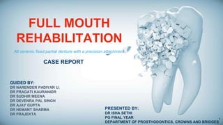
  • 1. FULL MOUTH REHABILITATION CASE REPORT GUIDED BY: DR NARENDER PADIYAR U. DR PRAGATI KAURANIDR DR SUDHIR MEENA DR DEVENRA PAL SINGH DR AJAY GUPTA DR HEMANT SHARMA DR PRAJEKTA PRESENTED BY: DR ISHA SETHI PG FINAL YEAR DEPARTMENT OF PROSTHODONTICS, CROWNS AND BRIDGES All ceramic fixed partial denture with a precision attachment 1
  • 3. Introduction01 Objectives of full mouth rehabilitation02 Reasons for full mouth rehabilitation03 Indications and contraindications04 CONTENTS Classifications of patients requiring FMR05 Etiology of worn dentition06 Diagnosis and treatment planning07 Vertical relation consideration08 Mandibular deprogramming 09 PART 1 3
  • 4. INTRODUCTION The personality of an individual is often judged by his looks. A beautiful smile always gives pleasure. However, the personality may be falsely interpreted by ugly and impaired teeth. 4
  • 5. INTRODUCTION “The time should be over where we are the dentists of the tooth or may be of two or three teeth at a time. Let us be the doctors of the mouth” McCollum 5
  • 6. INTRODUCTION Peter E. Dawson stated, ”Patient lose their teeth in two ways: either the teeth break down, other supporting structures break down” 6
  • 7. DEFINITION The term occlusal rehabilitation has been defined as the restoration of the functional integrity of the dental arches by use of inlays, crowns, bridges and partial dentures. DAWSON PE EVALUATION, DIAGNOSIS AND TREATMENT OF OCCLUSAL PROBLEMS, ST. LOUIS CV MOSBY, 2nd Edition, 164-68 Mouth Rehabilitation: Restoration of the form and function of the masticatory apparatus to as near normal as possible (GPT-9) Definition FULL MOUTH REHABILITATION The Glossary of Prosthodontic Terms, 9th Edition J Prosthet Dent 2017;117(5s) . 7
  • 9. Her teeth were straightened and the fangs were reshaped to give her a perfect smile. Teeth Whitening was also carried out on her stained teeth and she now sports beautiful porcelain veneers to give her that perfect celebrity smile . (May 23, 2014) Hollywood Stars Before & After Cosmetic Dentistry www.ewanbramley.com › Cosmetic Dentistry 9
  • 10. Introduction The word rehabilitation implies “To restore to the good condition or to restore to former privilege” The term ‘full mouth rehabilitation’ is used to indicate extensive and intensive restorative procedures in which the occlusal plane is modified in many aspects in order to accomplish “EQUILIBRATION”. Complete mouth rehabilitation is a dynamic functional endeavor and it embodies the correlation and integration of all component parts into one functioning unit. 10
  • 11. Introduction Planning and executing the restorative rehabilitation of a decimated occlusion is probably one of the most intellectually and technically demanding tasks facing a PROSTHODONTIST. The stakes are high and failure is costly. AHUJA.P., OCCLUSAL REHABILITATION. A CASE REPORT. JIPS 1999, 25- 11
  • 12. Objective Of Full Mouth Rehabilitation Our objective is to minimize these stresses so that they are not destructive. In order to prevent this stress from being destructive, the best thing to do is to distribute it evenly or as great area as possible, over as many teeth and as much tissue as possible, with the teeth providing a means by which the forces are distributed. All patients requiring full mouth rehabilitation have one problem in common: stress and strain. Usually the stress is due to malfunction or to poorly related parts of the oral mechanism. Irving Goldman: The goal of full mouth rehabilitation, J PROSTHET DENT 1951, vol 2, 246-251 12
  • 13. Reasons For Full Mouth Rehabilitation The most common reason for doing full mouth rehabilitation is to obtain and maintain the health of periodontal tissues. Temperomandibular joint disturbance is another reason. (Dawson, Lindhe & Nyman) Need for extensive dentistry as in case of missing teeth, worn down teeth and old fillings that need replacement. Esthetics, as in case of multiple anterior worn down teeth and missing teeth. Lucia W. O. : Modern Gnathological Concepts St.Louis: C.V.Mosby co.1961 13
  • 14. Indications Of Occlusal Rehabilitation Restore impaired occlusal function Preserve longevity of remaining teeth Maintain healthy periodontium Improve objectionable esthetics Eliminate pain and discomfort of teeth and surrounding structures. Lucia W. O. : Modern Gnathological Concepts St.Louis: C.V.Mosby co.1961 14
  • 16. Contraindications Of Full Mouth Rehabilitation Malfunctioning mouths that do not need extensive dentistry and have no joint symptoms should be best left alone. Prescribing a full mouth rehabilitation should not be taken as a preventive measure unless there is a definite evidence of tissue breakdown. In short, it can be concluded that : No pathology- No treatment. Lucia W. O. : Modern Gnathological Concepts St.Louis: C.V.Mosby co.1961 16
  • 17. Classifications of patients requiring FMR The patients were classified into three categories – Category 1 - Excessive wear with loss of vertical dimension. Category 2 - Excessive wear without loss of vertical dimension of occlusion but with space available. Category 3 - Excessive wear without loss of vertical dimension of occlusion but with limited space available Kenneth Turner & Donald Missirlian:Restoration of the extremely worn dentition, J PROSTHET DENT 1984, vol 52, 467-474 Classification by Turner and Missirlain (1984) 17
  • 18. CATEGORY 1 A typical patient in this category has few posterior teeth and unstable posterior occlusion. There is excessive wear of anterior teeth. Closest speaking space of 3 mm and interocclusal distance of 6 mm. There is some loss of facial contour that results in drooping of the corners of mouth. Kenneth Turner & Donald Missirlian: Restoration of the extremely worn dentition, J PROSTHET DENT 1984, vol 52, 467-474 18
  • 19. Category 1 Patients with dentinogenesis imperfecta with excessive occlusal attrition, around 35 years of age and appearing prognathic in centric occlusion also belongs to this category. Closest speaking space of 5 mm and inter occlusal distance of 9 mm indicates there is loss of occlusal vertical dimension with concomitant occlusal wear. Goud, Anil, and Saee Deshpande. "Prosthodontic rehabilitation of dentinogenesis imperfecta." Contemporary clinical dentistry 2.2 (2011): 138. (pictures) 19
  • 20. Category 2 -Patient has adequate posterior support and history of gradual wear. Closest speaking space of 1 mm and inter occlusal distance of 2-3 mm. -Continuous eruption has maintained occlusal vertical dimension leaving insufficient inter occlusal space for restorative material. -Manipulation of mandible into centric relation will often reveal significant anterior slide from centric relation to maximum intercuspation. Kenneth Turner & Donald Missirlian: Restoration of the extremely worn dentition, J PROSTHET DENT 1984, vol 52, 467-474 20
  • 21. Category 3 Posterior teeth exhibit minimal wear but anterior teeth show excessive gradual wear over a period of 20-25 years. Centric relation and centric occlusion are coincidental with closest speaking space 1mm and interocclusal distance 2- 3mm. It is most difficult to treat because vertical space must be obtained for restorative material. Kenneth Turner & Donald Missirlian: Restoration of the extremely worn dentition, J PROSTHET DENT 1984, vol 52, 21
  • 22. 22
  • 23. Classification by Breaker Group I Class I – Patients with collapse of vertical dimension of occlusion because of shifting of existing teeth caused by failure to replace missing teeth. Class II – Patients with collapse of vertical dimension of occlusion because of loss of all posterior teeth in one or both jaws with remaining teeth in unsatisfactory occlusal relationship. Class III – Patients with collapse of vertical dimension of occlusion because of excessive attritional wear of occlusal surfaces. Breaker S.C, Clinical procedures in occlusion Rehabilitation,W. B. Saunders, Philadelphia 1958 23
  • 24. Group II Class I – Patients with all or sufficient natural teeth present, with satisfactory occlusal relationship. Class II – Patients with limited teeth present but in satisfactory occlusal relationship requiring aid in the form of occlusal rims. Group III Patients requiring maxillofacial surgery of orthodontic treatment as an aid in restoring the lost vertical dimension. Group IV Patients in whom sectional treatment is required over extended periods of time because of status of health of the patient, age or economic factor. Breaker S.C, Clinical procedures in occlusion Rehabilitation,W. B. Saunders, Philadelphia 1958 24
  • 25. Etiology Of Extremely Worn Dentition Occlusal wear is most often attributed to attrition. Attrition is defined as ‘ the wearing away of one tooth surface by another tooth surface’. The causes for worn dentition are 1. Congenital abnormalities: • Amelogenesis imperfecta • Dentinogenesis imperfecta Bernard smith :Tooth wear : Etiology and diagnosis Gerodontology Text Book 1994 25
  • 26. Amelogenesis Impertecta Khodaeian, Niloufar, Mahmoud Sabouhi, and Ebrahim Ataei. "An Interdisciplinary Approach for Rehabilitating a Patient with Amelogenesis Imperfecta: A Case Report." Case reports in dentistry 2012 (2012). 26
  • 27. Goud, Anil, and Saee Deshpande. "Prosthodontic rehabilitation of dentinogenesis imperfecta." Contemporary clinical dentistry 2.2 (2011): 138. Dentogenesis Impertecta 27
  • 28. The causes for worn dentition are 2. Parafunctional occlusal habit 3. Abrasion 4. Erosion 5. Loss of posterior support: Posterior collapse that results from missing, tipped, rotated , broken down teeth, malposition and occlussal interference exerts undue force on anterior teeth resulting in teeth mobility and excessive wear of clinical crown. Bernard smith :Tooth wear : Etiology and diagnosis Gerodontology Text Book 1994, 88-102 28
  • 29. DIAGNOSIS AND TREATMENT PLANNING 29
  • 30. Complete mouth periapical radiographs and orthopentamograph Radiographs Dental history Behaviour evaluation Medical history The following aids should be used Computer imaging CBCT John Bowley, John Stockstill : A preliminary diagnostic and treatment protocol, D. Clin. North America1992, vol 36, 551-597 Photographs Clinical examination Diagnostic wax-up 30
  • 31. Diagnostic wax-up Before diagnostic wax-up, the occlusal discrepancies in centric and eccentric occlusion should be eliminated. Diagnostic preparation of gypsum stone teeth that will require prospective crowns is carried out. This will reveal any resistance or retention form problems caused by short axial walls. Thus planning of subgingival margins or surgical crown lengthening required can be done. Then wax is used to appropriately shape all crowns and final prosthesis is planned. This diagnostic wax-up can be used to prepare an elastomeric putty mould and used for temporization or sectioned through long axis of tooth to act as reduction guide intra-orally. John Bowley, John Stockstill : A preliminary diagnostic and treatment protocol, D. Clin. North America1992, vol 36, 551-597 31
  • 32. Treatment plan Pre- prosthetic phase Maintenance phase Prosthetic phase John Bowley, John Stockstill : A preliminary diagnostic and treatment protocol, D. Clin. North America1992, vol 36, 551-597) Comprehensive treatment plan must be established prior to start of the treatment . Communication and patient education are essential in order to match the dentist’s and patient’s definition of success. Treatment plan is divided into:- 32
  • 33. 1) Pre- prosthetic phase To develop proficiency in diagnosing the need of occlusal rehabilitation, all specialties (POEOP) be integrated in establishing an environment conducive to oral health. Harry Shrunik : Treatment Planning For Occlusal Rehabilitation, J PROSTHET DENT 1959, vol 9, 988-100 Periodontist Orthodontist Endodontist Prosthodontist Oral Surgeon 33
  • 34. 2) Prosthetic phase Prosthetic full mouth rehabilitation is divided into - Immediate treatment and Definitive treatment Harry Shrunik : Treatment Planning For Occlusal Rehabilitation, J PROSTHET DENT 1959, vol 9, 988-100 Immediate treatment In some cases like amelogenesis imperfecta in a child, postponing treatment until adulthood may cause adverse psychological effect and impair correct relationship between maxillary and mandibular teeth. Preformed nickel-chromium crowns are placed on first permanent molars and second deciduous molars to stabilize occlusion and halt attrition. Vertical dimension is not altered. As anterior teeth and premolars erupt, polycarbonate resin crowns are given. Second molar is fitted with nickel crown to preserve vitality. After all permanent teeth are erupted, these restorations serve as transitional treatment until adulthood. 34
  • 35. Definitive treatment Once all teeth have erupted and adulthood is reached, the size of pulp horns decreases compared to newly erupted teeth. A definitive treatment can then be planned. Harry Shrunik : Treatment Planning For Occlusal Rehabilitation, J PROSTHET DENT 1959, vol 9, 988-100 35
  • 36. Maintenance Phase Stimulate meticulous plaque control habits To monitor the dental health Identify incipient disease After placement and cementation of a prosthesis the patient treatment continues with carefully structured sequence of follow-up appointments. Recall schedule After maintaining adequate oral hygiene, patient is recalled at 1 month, 3 months, 6 and 12 months. After 1 year patient is recalled annually for check- up and prophylaxis. Follow ups Introduce any corrective measure if required Shetty et al, PHILOSOPHIES IN FULL MOUTH REHABILITATION – A SYSTEMATIC REVIEW , Int J Dent Case Reports,Nov-Dec 2013, Vol.3, ,No. 3 Adequate scaling is done periodically to maintain gingival health. Margins of restoration must be evaluated to detect secondary caries. Oral hygiene aids prescribed are tooth brushes, oral floss, interdental brush, oral irrigation devices and oral rinses. 36
  • 37. Prosthetic phase Diagnostic impression Facebow transfer Articulation 01 03 02 Encyclopedia of Biomedical Engineering, 2019 Articulator From Wikipedia, the free encyclopedia 37
  • 38. Determining the occlusal plane Simplified Occlusal Plane Analyzer Main Contents The average plane established by the incisal and occlusal surfaces of the teeth. Generally, it is not a plane but represents the planar mean of the curvature of these surfaces. Broadrick’s Occlusal Plane Analyzer (BOPA) Custom Made Occlusal Plane Analyzer The Glossary of Prosthodontic Terms, 9th Edition J Prosthet Dent 2017;117:05 Definition Various Occlusal Plane Analyser Dr. Lawson K Broadrick Availbility :- Broadrick flag Teledyne Water Pik Fort Collins Colo It is used for analyzing the Curve of Spee & developing an acceptable curve of Occlusion This simplified method reduces the time required for occlusal plane analysis because the analysis point for surveying the occlusal plane is already related to the condylar axis. Availability: Denar® Simplified Occlusal Plane Analyzer Whip Mix Corporation – West, CO 80525 38
  • 39. Preparation of the Analyzer Selection of anterior survey point Occlusal Plane Survey Line Determination of posterior survey point Broadrick’s Occlusal Plane Analyzer 1Step 2Step 3Step4Step Steps Gupta R, Luthra RP, Sheth H.H. Broadrick’s occlusal plane analyzer: A review. International Journal of Applied Dental Sciences 2019; 5(1): 95-98 39
  • 41. Can Vertical Dimension Be Altered? Out of the experience gained in occlusion of natural teeth has come an awareness that there are certain underlying treatment principles. These principles are so important that they cannot be overemphasized. Sicher (1949) and Silverman(1952). They concluded that as the teeth wear or become abraded, the teeth and alveolar bone elongate through growth to maintain the original vertical dimension with the maintenance of the same closest speaking space. However, occlusal wear may occur more rapidly than continuous eruption depending upon the etiology of the wear. Meyer Silverman : Vertical dimension must not be increased, J PROSTHET DENT 1952, v0l 2, pg 756-779 41
  • 42. Harry Kazis and Albert Kazis stated that treatment of reduced vertical dimension is not designed to increase the vertical dimension beyond the normal, but is intended to restore the amount of vertical dimension that has been lost. A young person will tolerate a greater correction of vertical dimension and become adjusted more easily to a reduction in the interocclusal distance as necessitated by the changes. Harry Kazis : Complete mouth rehabilitation through Fixed denture prosthodontics, J PROSTHET DENT 1969, vol 10, pg 296-303 42
  • 43. Meyer Silverman : Vertical dimension must not be increased, J PROSTHET DENT 1952, v0l 2, pg 756-779 Silverman (1956) said that closest speaking space can range from 0 to 10mm in different patients and that there is no average closest speaking space. But it is constant in an individual. Vertical dimension must not be increased beyond the normal for each patient. Increasing the vertical dimension only 1mm will cause discomfort to the patient . 43
  • 44. 44
  • 45. Joseph Landa : The freeway space and its significance in the rehabilitation of the masticatory apparatus,J PROSTHET DENT 1952, vol 2, pg 756-779 Landa (1955) stated that increasing the vertical dimension places the muscles of mastication and temperomandibular joint under strain. The crown to root ratio is also affected and hence ‘bite raising’ is contraindicated. The state of health of the temporomandibular joint structures, the neuromuscular reflexes of the masticatory mechanism, and the habitual postural position of the head and mandible should be taken into consideration in the determination of the vertical dimension of an individual case under treatment. 45
  • 46. Dawson PE Functional Occlusion. From TMJ to Smile Design, 1st edition (2009) Dawson (1974) stated that even when the teeth have grown down to the gum line the vertical dimension is not lost because of the eruption of the teeth along with the alveolar bone. Increase in vertical dimension interferes with the optimum length of the resting muscles which serve as a stimulus to produce hypertonicity. 46
  • 47. When it is not practical to restore severely worn dentition without restoring the vertical dimension, to obtain space for the restorative material, the dimension can be increased to 1- 1.5 mm. The potential problems of restoring the vertical dimension are clenching, muscle fatigue, soreness of teeth, muscles and joints, headache, intrusion of teeth, fracture of porcelain , occlusal instability due to shifting of restored teeth and continual wear. In such cases, checking and periodic occlusal adjustment must be done up to a year before normal stability returns. Dawson PE Functional Occlusion. From TMJ to Smile Design, 1st edition (2009) 47
  • 48. Carlsson et al : Effect of increasing vertical dimension on the masticatory system in subjects with natural teeth,J PROSTHET DENT 1979, vol 41, pg 284- 289 Carlsson et al (1979) increased the vertical dimension in natural dentition by cementing acrylic resin splints in lower canines, premolars and molars for 7 days. He found that subjects experienced moderate symptoms of discomfort initially but symptoms decreased later and no clinically demonstrable symptoms were found. A moderate increase in the vertical dimension of occlusion does not seem to be a hazardous procedure, provided that occlusal stability is established. 48
  • 49. Increasing occlusal vertical dimension — Why, When & How • VD is unrelated to temporomandibular disease (TMD) and there is no evidence to suggest that by changing VD one can treat TMD. However, VD can be increased or decreased for the best functional and aesthetic anterior contact in centric relation. Carlsson G E, Ingervall B, Kocak G. The effect of increasing vertical dimension on the masticatory system in subjects with natural teeth. J Prosthet Dent 1979; 41: 284-289. The vertical dimension of occlusion (VDO) is determined by the repetitive contracted length of the closing muscles, hence increase in VDO cannot be maintained as the jaw to jaw relationship will always return to the original dimension ie the MUSCLES always WIN. Kohno S ,Bando E. Die funktionelle anpassung der Kaumuskulatur Bei Starker Bissagbung (functional adaptation of masticatory muscles as a result of large increases in vertical dimension). Dtsch Zahnarztl ZI1983; 38: 759-764. 49
  • 50. Wear does not result in loss of VD, as the alveolar process lengthens to make up for this. But the position of the condyles does affect muscle length and hence the VDO. When looking at changes in VD it is paramount to mount the study casts in centric relation (CR). Dawson P E. Evaluation, diagnosis and treatment of occlusal problems. pp 280- 285. St Louis, MO: CV Mosby, 1989. 50
  • 51. Treatment options Reposition Restore Surgical osteotomy Orthogna thic surgery Equilibrate D. R. Bloom and J. N. Padayachy, Increasing occlusal vertical dimension — Why, When & How, BRITISH DENTAL JOURNAL VOLUME 200, NO.5 MARCH 2006 C 51
  • 52. Restore the lost Vertical dimension Grind opposing teeth Possible esthetic and pulpal problems Methods of Obtaining Space To Restore Worn Anterior Teeth May be required to increase axial wall height to aid in crown retention Crown Lengthening Rarely indicated but may be required where gross over- eruption has occurred Extraction/ Surgical Reposition ing Indicated only if majority of posterior teeth need full coverage restorations Robert Wassel : Tooth wear : Space creation with Dahl Appliance Gerodontology text book 1994,103-108 119/400 • Poyser, N. J., Porter, R. W. J., Briggs, P. F. A., Chana, H. S., & Kelleher, M. G. D. (2005). The Dahl Concept: past, present and future. British dental journal, 198(11), 669-676. 52
  • 53. VERTICAL DETERMINANTS There are four philosophies for condylar position when determining VD. All work on the basis of a canine protected occlusion. 1. Gnathological Involves use of fully adjustable articulators to determine condylar path from the hinge axis and setting this path for a 5 degree increase to ensure no posterior interferences D. R. Bloom and J. N. Padayachy, Increasing occlusal vertical dimension — Why, When & How, BRITISH DENTAL JOURNAL VOLUME 200, NO.5 MARCH 2006 C 53
  • 54. 4. Neuromuscular Based on the principles of muscle activity determined by electromyography. 2. Bioaesthetics Works via a fixed numerical value based on incisal relationship. Distance between gingival margins of 18-20 mm in an unworn class one occlusion, with upper incisal length of 12 mm, lower incisal length 10 mm, 4 mm overbite and 1 mm overjet. 3. Centric relation based Following the principles of P. Dawson whereby CR is defined as ‘when the heads of the condyles are in their most superior position within their sockets, lateral pterygoid muscle is relaxed and the elevator muscles are contracted with the disc properly aligned’ D. R. Bloom and J. N. Padayachy, Increasing occlusal vertical dimension — Why, When & How, BRITISH DENTAL JOURNAL VOLUME 200, NO.5 MARCH 2006 C 54
  • 55. POSSIBLE CLINICAL CONCERNS BEHIND CHANGING VD Stability When closing VD there is very little relapse; it may open by up to 1 mm within the first year and will then remain stable. Such a small amount is not detectable by the clinician or the patient. When opening the VD some patients can remain stable, others can relapse a little, and others a lot, but again this may go unnoticed dentally. D. R. Bloom and J. N. Padayachy, Increasing occlusal vertical dimension — Why, When & How, BRITISH DENTAL JOURNAL VOLUME 200, NO.5 MARCH 2006 C Joint or muscle pain This is not a problem, as altering VD does not produce pain of more than one to two weeks’ duration; any pain is a result of increased temporary muscle awareness Christensen J. Effect of occlusion raising procedures on the chewing system. Dent Pract Dent Rec 1970; 20: 233-238. 55
  • 56. Muscle activity Lindauer S J, Gay T, Rendell J. Effect of jaw opening on masticatory muscle EMG — force characteristics. J Dent Res 1993; 72: 51-55. The results of this study suggest that changes in masticatory muscle length resulting from vertical jaw opening cause alterations in muscle contractile properties, but the relative contributions of various masticatory muscles toward bite force production may also be affected by biomechanical factors and neural control adaptations. 56
  • 57. There can sometimes be a problem for the ‘S’ sounds. (Can be solved by creating space.) Generally this will be by shortening the lower incisors *- how depends on the lower incisor position when the ‘S’ sound is created: 1. If ‘S’ is generated with the lower incisors in the cingulum area of the upper incisors (ie behind and above the upper incisal tip), shortening the lower incisors will leave them out of contact when the teeth are in occlusion. For this reason the VD will then need to be reduced. 1. If ‘S’ is generated by the incisors being more edge-to edge the lower incisors can be reduced and the linguals of the upper incisors built out to maintain contact. Hammond R G, Beder O E. Increased vertical dimension and speech articulation errors. J Prosthet Dent 1984: 52: 401-406. Phonetics 57
  • 58. ANTERIOR DETERMINANTS OF VERTICAL DIMENSION When changing incisal position restoratively, it is paramount to do this in provisional restorations first. Provisional restorations can be modified in the mouth until all guidelines have been precisely followed and the patient completely happy. As ever, a diagnostic wax-up will aid in such treatment planning. . 1. Stable CR contacts. 2. Upper half of the labial surface. D. R. Bloom and J. N. Padayachy, Increasing occlusal vertical dimension — Why, When & How, BRITISH DENTAL JOURNAL VOLUME 200, NO.5 MARCH 2006 C 58
  • 59. 3. Lower half of labial surface 4.Incisal edge. 5.Anterior guidance D. R. Bloom and J. N. Padayachy, Increasing occlusal vertical dimension — Why, When & How, BRITISH DENTAL JOURNAL VOLUME 200, NO.5 MARCH 2006 C 6. Contour of the lingual surface from the centric stop to the gingival margin. 59
  • 61. CONTENTS Monolithic zirconia and Precision attachment (Brief Idea)02 Case report03 Conclusion and take away message 04 References 05 Definition of centric relation and muscle deprogramming01 PART 2 61
  • 62. > a maxiillomandibular relationship, > independent of tooth contact, > in which the condyles articulate in the anterior-superior position against the posterior slopes of the articular eminences; > in this position, the mandible is restricted to a purely rotary movement; > from this unstrained, physiologic, maxillomandibular relationship, the patient can make vertical, lateral or protrusive movements; > it is a clinically useful, repeatable reference position (GPT-9) Centric Relation Proper manipulation of mandible as in equilibration position when no bite record is taken 01 Manner of taking bite record for correct articulation of mounted models. 02 There are two aspects of taking centric relation DAWSON PE EVALUATION, DIAGNOSIS AND TREATMENT OF OCCLUSAL PROBLEMS, ST. LOUIS CV MOSBY, 2nd Edition, 164-68 62
  • 63. Mandibular Deprogramming Most patients have a reflex closure , an engram determined and guided by the teeth. Proprioceptive mechanism determines path of mandibular closure and is responsible for awareness of position of mandible in space. To enable the condyles to be placed in an unstrained position, the musculature must first be deprogrammed from its habitual closing pattern. DAWSON PE Functional occlusion. TMJ to smile design 1st edition (2009) 63
  • 64. Chinpoint Guidance method Guichet described this method. It places the condyles in most posterior and superior position which can result in trauma to TMJ. Hence use of this method is not advocated Bilateral Manipulation method Dawson introduced this method that guides the condyles into most superior position in the glenoid fossa. Unguided method Brill introduced a muscular position which allows patient’s natural muscle functions to position the mandible into centric relation position. Methods Available To Guide The Mandible Into Centric Relation DAWSON PE EVALUATION, DIAGNOSIS AND TREATMENT OF OCCLUSAL PROBLEMS, ST. LOUIS CV MOSBY, 2nd Edition, 164-68 64
  • 66. Anterior Bite Stops -Anterior jig prevents posterior teeth from occluding and thus disrupts the proprioceptive memory. -As the anterior stop is rigid on contact with lower incisor teeth, anterior resistance is created and a mandibular leverage is created with naturally braced tripod effect along with two condyles -Jig breaks the patient’s habitual closure pattern and acts as the third leg of the tripod by creating resistance while stopping the closure. Principles 1. The Pankey Jig by Dr. Keith Thornton 2. The Best Bite Appliance 3. Lucia Jig 4. The NTI 5. The Leaf Gauge by Dr. Hart Long 66
  • 67. Leaf Guage was first introduced by Dr. James .H. Long in 1973 It is the most useful and practical alternative to anterior jig. Leaf guage can be used for- – Centric relation interocclusal records – Occlusal equilibration – Relieve painful spasms of lateral pterygoid muscle. Previously they were made of unexposed X- ray films after developing to remove the emulsion coating. Clear film was then cut into 1 cm X 5 cm sections. Recently, leaf gauges of uniform 0.1mm thickness which are sequentially numbered are described. They are convenient and measure the exact vertical opening between the incisors. Leaf Guage 67
  • 68. Leaf Gauge Woelfel (1991) used leafguage wafer technique to record jaw relation Huffman (1987) advocated use of leaf guage for occlusal equilibration. Alber’s et al stated in 1997 that the use of cotton rolls for initial joint compression and retrusion followed by recording with leaf guage appears to be the best method for obtaining accuracy. . Williamson used leaf guage to deprogram the proprioceptive impulses from the periodontal membrane. McHorris advocated leaf guage for centric interocclusal records and relieving painful spasm of lateral pterygoid muscles. Golsen and Shaw recommended leaf guage in occlusal adjustment and for centric relation records Woelfel described a disposable leaf guage made of firm paper. Solomon and Shetty (1996) found obtaining centric relation with the use of leaf guage to be accurate compared to unguided technique and operator guided closure 68
  • 69. Arbitary number of leaves are placed at the maxillary anterior midline parallel to the lingual plane of central incisors. Patient is instructed to close on back teeth until lower incisors touch on back side of leaf guage. Leaves are added or subtracted until patient can barely feel a posterior tooth touch while closing firmly on leaf guage. Often the patient can feel a posterior tooth contact in 15- 52 seconds after the jaw is closed with a ‘half hard’ closing force. This procedure is repeated after adding a leaf guage until the patient can close for 2-5 minutes without feeling a posterior tooth contact. Procedure 69
  • 71. 71
  • 72. 72
  • 73. 73
  • 75. The desire to balance between functional stability and cosmetic appeal in partial dentures gave rise to the development of Precision Attachments The precision attachment is sometimes said to be a connecting link between fixed and removable partial denture as it incorporates features common to both types of construction. INTRODUCTION Harold W. Preiskel. Precision attachment in prosthodontics. Application of intracoronal and extracoronal attachments. Volume 1 75
  • 76. Winder “Winders design” Screw joint retention Parr (1886) Extracoronal socket attachment Stair Telescopic abutment restoration Ash (1912) Split bar attachment system Historical Background Harold W. Preiskel. Precision attachment in prosthodontics. Application of intracoronal and extracoronal attachments. Volume 1 76
  • 77. Late 19th century “T shaped” Precision Attachment (1906) “H shaped” Chayes Attachment (1912) First attachment to be available in the general market Dr.Herman, ES Chayes Harold W. Preiskel. Precision attachment in prosthodontics. Application of intracoronal and extracoronal attachments. Volume 1 77
  • 78. Definition Precision – quality or state of being precise Attachment – Mechanical device for the fixation, retention and stabilization of dental prosthesis. Precision Attachment (GPT-9) : A retainer consisting of a metal receptacle (matrix) and a closely fitting part (patrix); the matrix is usually contained within normal or expanded contours of the crown on the abutment tooth and the patrix is attached to a pontic or the removable partial denture framework. An interlocking device, one component of which is fixed to an abutment or abutments, and the other is integrated into a removable prosthesis to stabilize and/or retain it. chayes Ceka and dallabona Pin slot Harold W. Preiskel. Precision attachment in prosthodontics. Application of intracoronal and extracoronal attachments. Volume 1 78
  • 79. Mechanical device – Direct retainer • They are designed to replace occlusal rest, bracing arm, and retaining arm of the conventional clasp retained partial denture. • They function to retain, support and stabilize the removable partial denture. Harold W. Preiskel. Precision attachment in prosthodontics. Application of intracoronal and extracoronal attachments. Volume 1 79
  • 80. SYNONYMS OF ATTACHMENTS Matrix slot Male attachment Female attachment CryptInsert Key key Patrix Internal attachments, Frictional attachments, Parallel attachments, Slotted attachments, Key and Key way attachments Fitting part keyway Flange Recept acle Harold W. Preiskel. Precision attachment in prosthodontics. Application of intracoronal and extracoronal attachments. Volume 1 80
  • 81. CLASSIFICATION OF ATTACHMENTS Based on method of fabrication and the tolerance of fit I. Precision attachment (prefabricated types) II. Semi precision attachment (custom made / laboratory made types) (Prefabricated wax / plastic / nylon patterns) Harold W. Preiskel. Precision attachment in prosthodontics. Application of intracoronal and extracoronal attachments. Volume 1 81
  • 82. According to their relationship to the abutment teeth: Intracoronal (Internal attachment) Extracoronal (External attachment) Based on stiffness of the resulting joint: Rigid attachments Resilient attachments (Non rigid) Harold W. Preiskel. Precision attachment in prosthodontics. Application of intracoronal and extracoronal attachments. Volume 1 82
  • 83. Key and Keyway Interlocks Ball and socket Bar and clip / sleeve Hinge Telescopic Push button Latch Screw units Based on geometric configuration and design of the attachment Intra-dental attachments - Frictional - Magnetic Extra-dental attachments - Cantilever attachment - Bar attachment Gerardo Beccera and others (1987) Harold W. Preiskel. Precision attachment in prosthodontics. Application of intracoronal and extracoronal attachments. Volume 1 83
  • 84. ADVANTAGES Improved esthetics and elevated psychological acceptance Mechanical advantage * Direct the forces along the long axis of the teeth / more apically * Force application closer to the fulcrum of the tooth - Reduces Non axial loading -Decreases Torquing forces -Rotational movement of the abutment *In Distal extension base cases – “Broken stress philosophy” -Reduced stress to the abutment -Stress free rotational/vertical movements *Cross arch load transfer and prosthesis stabilization -Less liable to fracture than clasp Harold W. Preiskel. Precision attachment in prosthodontics. Application of intracoronal and extracoronal attachments. Volume 1 84
  • 85. DISAVANTAGE *Complexity of design, procedures for fabrication & clinical treatment *Minimum occlusogingival abutment height (4-6mm) -To incorporate attachment without overcontouring -Enough length of parallel contact *Expensive -Complexity of laboratory and clinical procedure -Attachment maintenance (repair or periodic replacement) *Wearing of attachment components *Require high technical expertise – Dentist and laboratory technician *Requires aggressive tooth preparation *Cooperation and manual dexterity on the part of the patient -Difficult to insert and remove -Visually or manually challenged patient Harold W. Preiskel. Precision attachment in prosthodontics. Application of intracoronal and extracoronal attachments. Volume 1 85
  • 86. Indications Removable Prosthodontics As a retainer in a removable tooth supported partial denture - 4 large well rounded abutments are available - For esthetic concern in the anterior part of the mouth Stress Breakers - Free end saddles/Distal Extension Base cases (DEB) - When cantilevered pontic is to be used as abutment For movable joints in sectional dentures Periodontal involvement of the tooth - Contraindicates rigid FPDs - Most efficient bilateral bracing and support are essential Divergent abutment teeth with high survey lines (parallel path of placement.)Harold W. Preiskel. Precision attachment in prosthodontics. Application of intracoronal and extracoronal attachments. Volume 1 86
  • 87. Implant prosthodontics -Implant supported over denture -They are used for connection between the tooth and the implant Fixed Prosthodontics - As a connector in fixed partial denture construction (long span bridges) -To overcome alignment problems where abutments have differing path of withdrawal Harold W. Preiskel. Precision attachment in prosthodontics. Application of intracoronal and extracoronal attachments. Volume 1 87
  • 88. CONTRAINDICATIONS *Poor periodontal support. *Poor crown to root ratio *Poor oral hygiene habits *Abnormally high carious rate *Inadequate space / room to employ the attachment *Compromised endodontic and restorative conditions Harold W. Preiskel. Precision attachment in prosthodontics. Application of intracoronal and extracoronal attachments. Volume 1 88
  • 89. Case Report Full Mouth Rehabilitation of Patient with Bruxism using Tooth-supported Monolithic Zirconia fixed prosthesis and precision attachment. 89
  • 90. Summary Full mouth rehabilitation in patients with bruxism is often challenging. Bruxism results in severely worn dentition and often loss in Vertical Dimension of Occlusion (VDO). This case report is about a patient having bruxism with generalized attrition having minimal loss of vertical dimension in occlusion. After complete mouth root canal treatment, patient was kept on interim restorations for a month. A multidisciplinary approach was followed for permanent rehabilitation with fixed monolithic zirconia crowns and precision attachment for edentulous area. A review after 3 months revealed a well functional and esthetic prosthesis. Chief complaint Patient’s chief complaint was of sensitivity in upper and lower teeth region. Patient also complaint of bad look of teeth. History (Dental/Medical) Patient did not report to any dentist before, lost his teeth in upper right teeth region 5 years back as it got loose. Medical history- NA Personal history Patient has a habit of grinding teeth while at the job. Non smoker, non alcoholic. Extraoral examination Patient had no symptoms of TMJ disorders or any lymph node abnormality. There were no sign of swelling or any other extraoral abnormality. Competent lips Case History 90
  • 91. Soft tissue Oral hygiene – Good Gingiva – reddish pink, firm, resilient, no inflammation, stippling visible. No periodontal pockets Missing 15,16,17,18 24 35, 37, 38 47 Hard tissue Generalized severe attrition Grossly decayed- 25,36 category 2 of tooth wear Other findings Generalized sensitivity, Generalized TOP positive, Class I Occlusion (dental & skeletal) Type 1 Dawson Classification. Intra oral examination L R R L 91
  • 92. Facebow transfer 1. Diagnostic impression: Alginate impression was made of upper and lower arch. 1. Facebow transfer 1. Inter-occlusal bite registration in centric and protrusive using Alu Wax. 1. Mounting of upper and lower casts on semi-djustable articulator. 1. Occlusal plane analysis Diagnostic Procedures 92
  • 93. Diagnostic wax up 1. All the occlusal discrepancies were removed and desired mockup preparation was done on the casts. 1. The patient was shown the diagnostic wax-up over the articulator for the acceptance and consent was taken. 1. The putty index was made for upper and lower diagnostic waxups for further assistance in various clinical procedures. Note – there was no need for increase in the vertical dimension as the patient was under category 2 classification of T&M. Diagnostic Procedures 93
  • 94. Treatment planning Based on the case history, oral examinations, radiographic examinations, CBCT, diagnosis and patient’s own expectations and requirements, following treatment planning was proposed- 1. Endodontic treatment for full mouth was advised. 1. All ceramic restorations for full mouth rehabilitation except 13 and 14 in which PFM crowns were planned so that to give precision attachment was advised. Post and core was planned for 21 ad 25. 1. For the edentulous region, the CBCT was advised as patient wanted a fixed restoration but the CBCT reports showed lack of available bone for implant so precision attachment was advised. 1. Followed by maintenance follow ups for one year was done as explained earlier. 94
  • 102. Final prosthesis at lab Pictures sent by ILLUSION Lab, Bombay Die cutting and die ditching 102
  • 103. Final prosthesis Different views of the prosthesis in centric relation 103
  • 105. Precision attachment 105(OT CAP, Rhein 83 Inc, USA)
  • 106. Pre and post operative pictures Patient was well satisfied with the prosthesis. Evaluation of all the crowns individually was done for any remaining cement. All the oral hygiene measures were described to the patient along with the maintenance of the removable part of the rehabilitation. Final appointment Was done after 24 hours of cementation 106
  • 107. As the goal of medicine is to increase the life span of the functioning individual, the goal of dentistry is to increase the life span of the functioning dentition. “You are never fully dressed without a smile”. Conclusion 107
  • 108. References 1. Dawson PE, Evaluation, Diagnosis and Treatment of Occlusal Problems, St. LOUIS CV MOSBY, 2nd edition, 164-68. 2. AHUJA.P., OCCLUSAL REHABILITATION. A CASE REPORT. JIPS 1999, 25-28 3. Irving Goldman: The goal of full mouth rehabilitation , J Prosth Dent 2(2) : 246 -51, 1952 4. Mann A W, Pankey L D: The Pankey Mann philosophy of occlusal rehabilitation, Dent Clin North Am 7: 621-38 , 1963 5. Mann A W, Pankey L D: Oral Rehabilitation, J Prosth Dent 10: 135-62 ,1960 6. Schyuler C H : Factors in Occlusion applicable to restorative dentistry , J Prosth Dent 3 : 722- 82 , 1953 234/240 235 7. Shetty et al, PHILOSOPHIES IN FULL MOUTH REHABILITATION – A SYSTEMATIC REVIEW , Int J Dent Case Reports,Nov-Dec 2013, Vol.3, ,No. 3 8. Hobo S : Twin Table technique for occlusal rehabilitation : Part I – Mechanism of Anterior guidance , J Prosth Dent 66 (3) : 299-303 , 1991 9. Hobo S: Twin Table technique for Occlusal rehabilitation: Part II – Clinical procedure , J Prosth Dent 66 (4) : 471- 77 , 1991 10. Hobo S: Oral rehabilitation . Clinical determination of Occlusion. Quintessence publication, London. 11. Kazis Harry: Complete Mouth Rehabilitation through restoration of lost vertical dimension , J.A.D.A 37 : 19, 1948 12. Kazis Harry: Functional aspects of complete mouth rehabilitation. J Prosth Dent 4 (6): 833-842, 1954 235/240 13. Harry Kazis, Albert Kazis : Complete Mouth Rehabilitation through fixed partial denture Prosthodontics. J Prosth Dent 10 (2): 296-303 , 1960 14. Joseph. S. Landa: An analysis of current practices in mouth rehabilitation. J Prosth Dent 5(4):527-37, 1955 15. The Glossary of Prosthodontic Terms, 9th Edition J Prosthet Dent 2017;115:05 16. Rosenstiel SF, Land MF, Fujimoto J. Contemporary fixed prosthodontics. 3rd ed. St. Louis: Elsevier; 2000. p. 46-64. 17. Shillingburg HT, Hobo S, Whitsett LD, Jacobi R, Brackett SE. Fundamentals of fixed prosthodontics. 3rd ed. Chicago: Quintessence; 1997.p. 85-103, 191-2. 18. V Rangarajan, Textbook Of Prosthodontics, pg 470 19. Joseph E. Ewing, Fixed Parial Prosthesis, 2nd Edition, 14-20. 236/240 108