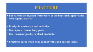
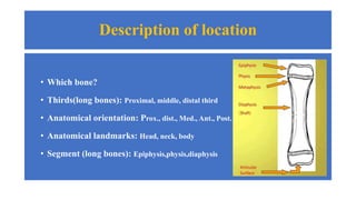
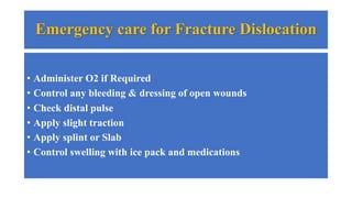
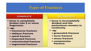
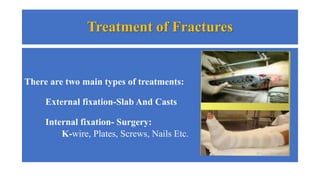
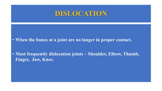
FRACTURE DISLOCATION
FRACTURE
• Bones form the skeletal frame work of the body and supports the
body against activity.
• It helps in movement and activities.
• Bones protect some body parts.
• Bone marrow produces blood products.
• Fractures occur when bone cannot withstand outside forces.
Description of location
• Which bone?
• Thirds(long bones): Proximal, middle, distal third
• Anatomical orientation: Prox., dist., Med., Ant., Post.
• Anatomical landmarks: Head, neck, body
• Segment (long bones): Epiphysis,physis,diaphysis
Signs and Symptoms of a Fracture
• Pain and tenderness
• Loss of function
• A wound may present
• Deformity
• Abnormal Mobility
• Loss of Transmitted Mobility
• Swelling and bruising
Emergency care for Fracture Dislocation
• Administer O2 if Required
• Control any bleeding & dressing of open wounds
• Check distal pulse
• Apply slight traction
• Apply splint or Slab
• Control swelling with ice pack and medications
Diagnosing Bone Fractures
• X-ray of injured area.
• Some fractures are difficult to see in an X- Ray, so a CT scan, MRI
or other bone scans are used.
Types of Fractures
Complete Fractures
Incomplete Fractures
Treatment of Fractures
There are two main types of treatments:
External fixation-Slab And Casts
Internal fixation- Surgery:
K-wire, Plates, Screws, Nails Etc.
DISLOCATION
• When the bones at a joint are no longer in proper contact.
• Most frequently dislocation joints – Shoulder, Elbow, Thumb,
Finger, Jaw, Knee.
Sign of a Dislocation
• Deformity or abnormal appearance.
• Pain and tenderness aggravated by movement.
• Loss of normal function.
• Joint may be locked in one position.
• Swelling of joint.
General Treatment Principles
• Stop the activity.
• Survey the injured area.
• First aid if qualified.
• Determine if additional medical attention is necessary.
RICE
• REST
• IMMOBILIZE
• COLD
• ELEVATION
Shoulder Dislocation
• Take a past medical history
• Clinical examination
• X-ray
• Several method for reduction scapular rotation
Anterior Dislocation, Right shoulder
Glenohumeral Reductions
THANK YOU

FRACTURES & DISLOCATIONS general overview.pptx

  • 1.
  • 2.
    FRACTURE • Bones formthe skeletal frame work of the body and supports the body against activity. • It helps in movement and activities. • Bones protect some body parts. • Bone marrow produces blood products. • Fractures occur when bone cannot withstand outside forces.
  • 3.
    Description of location •Which bone? • Thirds(long bones): Proximal, middle, distal third • Anatomical orientation: Prox., dist., Med., Ant., Post. • Anatomical landmarks: Head, neck, body • Segment (long bones): Epiphysis,physis,diaphysis
  • 4.
    Signs and Symptomsof a Fracture • Pain and tenderness • Loss of function • A wound may present • Deformity • Abnormal Mobility • Loss of Transmitted Mobility • Swelling and bruising
  • 5.
    Emergency care forFracture Dislocation • Administer O2 if Required • Control any bleeding & dressing of open wounds • Check distal pulse • Apply slight traction • Apply splint or Slab • Control swelling with ice pack and medications
  • 6.
    Diagnosing Bone Fractures •X-ray of injured area. • Some fractures are difficult to see in an X- Ray, so a CT scan, MRI or other bone scans are used.
  • 7.
  • 8.
  • 9.
  • 10.
    Treatment of Fractures Thereare two main types of treatments: External fixation-Slab And Casts Internal fixation- Surgery: K-wire, Plates, Screws, Nails Etc.
  • 11.
    DISLOCATION • When thebones at a joint are no longer in proper contact. • Most frequently dislocation joints – Shoulder, Elbow, Thumb, Finger, Jaw, Knee.
  • 12.
    Sign of aDislocation • Deformity or abnormal appearance. • Pain and tenderness aggravated by movement. • Loss of normal function. • Joint may be locked in one position. • Swelling of joint.
  • 13.
    General Treatment Principles •Stop the activity. • Survey the injured area. • First aid if qualified. • Determine if additional medical attention is necessary.
  • 14.
  • 15.
    Shoulder Dislocation • Takea past medical history • Clinical examination • X-ray • Several method for reduction scapular rotation
  • 16.
  • 17.
  • 18.