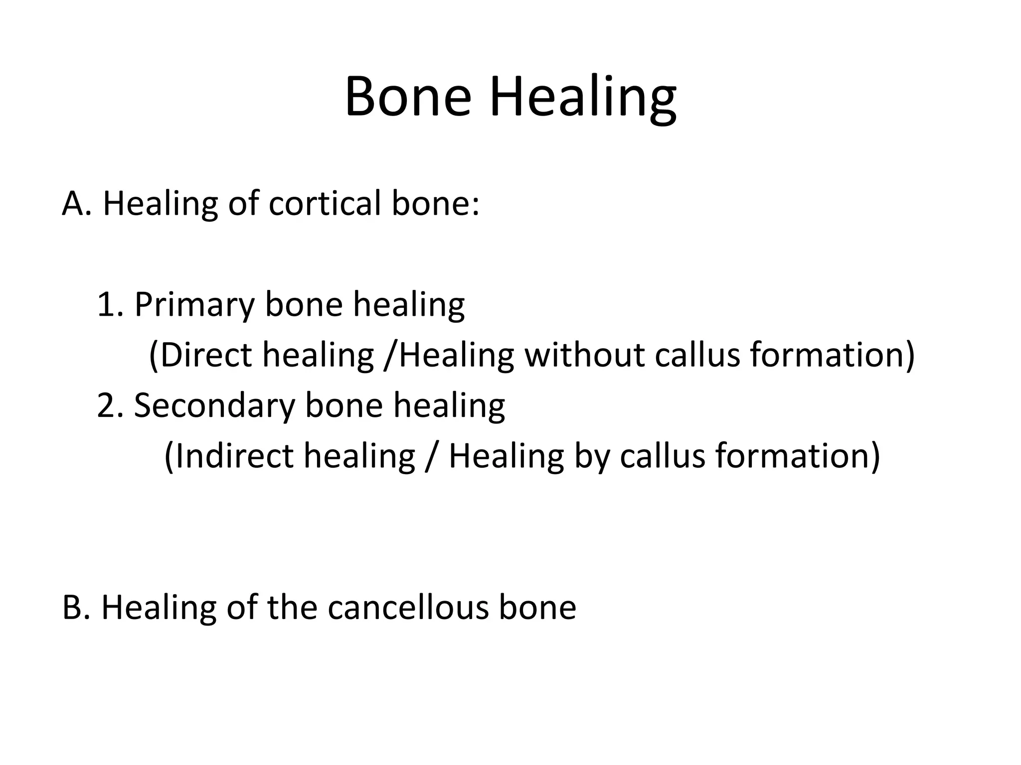
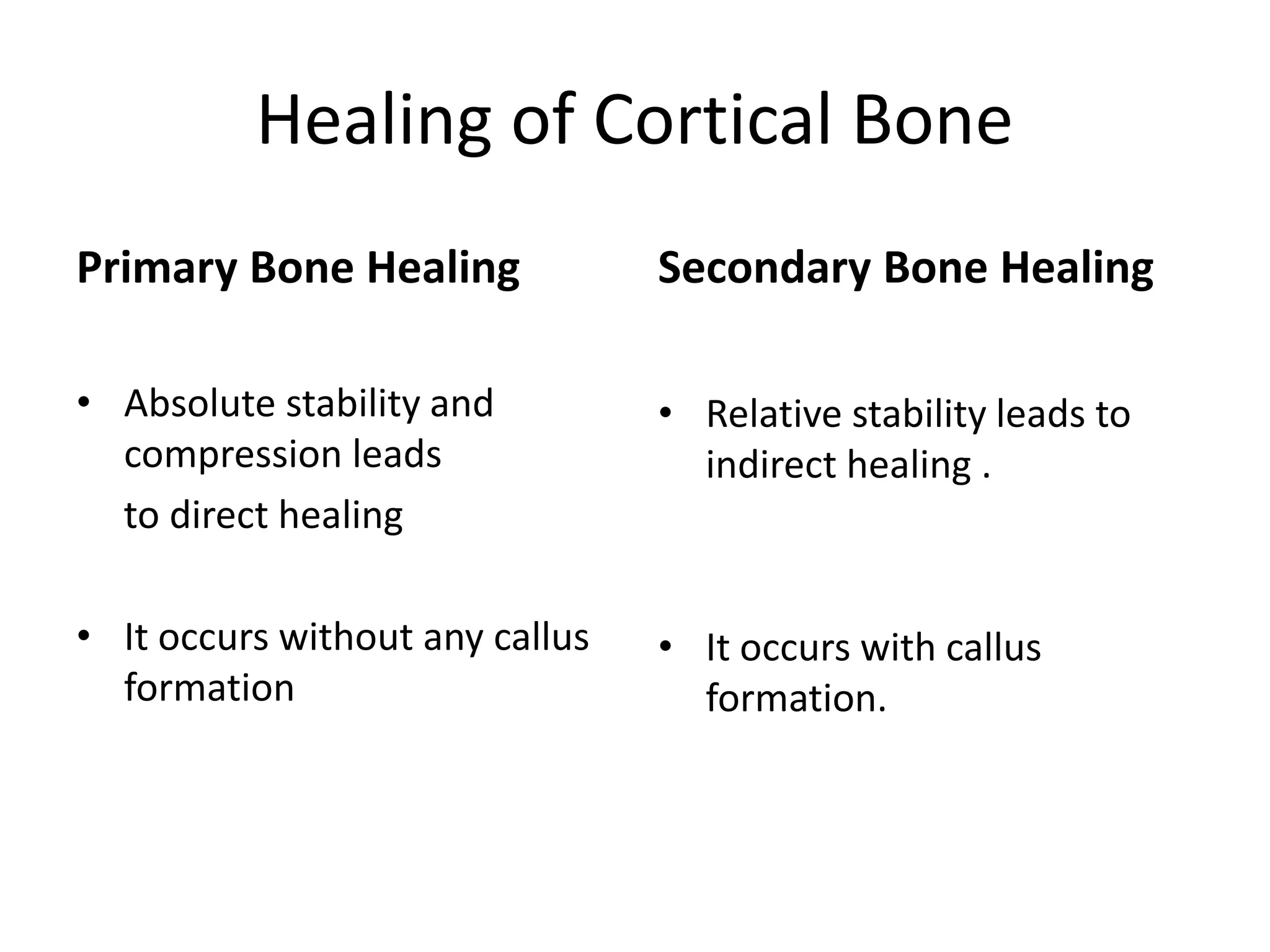
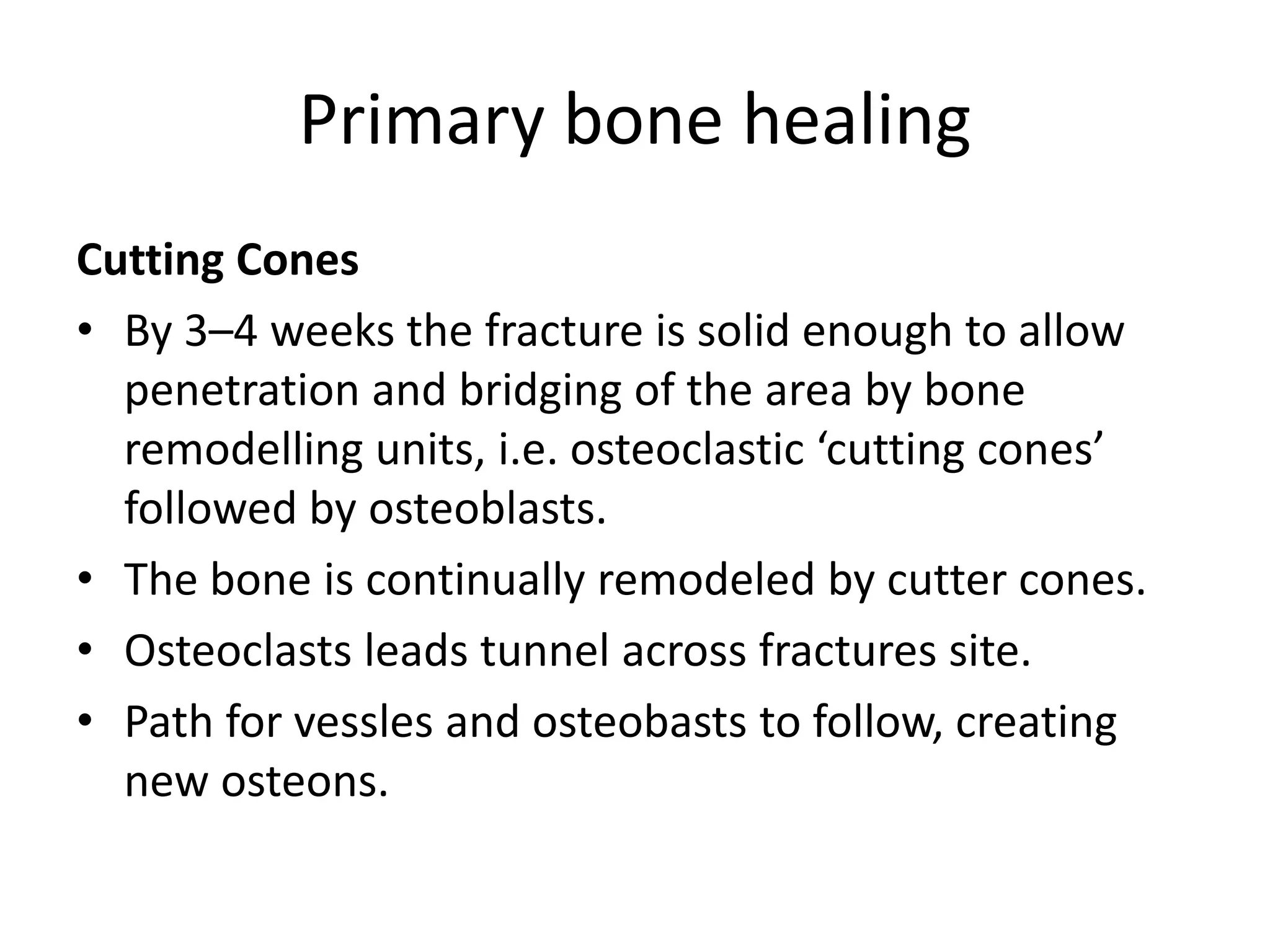
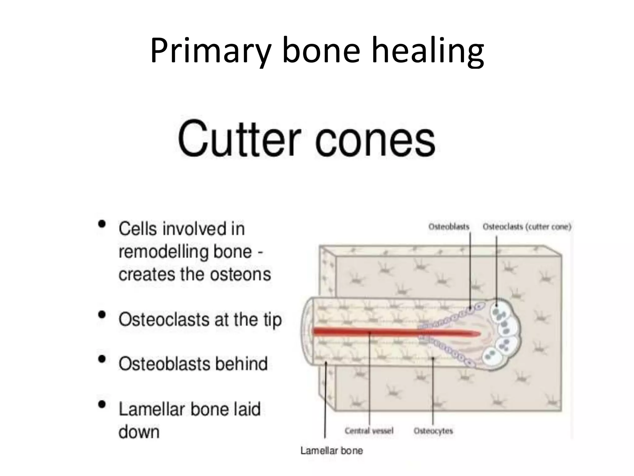
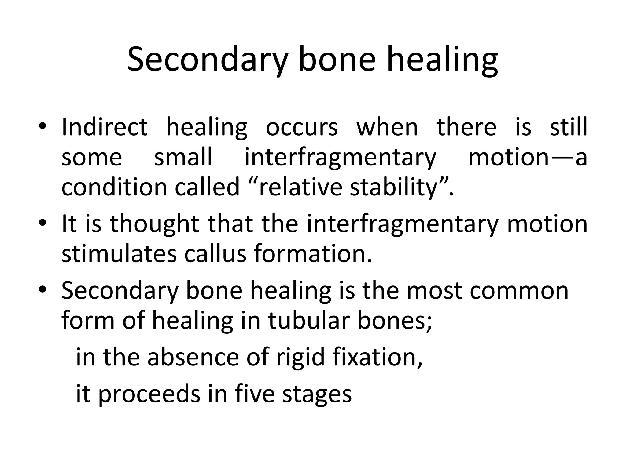
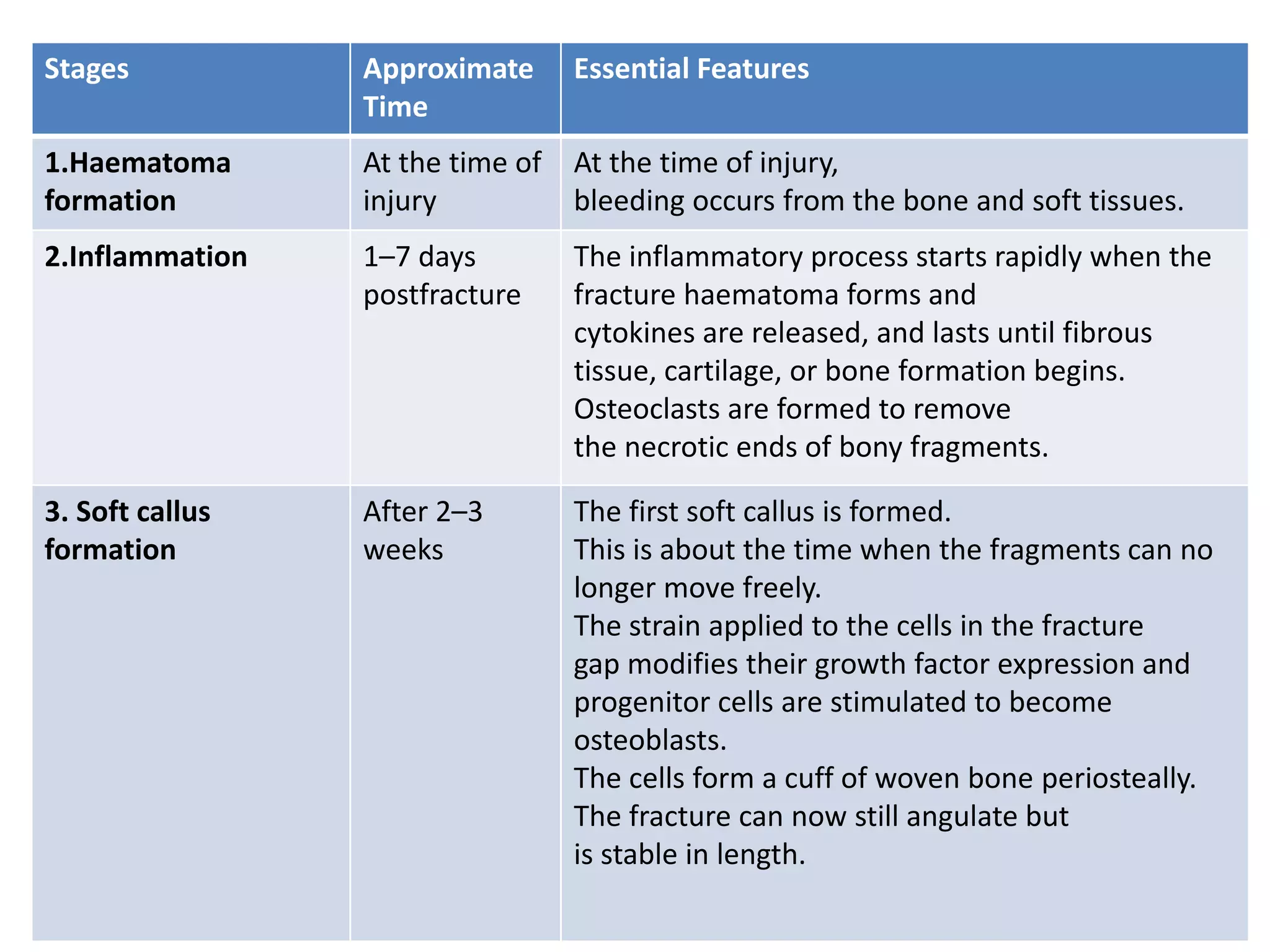
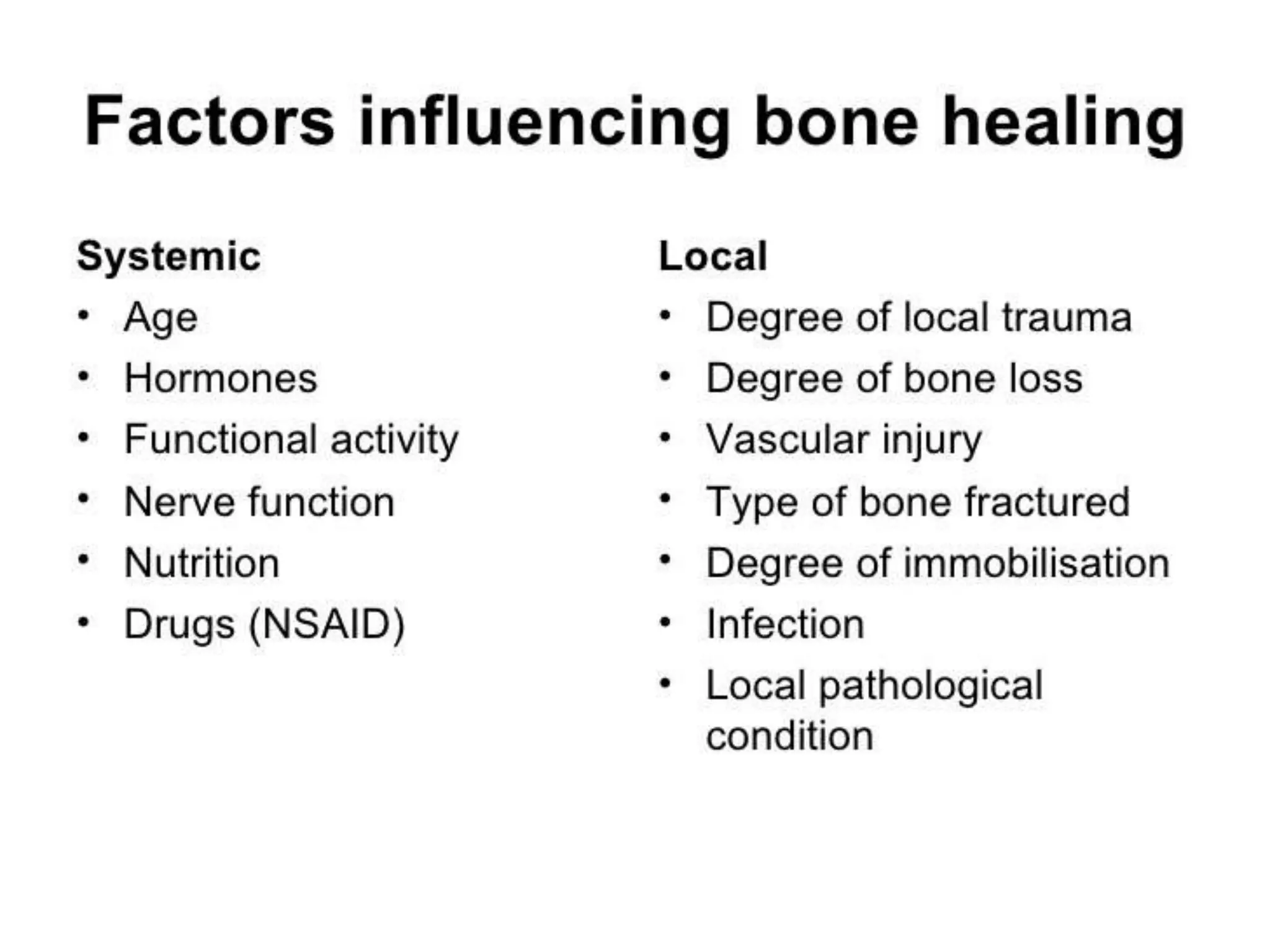
2. Bone healing:
Bone healing is characterized by a process of
new bone formation with fusion of the bone
fragments.
3. Bone Healing
A. Healing of cortical bone:
1. Primary bone healing
(Direct healing /Healing without callus formation)
2. Secondary bone healing
(Indirect healing / Healing by callus formation)
B. Healing of the cancellous bone
4. Healing of Cortical Bone
Primary Bone Healing
• Absolute stability and
compression leads
to direct healing
• It occurs without any callus
formation
Secondary Bone Healing
• Relative stability leads to
indirect healing .
• It occurs with callus
formation.
5. Primary bone healing
• Direct fracture healing occurs:
Under conditions of absolute fracture stability as
provided by
lag screws,
compression plate and
TBW.
By direct osteonal remodeling.
Without callus formation.
• If the fracture site is absolutely stable – for example, an impacted
fracture in cancellous bone, or a fracture held by a metal plate with
absolute stability – there is no stimulus for callus.
• Osteoblastic new bone formation occurs directly between the
fragments.
6. Primary bone healing
Contact Healing
• Exposed fracture surfaces are in intimate contact
and held from the outset with absolute stability,
internal bridging may occasionally occur without
any intermediate stages.
Gap Healing
• It occurs even if there is a minimal gap.
• This is called gap healing.
• Gaps between the fracture surfaces are invaded by new
capillaries and osteoprogenitor cells growing in from the
edges, and new bone is laid down on the exposed surface
7. Primary bone healing
Cutting Cones
• By 3–4 weeks the fracture is solid enough to allow
penetration and bridging of the area by bone
remodelling units, i.e. osteoclastic ‘cutting cones’
followed by osteoblasts.
• The bone is continually remodeled by cutter cones.
• Osteoclasts leads tunnel across fractures site.
• Path for vessles and osteobasts to follow, creating
new osteons.
8. Primary bone healing
• An osteon is a basic construction unit also called
haversian system.
• Each osteon has a central canal, containing blood
vessels and a small amount of connective tissue with
interconnecting channels surrounded by concentric
layers of bone, the laminae.
• There are no osteons in cancellous bone.
• Due to the activity of cutter cones, tunnels are cut
through the compact bone, resulting in the creation of
new haversian osteons in their wake which bridges the
fractures.
10. Primary bone healing
• With rigid metal fixation,however, the absence of
callus means that there is a long period during
which the bone depends entirely upon the metal
implant for its integrity, increasing the risk of
implant failure.
• Moreover, the implant diverts stress away from
the bone, which may become osteoporotic
and may not recover fully until the metal is
removed.
11. Secondary bone healing
Healing by callus, though less direct (indirect
healing) has distinct advantages:
it ensures mechanical strength while
the bone ends heal, and
with increasing stress the callus grows
stronger and stronger
(according to Wolff’s law)
12. Secondary bone healing
• Indirect healing occurs when there is still
some small interfragmentary motion—a
condition called “relative stability”.
• It is thought that the interfragmentary motion
stimulates callus formation.
• Secondary bone healing is the most common
form of healing in tubular bones;
in the absence of rigid fixation,
it proceeds in five stages
14. Stages Approximate
Time
Essential Features
1.Haematoma
formation
At the time of
injury
At the time of injury,
bleeding occurs from the bone and soft tissues.
2.Inflammation 1–7 days
postfracture
The inflammatory process starts rapidly when the
fracture haematoma forms and
cytokines are released, and lasts until fibrous
tissue, cartilage, or bone formation begins.
Osteoclasts are formed to remove
the necrotic ends of bony fragments.
3. Soft callus
formation
After 2–3
weeks
The first soft callus is formed.
This is about the time when the fragments can no
longer move freely.
The strain applied to the cells in the fracture
gap modifies their growth factor expression and
progenitor cells are stimulated to become
osteoblasts.
The cells form a cuff of woven bone periosteally.
The fracture can now still angulate but
is stable in length.
15. Stages Approximate Time Essential Features
4.Hard callus
formation
3–4 months When the fracture ends are linked
together, the hard callus starts and lasts
until the fragments are firmly united (3–4
months).
Bone callus forms at the periphery of the
fracture and progressively moves centrally.
5.Remodelling Few months to several
years.
The woven bone is slowly replaced
by lamellar bone.
18. Healing of cancellous bone:
• Has a different pattern.
• Bone is of uniform spongy texture and has no
medullary cavity so that there is large area of contact
between the trabeculae, has rich blood supply.
• Heals by creeping substitution i.e. new blood vessels
invade the trabecula and bone opposition takes place
directly on the surface of trabeculum.
• Union occur directly between the bony trabeculae
• Haematoma formation
• Inflammation and cellular proliferation
• Mature osteoblasts lay down woven bone in the
intercellular matrix
• Fracture unites
21. Delayed union
• Union is considered delayed when healing has not
advanced at the average rate for the location and the
type of fracture but healing is still possible (usually 3-
6 months).
• Often can be treated successfully by cast that allows
as much function as possible and can be continued 4
to 12 additional weeks.
• Electrical stimulation and external ultrasound are
other non operative options.
• Treatment of the cause.
22. • Open reduction to remove the interposed tissue
and to appose the widely separated fractures if
it was due to poor reduction.
• If planned for ORIF with plate and screws, then
use the bone grafts.
• In closed intramedullary nailing, graft usually
are not needed but if open nailing is done to
achieve reduction, a graft usually is added.
23. Malunion
• Is the one when healing
of the fracture
fragments occur in non
anatomical position.
• Generally are caused by
the inaccurate
reduction or ineffective
immobilization during
healing.
24. Malunion can impair function in several ways:
• Abnormal joint surface can cause irregular load
transfer and arthritis of the joint in lower
extremities.
• Rotation and angulation can interfere with the
proper balance or gait in lower extremities and
positioning in upper extremities.
• Overriding of the fragment and the bone loss can
result in perceptible shortening.
• Movements of the neighboring joints can be
blocked.
25. • Objective of surgery of malunion is to restore the
function.
• Surgery rarely justified for the cosmetic reason
alone.
• Operative treatment of malunion of most of the
fractures should not be considered until 6 to 12
months after the fracture has occurred.
• In intra-articular fractures surgery is required
earlier to restore the satisfactory function.
• Degree of osteoporosis and soft tissue atrophy
should be evaluated before surgery.
• Various corrective surgery can be done.
26. Non- union
• No clear criteria.
• US FDA defined nonunion as “established when
a minimum of 9 months has lapsed since injury
and the fracture shows no visible signs of
healing for last three months.”
• Causes:
• Aseptic non-union:
Mechanical instability, Impired vascularity
• Septic non-union: Infected osteosynthesis
27. • Nonunion of long bones has been found common
when fractures are:
1. Open
2. Infected
3. Segmental, usually the middle fragment
4. Insufficient immobilization
5. Treated by ill advised open reduction
6. Distracted either by traction or plate and screws
7. Irradiated bone
In case of tibia, delay in weight bearing, an intact
fibula and fracture of the distal third of tibia are more
important factors in the development of nonunion.