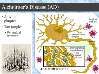
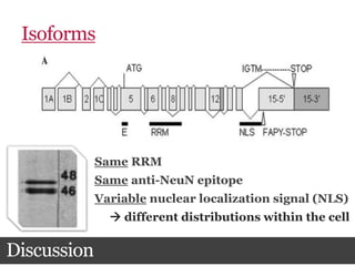
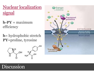
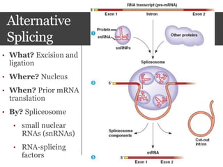
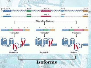
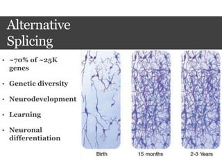
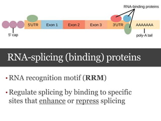
Subcellular Distribution of RNA-splicing
Factor NeuN/Fox-3 in Alzheimer’s
Braeden L. Lovett; Dr. Amanda Brown
Johns Hopkins, Department of Neuroimmunology
November 21st, 2014
1992
Neuronal
Nuclei (NeuN)
• mAB 60 (anti-
NeuN)
• 46-48 kDa
• Neuron-specific
• Largely nuclear
• Temporally-
regulated
2009
NeuN=Fox-3
• SDS-PAGE
• Mass spectrometry
• Peptide sequence analysis
• Doublet of doublets
Genetics Interlude
Alternative
Splicing
• What? Excision and
ligation
• Where? Nucleus
• When? Prior mRNA
translation
• By? Spliceosome
• small nuclear
RNAs (snRNAs)
• RNA-splicing
factors
Isoforms
Alternative
Splicing
• ~70% of ~25K
genes
• Genetic diversity
• Neurodevelopment
• Learning
• Neuronal
differentiation
RNA-splicing (binding) proteins
• RNA recognition motif (RRM)
• Regulate splicing by binding to specific
sites that enhance or repress splicing
RNA-splicing
Factors
• Nuclear/cytopla
smic shuttling
• mRNA transport
• mRNA
stabilization
Fox Family of RNA-Splicing Factors
• Fox-1, Fox-2, Fox-3 contain conserved RRM
• Members & isoforms capable of cross- and
auto-regulation
Fox Family of RNA-Splicing Factors
• Fox-1 & Fox-2
• Autism spectral disorder, bipolar disorder,
schizophrenia, epilepsy, attention deficit
hyperactivity disorder, mental retardation
Fox-3/Rbfox-3
• Fox-2
• Numb protein
• abnormal cerebellar
development
• NMHC-B
• Amyotrophic lateral sclerosis
(ALS)
• Mutation
• Epilepsy
Impacts on
splicing activity
• Phosphorylation/dephosphorylati
on
• Influences splicing activity
• Influences location  splicing activity
• Chemical stressors  cytoplasm
• Arsenite (RBM4)
• Osmotic shock (hn-RNP)
• Actinomycin-D (hn-RNP)
Could some type of stress
affect the location of Fox-
3?
Fox-3 as a Phosphoprotein
1. + Alkaline phosphatases
2. + Antibodies to
phospho-Y, phospho-T,
phospho-S
Detection
Fox-3 as a Phosphoprotein
3. Multiple isoelectric
points (pI) of increasing
acidity
Isoelectric
focusing
• pH
gradient
(-)
,
[H+]
Alzheimer’s Disease (AD)
• Amyloid
plaques
• Tau tangles
• Pyramidal
neurons
Where is Fox-3 located in
Alzheimer’s?
Materials and methods
OCC
MFG
2 controls (N = 4)
9 AD (N = 13)
Materials and methods
Formalin fixation
• Purpose: Prevent
autolysis by proteases
 preservation
• Covalent bonds 
rigidity = stability
Materials and methods
Paraffin embedding
• Purpose: To allow fine
sectioning
Materials and methods
1. Deparaffinization
• Histo-Clear
2. Rehydration
• Decreasing dilutions of EtOH
3. Proteinase K solution
4. Blocking buffer
• 10% goat serum,
• 0.3% Triton X-100
• TBS
Triton-x
Materials and methods
5. 1:100 Rabbit anti-NeuN
antibody
6. 1:500 Goat-anti-rabbit
alkaline phosphatase
antibody
7. Alkaline phosphatase
substrate solution
8. 1:4 Hematoxylin QS
9. Dehydration (increasing
dilutions of EtOH)
1°
2°
C
N N+CN
C
N+C
Nuclear (N)
• Blue + red  dark red/purple nuclei
Both (N+C)
• Dark red/purple nuclei + red cell soma
Cytoplasmic (C)
• Blue nuclei + red cell soma
Glial
~200-300
pyramidal
neurons
0
10
20
30
40
50
60
70
80
90
100
Control AD Control AD Control AD
Nuclear N+C Cytoplasmic
PercentofCellCount
Expression Type
N=13
*
* *
N=4
N=4
N=4
N=13
N=13
*p<0.04
Median
Age/Tau Tangles
Discussion
Post Mortem Delay (PMD)
Discussion
PMD
Autolysis
Isoforms
Discussion
Same RRM
Same anti-NeuN epitope
Variable nuclear localization signal (NLS)
 different distributions within the cell
Discussion
Nuclear localization
signal
h-PY = maximum
efficiency
h= hydrophobic stretch
PY=proline, tyrosine
Discussion
Karyopherin β2
Fox3v3 
h-PY
X-PY
X-XX  exclusively cytoplasmic
Fox3v1   exclusively
nuclear
Fox3v2 
predominantly
nuclear
Discussion
Disease
Progression
Limitations/Future Work
• Small sample size
• Age
• Isoforms
• Subjectivity *
• Slide preparation/immunohistochemistry
Acknowledgements
• Department of Pathology, Johns Hopkins
• Dr. Amanda Brown
• Dr. R. Smith
• Family and friends
• Eric Bowman, Peter Merkel, Mary Yates
Questions?
• Mullen, R.J.; Buck, C.R., Smith, A.M. NeuN, a neuronal specific nuclear protein in vertebrates. Development, 1992, 116.
201-211.
• Kim, K.K.; Adelstein R.S.; Kawamoto, S. Identification of Neuronal Nuclei (NeuN) as Fox-3, a New Member of the Fox-1
Gene Family of Splicing Factors. J. Biol. Chem. 2009, 284, 31052-31061.
• Lukong, K.E.; Chang, K.; Khandjian, E.W.; Richard, S. RNA-binding proteins in human genetic disease. Trends in
Genetics, 2008, 24, 416-425.
• Misteli, T. RNA splicing: what has phosphorylation got to do with it? Current Biology, 1999, 9, R198-R200.
• Nakahata, S.; Kawamoto, S. Tissue-dependent isoforms of mammalian Fox-1 homologs are associated with tissue-specific
splicing activities. Nucleic Acids Res., 2005, 33, 2087-2089.
• Lin, J.; Hsu, M; Tarn, W. Cell stress modulates the function of splicing regulatory protein RBM4 in translation control.
PNAS, 2007, 104, 2235-2240.
• Oordt, W.H.; Diaz-Meco, M.T.; Lozano, J.; Krainer, A.R.; Moscat, J.; Caceres, J.F. The MKK-p38-signaling cascade alters
the subcellular distribution of hnRNP A1 and modulates alternative splicing regulation. J. Cell Biol., 2000, 149, 307-316.
• Xu, Y.; Gendron, T.F.; Petrucelli, L. Wild-type human TDP-43 expression causes TDP-43 phosphorylation, mitochondrial
aggregation, motor deficits, and early mortality in transgenic mice. J. Neurosci., 2010, 30, 10851-10859.
• Polymenidou, M.; Lagier-Tourenne, C.; Hutt, K.R.; Bennett, C.F.; Cleveland, D.W.; Yeo, G.W. Misregulated RNA
processing in amyotrophic lateral sclerosis. Brain Res., 2012, 1462, 3-15.
• Lind, D.; Franken, S.; Kappler, J.; Jankowski, J.; Schilling, K. Characterization of the neuronal marker NeuN as a multiply
phosphorylated antigen with discrete subcellular localization. J. Neuro. Res., 2005, 79, 295-302.
• Hardie, D.G. Protein phosphorylation: A practical approach. 2nd ed. Oxford University Press. 1999.
• Delom, F.; Chevet, E. Phosphoprotein analysis: from proteins to proteomes. Proteome Science, 2006, 4, 1-12.
• Dredge, B.K.; Jensen, K.B. NeuN/Rbfox3 Nuclear and Cytoplasmic Isoforms Differentially Regulate Alternative Splicing
and Nonsense-Mediated Decay of Rbfox2. PLoS ONE, 2011, 6, 1-12.
• Fogel, B.L.; Wexler, E.; Geschwind, D.H. RBFOX1 regulates both splicing and transcriptional networks in human neuronal
development. Hum Mol Genet., 2012, 21, 4171-4186.
• Bhalla, K.; Phillips, H.A.; Crawford, K.; McKenzie O.L.D.; Mulley, J.C.; Eyre, H.; Gardner A.E.; Kremmidiotis, G.; Callen,
D.F. The de novo chromosome 16 translocations of two patients with abnormal phenotypes (mental retardation and
epilepsy) disrupt the A2BP1 gene. J Hum Genet, 2005, 49, 308-311.
References
• Salawu, F.K.; Umar, J.T.; Olokoba, A.B. Alzheimer’s disease: A review of recent developments. Ann. Afr. Med., 2011, 10,
73-79.
• Pearson, R.C.; Esiri, M.M.; Hioms, R.W.; Wilcock, G.K.; Powell, T.P. Anatomical correlates of the distribution of the
pathological changes in the neocortex in Alzheimer disease. Proc. Natl. Acad. Sci., 1985, 82, 4531-4534
• Buee, L.; Bussiere, T.; Buee-Scherrer, V.; Delacourte, A.; Hof, P.R. Tau protein isoforms, phosphorylation and role in
neurodegenerative disorders. Brain Research Reviews, 2000, 33, 95-130.
• Mirra, S.S.; Heymand, A.; Mckeel, M.D.; Sumi, S.M.; Crain, B.J., Brownlee, L.M.; Vogel, F.S.; Hughes, J.P., Van Belle,
G.; Berg, L. The Consortium to Establish a Registry for Alzheimer’s Disease (CERAD), Part II. Standarization of the
neuropathologic assessment of Alzheimer’s disease. Neurology, 1991, 41, 479-486.
• Unal-Cevik, I.; Kilinc, M.; Gursoy-Ozdemir, Y.; Gurer, G.; Dalkara, T. Loss of NeuN immunoreactivity after cerebral
ischemia does not indicate neuronal cell loss: a cautionary note. Brain Res, 2004, 1015, 169-174.
• Maxeiner, S.; Glassmann, A.; Kao, H.; Schilling, K. The molecular basis of the specificity and cross-reactivity of the
NeuN epitope of the neuron-specific splicing regulator, Rbfox3. Histochem. Cell. Biol., 2014, 141, 43-55.
• Lee, B.J. Cansizuglu A.E.; Suel, K.E.; Louis, T.H.; Zhang, Z.; Chook, Y.M. Rule for nuclear localization sequence
recognition by KaryopherinB2. Cell, 2006, 126, 543-558.
• Pollard, V.W.; Michael, W.M.; Nakielny, S.; Siomi, M.C.; Wang, F.; Dreyfuss, G. A novel receptor-mediated nuclear
protein import pathway. Cell, 1996, 86, 985-994.
• Minati, L.; Edginton, T.; Bruzzone, M.G.; Giaconne, G. Current concepts in Alzheimer’s disease: A multidisciplinary
review. AM. J. ALZHEIMERS DIS. OTHER DEMEN., 2008, 24, 95-121.
• Gehman, L.T.; Meera, P.; Black, D.L. The splicing regulator Rbfox2 is required for both cerebellar development and
mature motor function. Genes Dev., 2012, 26, 445-460.
• Lal, D.; Reinthaler, E.M.; Altmuller, J.; Toliat, M.R.; Thiele, H.; Nurnberg, P.; Lerche, H.; Muhle, H.; Sander, T.;
Zimprich, F.; Neubauer, B.A. Rbfox1 and Rbfox3 mutations in Rolandic epilepsy. PLoS ONE, 2013, 8, 1-6.
• Kim, K.K.; Kim, Y.C.; Adelstein, R.S.; Kawamoto, S. Fox-3 and PSF interact to activate neural cell-specific alternative
splicing. Nucleic Acids Research, 2011, 39, 3064-3078.
• Ma, X.; Kawamoto, S.; Adelstein, R.S. Function of the neuron-specific alternatively spliced isoforms of nonmuscle
myosin II-B during mouse brain development. Mol. Biol. Cell., 2006, 17, 2138-2149.
• Kim, K.K.; Nam, J.; Mukouyama, Y.; Kawamoto, S. Rbfox3-regulated alternative splicing of Numb promotes neuronal
differentiation during development. J. Cell Biol., 2013, 200, 443-458.
References
References
• http://www.wired.com/category/science-blogs/brainwatch/Age
• http://lansing.legalexaminer.com/head-brain-injuries/eternal-sunshine-of-the-spotless-mouse-mind-mit-
research-shows-promise-for-ptsd/
• http://www.themarysue.com/mouse-embryo-grows/
• http://www.brainhq.com/media-gallery/detail/161/94
• http://www.bio.utexas.edu/faculty/sjasper/bio212/proteins.html
• http://www.photonics.com/Article.aspx?AID=35734
• http://en.wikipedia.org/wiki/Alternative_splicing
• http://jcs.biologists.org/content/123/7/1020/F9.expansion.html
• http://nikhilthegrizzlybear.blogspot.com/2014/05/child-abuse-neglect-and-stress-in-first.html
• http://cnx.org/contents/185cbf87-c72e-48f5-b51e-f14f21b5eabd@9.3:47
• http://glossary.periodni.com/download_image.php?name=zwitterion.png&source=zwitterion
• http://www.nia.nih.gov/alzheimers/publication/dementias/types-dementia
• http://society6.com/product/pyramidal-neuron-forest_print#1=45
• http://www.alz.org/braintour/progression.asp
• https://www.etsy.com/market/chemistry_cat
• http://www.sciencedirect.com/science?_ob=GatewayURL&_origin=CELLPRESS&_urlversion=4&_method
=citationSearch&_version=1&_piikey=S0092867409000105&md5=6f60ad579fd65891d0d35daba66b4359
• http://en.wikipedia.org/wiki/Inferior_frontal_gyrus
• http://www.koofers.com/flashcards/bio-exam-1-11/review
• http://commons.wikimedia.org/wiki/File:Triton_X-100.png
• http://en.wikipedia.org/wiki/Immunohistochemistry
• http://hhmi.mcdb.ucsb.edu/course/mcdb-109l-tau-hyperphosphorylation
• http://www.cancerdxpathology.org.au/cancer-pathology.html
• http://www.dreamstime.com/royalty-free-stock-photography-push-pins-interconnected-concept-network-
image21111287
• http://homepages.uconn.edu/~mb2225vc/MCB_2225/Fixation.html

FINALBIOCHEMCAPSTONEPP

  • 1.
    Subcellular Distribution ofRNA-splicing Factor NeuN/Fox-3 in Alzheimer’s Braeden L. Lovett; Dr. Amanda Brown Johns Hopkins, Department of Neuroimmunology November 21st, 2014
  • 2.
  • 4.
    Neuronal Nuclei (NeuN) • mAB60 (anti- NeuN) • 46-48 kDa • Neuron-specific • Largely nuclear • Temporally- regulated
  • 5.
  • 6.
    NeuN=Fox-3 • SDS-PAGE • Massspectrometry • Peptide sequence analysis • Doublet of doublets
  • 7.
  • 9.
    Alternative Splicing • What? Excisionand ligation • Where? Nucleus • When? Prior mRNA translation • By? Spliceosome • small nuclear RNAs (snRNAs) • RNA-splicing factors
  • 10.
  • 11.
    Alternative Splicing • ~70% of~25K genes • Genetic diversity • Neurodevelopment • Learning • Neuronal differentiation
  • 12.
    RNA-splicing (binding) proteins •RNA recognition motif (RRM) • Regulate splicing by binding to specific sites that enhance or repress splicing
  • 13.
  • 14.
    Fox Family ofRNA-Splicing Factors • Fox-1, Fox-2, Fox-3 contain conserved RRM • Members & isoforms capable of cross- and auto-regulation
  • 15.
    Fox Family ofRNA-Splicing Factors • Fox-1 & Fox-2 • Autism spectral disorder, bipolar disorder, schizophrenia, epilepsy, attention deficit hyperactivity disorder, mental retardation
  • 16.
    Fox-3/Rbfox-3 • Fox-2 • Numbprotein • abnormal cerebellar development • NMHC-B • Amyotrophic lateral sclerosis (ALS) • Mutation • Epilepsy
  • 17.
    Impacts on splicing activity •Phosphorylation/dephosphorylati on • Influences splicing activity • Influences location  splicing activity • Chemical stressors  cytoplasm • Arsenite (RBM4) • Osmotic shock (hn-RNP) • Actinomycin-D (hn-RNP)
  • 18.
    Could some typeof stress affect the location of Fox- 3?
  • 19.
    Fox-3 as aPhosphoprotein 1. + Alkaline phosphatases 2. + Antibodies to phospho-Y, phospho-T, phospho-S Detection
  • 20.
    Fox-3 as aPhosphoprotein 3. Multiple isoelectric points (pI) of increasing acidity Isoelectric focusing • pH gradient (-) , [H+]
  • 21.
    Alzheimer’s Disease (AD) •Amyloid plaques • Tau tangles • Pyramidal neurons
  • 22.
    Where is Fox-3located in Alzheimer’s?
  • 23.
  • 24.
    2 controls (N= 4) 9 AD (N = 13) Materials and methods
  • 25.
    Materials and methods Formalinfixation • Purpose: Prevent autolysis by proteases  preservation • Covalent bonds  rigidity = stability
  • 26.
    Materials and methods Paraffinembedding • Purpose: To allow fine sectioning
  • 27.
    Materials and methods 1.Deparaffinization • Histo-Clear 2. Rehydration • Decreasing dilutions of EtOH 3. Proteinase K solution 4. Blocking buffer • 10% goat serum, • 0.3% Triton X-100 • TBS Triton-x
  • 28.
    Materials and methods 5.1:100 Rabbit anti-NeuN antibody 6. 1:500 Goat-anti-rabbit alkaline phosphatase antibody 7. Alkaline phosphatase substrate solution 8. 1:4 Hematoxylin QS 9. Dehydration (increasing dilutions of EtOH) 1° 2°
  • 29.
    C N N+CN C N+C Nuclear (N) •Blue + red  dark red/purple nuclei Both (N+C) • Dark red/purple nuclei + red cell soma Cytoplasmic (C) • Blue nuclei + red cell soma Glial ~200-300 pyramidal neurons
  • 30.
    0 10 20 30 40 50 60 70 80 90 100 Control AD ControlAD Control AD Nuclear N+C Cytoplasmic PercentofCellCount Expression Type N=13 * * * N=4 N=4 N=4 N=13 N=13 *p<0.04 Median
  • 31.
  • 32.
    Post Mortem Delay(PMD) Discussion PMD Autolysis
  • 33.
    Isoforms Discussion Same RRM Same anti-NeuNepitope Variable nuclear localization signal (NLS)  different distributions within the cell
  • 34.
    Discussion Nuclear localization signal h-PY =maximum efficiency h= hydrophobic stretch PY=proline, tyrosine
  • 35.
    Discussion Karyopherin β2 Fox3v3  h-PY X-PY X-XX exclusively cytoplasmic Fox3v1   exclusively nuclear Fox3v2  predominantly nuclear
  • 36.
  • 37.
    Limitations/Future Work • Smallsample size • Age • Isoforms • Subjectivity * • Slide preparation/immunohistochemistry
  • 38.
    Acknowledgements • Department ofPathology, Johns Hopkins • Dr. Amanda Brown • Dr. R. Smith • Family and friends • Eric Bowman, Peter Merkel, Mary Yates
  • 39.
  • 40.
    • Mullen, R.J.;Buck, C.R., Smith, A.M. NeuN, a neuronal specific nuclear protein in vertebrates. Development, 1992, 116. 201-211. • Kim, K.K.; Adelstein R.S.; Kawamoto, S. Identification of Neuronal Nuclei (NeuN) as Fox-3, a New Member of the Fox-1 Gene Family of Splicing Factors. J. Biol. Chem. 2009, 284, 31052-31061. • Lukong, K.E.; Chang, K.; Khandjian, E.W.; Richard, S. RNA-binding proteins in human genetic disease. Trends in Genetics, 2008, 24, 416-425. • Misteli, T. RNA splicing: what has phosphorylation got to do with it? Current Biology, 1999, 9, R198-R200. • Nakahata, S.; Kawamoto, S. Tissue-dependent isoforms of mammalian Fox-1 homologs are associated with tissue-specific splicing activities. Nucleic Acids Res., 2005, 33, 2087-2089. • Lin, J.; Hsu, M; Tarn, W. Cell stress modulates the function of splicing regulatory protein RBM4 in translation control. PNAS, 2007, 104, 2235-2240. • Oordt, W.H.; Diaz-Meco, M.T.; Lozano, J.; Krainer, A.R.; Moscat, J.; Caceres, J.F. The MKK-p38-signaling cascade alters the subcellular distribution of hnRNP A1 and modulates alternative splicing regulation. J. Cell Biol., 2000, 149, 307-316. • Xu, Y.; Gendron, T.F.; Petrucelli, L. Wild-type human TDP-43 expression causes TDP-43 phosphorylation, mitochondrial aggregation, motor deficits, and early mortality in transgenic mice. J. Neurosci., 2010, 30, 10851-10859. • Polymenidou, M.; Lagier-Tourenne, C.; Hutt, K.R.; Bennett, C.F.; Cleveland, D.W.; Yeo, G.W. Misregulated RNA processing in amyotrophic lateral sclerosis. Brain Res., 2012, 1462, 3-15. • Lind, D.; Franken, S.; Kappler, J.; Jankowski, J.; Schilling, K. Characterization of the neuronal marker NeuN as a multiply phosphorylated antigen with discrete subcellular localization. J. Neuro. Res., 2005, 79, 295-302. • Hardie, D.G. Protein phosphorylation: A practical approach. 2nd ed. Oxford University Press. 1999. • Delom, F.; Chevet, E. Phosphoprotein analysis: from proteins to proteomes. Proteome Science, 2006, 4, 1-12. • Dredge, B.K.; Jensen, K.B. NeuN/Rbfox3 Nuclear and Cytoplasmic Isoforms Differentially Regulate Alternative Splicing and Nonsense-Mediated Decay of Rbfox2. PLoS ONE, 2011, 6, 1-12. • Fogel, B.L.; Wexler, E.; Geschwind, D.H. RBFOX1 regulates both splicing and transcriptional networks in human neuronal development. Hum Mol Genet., 2012, 21, 4171-4186. • Bhalla, K.; Phillips, H.A.; Crawford, K.; McKenzie O.L.D.; Mulley, J.C.; Eyre, H.; Gardner A.E.; Kremmidiotis, G.; Callen, D.F. The de novo chromosome 16 translocations of two patients with abnormal phenotypes (mental retardation and epilepsy) disrupt the A2BP1 gene. J Hum Genet, 2005, 49, 308-311. References
  • 41.
    • Salawu, F.K.;Umar, J.T.; Olokoba, A.B. Alzheimer’s disease: A review of recent developments. Ann. Afr. Med., 2011, 10, 73-79. • Pearson, R.C.; Esiri, M.M.; Hioms, R.W.; Wilcock, G.K.; Powell, T.P. Anatomical correlates of the distribution of the pathological changes in the neocortex in Alzheimer disease. Proc. Natl. Acad. Sci., 1985, 82, 4531-4534 • Buee, L.; Bussiere, T.; Buee-Scherrer, V.; Delacourte, A.; Hof, P.R. Tau protein isoforms, phosphorylation and role in neurodegenerative disorders. Brain Research Reviews, 2000, 33, 95-130. • Mirra, S.S.; Heymand, A.; Mckeel, M.D.; Sumi, S.M.; Crain, B.J., Brownlee, L.M.; Vogel, F.S.; Hughes, J.P., Van Belle, G.; Berg, L. The Consortium to Establish a Registry for Alzheimer’s Disease (CERAD), Part II. Standarization of the neuropathologic assessment of Alzheimer’s disease. Neurology, 1991, 41, 479-486. • Unal-Cevik, I.; Kilinc, M.; Gursoy-Ozdemir, Y.; Gurer, G.; Dalkara, T. Loss of NeuN immunoreactivity after cerebral ischemia does not indicate neuronal cell loss: a cautionary note. Brain Res, 2004, 1015, 169-174. • Maxeiner, S.; Glassmann, A.; Kao, H.; Schilling, K. The molecular basis of the specificity and cross-reactivity of the NeuN epitope of the neuron-specific splicing regulator, Rbfox3. Histochem. Cell. Biol., 2014, 141, 43-55. • Lee, B.J. Cansizuglu A.E.; Suel, K.E.; Louis, T.H.; Zhang, Z.; Chook, Y.M. Rule for nuclear localization sequence recognition by KaryopherinB2. Cell, 2006, 126, 543-558. • Pollard, V.W.; Michael, W.M.; Nakielny, S.; Siomi, M.C.; Wang, F.; Dreyfuss, G. A novel receptor-mediated nuclear protein import pathway. Cell, 1996, 86, 985-994. • Minati, L.; Edginton, T.; Bruzzone, M.G.; Giaconne, G. Current concepts in Alzheimer’s disease: A multidisciplinary review. AM. J. ALZHEIMERS DIS. OTHER DEMEN., 2008, 24, 95-121. • Gehman, L.T.; Meera, P.; Black, D.L. The splicing regulator Rbfox2 is required for both cerebellar development and mature motor function. Genes Dev., 2012, 26, 445-460. • Lal, D.; Reinthaler, E.M.; Altmuller, J.; Toliat, M.R.; Thiele, H.; Nurnberg, P.; Lerche, H.; Muhle, H.; Sander, T.; Zimprich, F.; Neubauer, B.A. Rbfox1 and Rbfox3 mutations in Rolandic epilepsy. PLoS ONE, 2013, 8, 1-6. • Kim, K.K.; Kim, Y.C.; Adelstein, R.S.; Kawamoto, S. Fox-3 and PSF interact to activate neural cell-specific alternative splicing. Nucleic Acids Research, 2011, 39, 3064-3078. • Ma, X.; Kawamoto, S.; Adelstein, R.S. Function of the neuron-specific alternatively spliced isoforms of nonmuscle myosin II-B during mouse brain development. Mol. Biol. Cell., 2006, 17, 2138-2149. • Kim, K.K.; Nam, J.; Mukouyama, Y.; Kawamoto, S. Rbfox3-regulated alternative splicing of Numb promotes neuronal differentiation during development. J. Cell Biol., 2013, 200, 443-458. References
  • 42.
    References • http://www.wired.com/category/science-blogs/brainwatch/Age • http://lansing.legalexaminer.com/head-brain-injuries/eternal-sunshine-of-the-spotless-mouse-mind-mit- research-shows-promise-for-ptsd/ •http://www.themarysue.com/mouse-embryo-grows/ • http://www.brainhq.com/media-gallery/detail/161/94 • http://www.bio.utexas.edu/faculty/sjasper/bio212/proteins.html • http://www.photonics.com/Article.aspx?AID=35734 • http://en.wikipedia.org/wiki/Alternative_splicing • http://jcs.biologists.org/content/123/7/1020/F9.expansion.html • http://nikhilthegrizzlybear.blogspot.com/2014/05/child-abuse-neglect-and-stress-in-first.html • http://cnx.org/contents/185cbf87-c72e-48f5-b51e-f14f21b5eabd@9.3:47 • http://glossary.periodni.com/download_image.php?name=zwitterion.png&source=zwitterion • http://www.nia.nih.gov/alzheimers/publication/dementias/types-dementia • http://society6.com/product/pyramidal-neuron-forest_print#1=45 • http://www.alz.org/braintour/progression.asp • https://www.etsy.com/market/chemistry_cat • http://www.sciencedirect.com/science?_ob=GatewayURL&_origin=CELLPRESS&_urlversion=4&_method =citationSearch&_version=1&_piikey=S0092867409000105&md5=6f60ad579fd65891d0d35daba66b4359 • http://en.wikipedia.org/wiki/Inferior_frontal_gyrus • http://www.koofers.com/flashcards/bio-exam-1-11/review • http://commons.wikimedia.org/wiki/File:Triton_X-100.png • http://en.wikipedia.org/wiki/Immunohistochemistry • http://hhmi.mcdb.ucsb.edu/course/mcdb-109l-tau-hyperphosphorylation • http://www.cancerdxpathology.org.au/cancer-pathology.html • http://www.dreamstime.com/royalty-free-stock-photography-push-pins-interconnected-concept-network- image21111287 • http://homepages.uconn.edu/~mb2225vc/MCB_2225/Fixation.html

Editor's Notes

  • #3 so let me take you back to 1992 when NeuN/Fox-3 was discovered
  • #4 in 1992, mullen et al. was looking for antibodies that are specific to the vertebrate nervous system, and came upon NeuN
  • #5 furthermore, these bands were expressed exclusively by neurons in both the CNS and PNS. and were expressed predominantly in the nucleus, with lighter staining in the cytoplasm. for this reason, he called the antigen of mAB 60, Neuronal nuclei, or NeuN. He hypothesized that NeuN could play some sort of role in neuro development for a couple of reasons. Its expression was temporally regulated. meaning its expression increased over the course of development of developing mice embryos. and were only expressed by differentiated neuronal cells. second, he hypothesized NeuN to be a DNA-binding protein, because it
  • #6 now lets fast forward to 2009. for years, NeuN has been used as a robust marker for postmitotic neurons in tissue culture even though the exact identity of NeuN, that Is the antigen of anti-NeuN, was not identified. during these years, many disoveries were being made in the molecular genetis world…..
  • #7  so finally in 2009, kim et al. duplicated the experiments of Mullen. she extracted brains from mice in the same function, and subjected nuclear extracts to SDS-PAGE, where she obtained the same molecular weight bands. She then subjected these bands to mass spectrometric analysis which helps to elucidate the relative abundance of amino acids that make up a structure. she then sent fragments digested by trypsin, and found the fragments to be most similar to Fox-1 and Fox-2, members of the Fox family of RNA-splicing factors. furthermore, she characterized the bands as a doublet of doublets. wherein each band contains part of Fox-3, or different isoforms which are different versions of the same protein which I will discuss in further detail doublet of doublets… both bands contain Fox-3… due to presence of isoforms or different versions of the same protein
  • #8 so before I can discuss what RNA-splicing factors are, we need to have a brief genetics interlude
  • #9 central dogma states that DNA is first replicated in the nucleus, and replicated strands are used to make complementantry strands of pre-messenger RNA (or pre-mRNA). several modifications of pre-mRNA are made, and after these are done, mRNA is transported into the cytoplasm where it is translated into a peptide sequence. one of the modifications that occurs before mRNA is ready to be transported is alternative splicing
  • #10 so alternative splicing occurs in the nucleus prior to mRNA transport and translation. it occurs by a complex of macromolecules called the spliceosome in the nucleus. Basically, the spliceosome includes all these snRNAS which are involved in the splicing of mRNA and the stabilization of mRNA… it is only after all components have assembled that altnerative splicing can take place. RNA splicing: the process by which introns are removed from the pre-mRNA. ● Alternative splicing: some pre-mRNAs can be spliced in more than one way , generating alternative mRNAs. 60% of the human genes are spliced in this manner. The 5' and 3' splice sites and the branch sequence are recognized by components of the splicing apparatus that assemble to form a large complex. This complex brings together the 5' and 3' splice sites before any reaction occurs, explaining why a deficiency in any one of the sites may prevent the reaction from initiating. The complex assembles sequentially on the pre-mRNA, and several intermediates can be recognized by fractionating complexes of different sizes. Splicing occurs only after all the components have assembled in the most simplest mechanism, introns (non-coding regions of DNA) are excised or spliced out, and exons (coding regions ligated)…. in reality, however, many modes of alternative splicing
  • #11 many modes of alternative splicing, not as easy as take out introns. and put exons together…. some exons can be left in, some taken out, even introns can be left in…. but the results are the same  isoforms, or different protein versions derived from a single mRNA transcript
  • #12 so what is the significance of alternative splicing? well its is ubiquitous, about 70% of 25K genes use alternative splicing. it is especially important in the brain, during neurodevelopment, learning, neuronal differentiation etc
  • #13 so I mentioned RNA splicing or binding proteins or factors they do not do the splicing but they in the spliceosome, can bind to regions that enhance or repress splicing of a pre-mRNA transcript.
  • #14 furthermore, they are said to be able to shuttle between the nucleus and the cytoplasm, where they can assist in transport, as well as RNA stabilization when in the cytoplasm. the ability to shuttle may explain why Fox-3 expression is lightly apparent in the cytoplasm resported by Mullen and others
  • #15 So Fox-3, as mentioned before, is part of the Fox family of RNA splicing factors, along with Fox-1, Fox-2. They all share a conserved RNA recognition motif, or a region that recognized a region on pre-mRNA where they can bind and either repress or enhance alternative splicing. The Fox family is very complex. Each homolog exists as multiple isoforms themselves, and the members and the isoforms are capable of regulating the activity and expression of each other, as well as autoregulating the expression of their own mRNA expression
  • #16 Because Fox-3 was only recently discovered, not much is known. More is know about Fox-1 and Fox-2. mutations or defects in their functioning are associated with …….
  • #17 so what we know so far. first, Fox-3 is capable of regulating the expression of Fox-2. so a defect in Fox-3 can affect the functioning of fox-2, and therefore it is indirectly related to all the neurocognitive and developmental disorders. Fox-3 is involved in the altnerative splicing of the numb protein. Defects in alternative splicing of Numb leads to abnormal cerebellar development, reduced dendritic branching in mice. Fox-3 interacts with PSF to splice a protein NMHC-B. defects in this process are associated with amylotropic lateral sclerosis. a mutation in Fox-3 has also recently been linked to epilepsy
  • #18 So how can processes affect splicing activity? spliceosome function highly dependent on phosphorylation events. chemical stressors such as arsenite, osmotic shock, UV-C light have been shown to induce a phosphorylation-dependent response in other kinds of RNA-splicing factors, which alters their location, and in turn, their splicing activity alternative splicing by the spliceosome involves mutliple phosphorylation events that influence its activity Steps with a known requirement for phosphorylation are indicated in green, steps with a known requirement for dephosphorylation are indicated in red.
  • #19 so we wanted to see if the cellular environment, especially that of intracellular tau tangles in pyramidal neeurons could be associated with altered distribution of Fox-3, as it is classically considered to be largely nuclear.
  • #20 so what we know so far. first, Fox-3 is capable of regulating the expression of Fox-2. so a defect in Fox-3 can affect the functioning of fox-2, and therefore it is indirectly related to all the neurocognitive and developmental disorders. Fox-3 is involved in the altnerative splicing of the numb protein. Defects in alternative splicing of Numb leads to abnormal cerebellar development, reduced dendritic branching in mice. Fox-3 interacts with PSF to splice a protein NMHC-B. defects in this process are associated with amylotropic lateral sclerosis. a mutation in Fox-3 has also recently been linked to epilepsy
  • #21 so what we know so far. first, Fox-3 is capable of regulating the expression of Fox-2. so a defect in Fox-3 can affect the functioning of fox-2, and therefore it is indirectly related to all the neurocognitive and developmental disorders. Fox-3 is involved in the altnerative splicing of the numb protein. Defects in alternative splicing of Numb leads to abnormal cerebellar development, reduced dendritic branching in mice. Fox-3 interacts with PSF to splice a protein NMHC-B. defects in this process are associated with amylotropic lateral sclerosis. a mutation in Fox-3 has also recently been linked to epilepsy
  • #22 AD is characterized by histopathologic markers amyloid plaques formed extracellularly, and intracellular tau tangles
  • #23 so we wanted to see if the cellular environment, especially that of intracellular tau tangles in pyramidal neeurons could be associated with altered distribution of Fox-3, as it is classically considered to be largely nuclear.
  • #24 so we obtained samples from two regions of the brain affected by AD, one from the middle frontal gyrus of the frontal lobe, and another from the occipital lobe.
  • #25 we had two controls, and 9 AD patients, so we had a total of 4 samples for the controls, and 13 for the AD. one sample had to be excluded because the tissue morphology prevented sound analysis of Fox-3 expression. all were white, about half were female, and half were male in AD, and all controls were female. CERAD and BRAAK are measures of the degree of AD. all had similar scoring. PMD is the time after death of tissue fixation. as you can see, they had various times
  • #26 remove water from tissues and replace with a medium that solidifies to allow thin sections to be cut http://www.jbc.org/content/279/8/6235.long schiff bases  methylene bridges so first, although this wasn’t performed by us, the tissue was fixated in formalin, which is a solution of formaldehyde gas in methanol. formalin creates methylene bridges between lysine amino acids that stabilize the tissue, and prevent autolysis by proteolytic enzymes. the convalent bonds create stability Formalin is produced by dissolving the gas in water until the desired concentration is reached, up to a maximum of 40%. Since water is the other product of the reaction, the process can produce formalin directly. Formaldehyde in aqueous solution is unstable and tends to polymerize, forming larger molecules that are insoluble and therefore precipitate out of the solution. For this reason, methanol, which prevents polymerization, is added to the solution.
  • #27 tissues were then embedded in paraffin, which is a wax, to allow for fine sectioning of the brain into very thin slices
  • #28 slides were warmed. Histoclear which IS ????? removes the paraffin. the tissues were then rehydrated in a series of graded dilutions to prepare for the staining processes. proteinase K solution was used to break the aldehydic cross-links formed by the fixation step, along with antigen retrieval buffer, that may mask the Fox-3 epitope blocking buffer was then used to prevent non-specific binding between ??????? triton is a ????? min (BSA) is a common procedure used to reduce non-specific hydrophobic binding triton- nonionic detergent, facilitating lysis of cells and extraction of soluble protein, often in native form. Using certain buffer conditions, various detergents effectively penetrate between the membrane bilayers at concentrations sufficient to form mixed micelles with isolated phospholipids and membrane proteins; Nondenaturing detergents such as Triton X-100 have rigid and bulky nonpolar heads that do not penetrate into water-soluble proteins; consequently, they generally do not disrupt native interactions and structures of water-soluble proteins and do not have cooperative binding properties. The main effect of nondenaturing detergents is to associate with hydrophobic parts of membrane proteins, thereby conferring miscibility to them. histoclear is a less toxic form of xylene. xylene is miscible with paraffin so replaces paraffin, has same refractive index as the protein so is clear xylene used to remove paraffin Using normal serum makes sense, because it will contain a mixture of antibodies. They won't specifically recognize your target antigen, since they come from a non-immunized animal. However, they will stick to anything that tends to bind Ab's non-specifically.
  • #29 Immunohistochemistry alkaline phosphatase are commonly used to catalyse reactions that give a coloured or chemiluminescent product. take a primary antibody secondary antibody then a fluorescing solution that interacts with the secondary antibody, which gives it color, and allows us to detect the presence of bound anti-NeuN. anti-NeuN stains red a counter-stain was applied. hematoxylin, with ambiguously stains all nuclei blue, nuclei of neurons and glial cells
  • #31 scored 200-300 pyramidal neurons box- represents 50% of distribution with center line representing the median
  • #32 AD most often occurs in the elderly with its risk of development doubling every five years after the age of 65
  • #33 http://www.scielo.cl/pdf/ijmorphol/v27n1/art36.pdf
  • #36 highly negative binding pocket, so positively charged groups, tyrosine is positive at pH 7, and proline is neutral