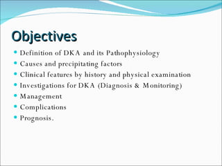
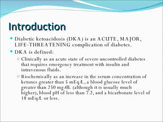
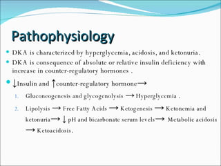
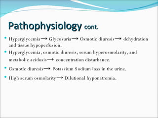
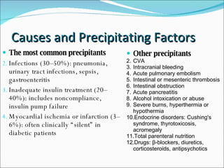
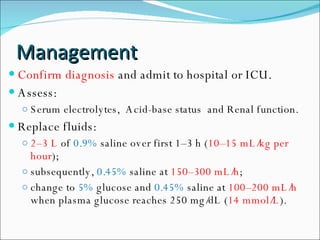
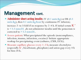
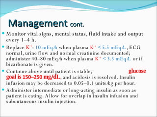
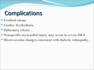
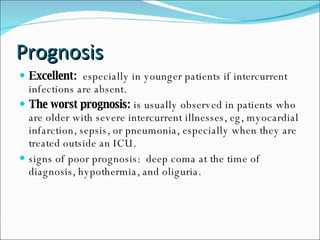
إعداد و تقديم طلاب سنة سادسة  سمهر العلي – معاذ عيّاد – يزيد جبريل
Objectives Definition of DKA and its Pathophysiology Causes and precipitating factors Clinical features by history and physical examination Investigations for DKA (Diagnosis & Monitoring) Management  Complications  Prognosis.
Introduction Diabetic ketoacidosis (DKA) is an ACUTE, MAJOR, LIFE-THREATENING complication of diabetes.  DKA is defined:  Clinically as an acute state of severe uncontrolled diabetes that requires emergency treatment with insulin and intravenous fluids.  Biochemically as an increase in the serum concentration of ketones greater than 5 mEq/L, a blood glucose level of greater than 250 mg/dL (although it is usually much higher), blood pH of less than 7.2, and a bicarbonate level of 18 mEq/L or less.
Pathophysiology DKA is characterized by hyperglycemia, acidosis, and ketonuria. DKA is consequence of absolute or relative insulin deficiency with increase in counter-regulatory hormones . ↓ Insulin and  ↑ counter-regulatory hormone -> Gluconeogenesis and glycogenolysis   ->   Hyperglycemia . Lipolysis  ->   Free Fatty Acids   ->  Ketogenesis  ->  Ketonemia and ketonuria ->   ↓   pH and bicarbonate serum levels ->  Metabolic   acidosis   ->  Ketoacidosis.
Pathophysiology  cont. Hyperglycemia ->  Glycosuria ->  Osmotic diuresis ->  dehydration and tissue hypoperfusion. Hyperglycemia, osmotic diuresis, serum hyperosmolarity, and metabolic acidosis ->  concentration disturbance. Osmotic diuresis ->  Potassium Sodium loss in the urine. High serum osmolarity ->  Dilutional hyponatremia.
Causes and Precipitating Factors The most common precipitants Infections (30–50%): pneumonia, urinary tract infections, sepsis, gastroenteritis  Inadequate insulin treatment (20–40%): includes noncompliance, insulin pump failure  Myocardial ischemia or infarction (3–6%): often clinically “silent” in diabetic patients  Other precipitants CVA Intracranial bleeding Acute pulmonary embolism  Intestinal or mesenteric thrombosis  Intestinal obstruction  Acute pancreatitis  Alcohol intoxication or abuse  Severe burns, hyperthermia or hypothermia  Endocrine disorders: Cushing's syndrome, thyrotoxicosis, acromegaly  Total parenteral nutrition  Drugs: β-blockers, diuretics, corticosteroids, antipsychotics
Clinical Features Symptoms: Polydypsia. Polyuria. Hyperglycemia. Nausea, lethargy, anorexia, weakness. Abdominal pain. Reduced motility of GI. Vomiting. Signs: Dehydration:  Dry skin and mucous . Orthostatic hypotension.  Tachycardia.  Reduced JVP. Reduced mental function Ketosis:  Sweet odor  Kussmaul breathing
Diagnosis
Investigations Glucose level. Serum Ketones. Acid-base status: pH, Serum bicarbonate and Anion gap. Electrolytes: Na  + K +  Cl  -  Mg  +2 ECG CBC, WBC. Urinalysis. Cardiac markers, Liver enzymes and Amylase. Chest X-Ray. Blood and urine culture.
Management Confirm diagnosis  and admit to hospital or ICU. Assess: Serum electrolytes,  Acid-base status  and Renal function. Replace fluids:  2–3 L  of  0.9%  saline over first 1–3 h ( 10–15 mL/kg per hour );  subsequently,  0.45%  saline at  150–300 mL/h ;  change to  5%  glucose and  0.45%  saline at  100–200 mL/h  when plasma glucose reaches 250 mg/dL ( 14 mmol/L ).
Management  cont. Administer short acting insulin : IV ( 0.1 units/kg ) or IM ( 0.3 units/kg ), then  0.1 units/kg/hour  by continuous IV infusion; increase 2- to 3-fold if no response by 2–4 h. If initial serum  K +   is  < 3.3 mmol/L  ,do not administer insulin until the potassium is corrected to  > 3.3 mmol/L . Assess patient : What precipitated the episode (noncompliance, infection, trauma, infarction, cocaine)? Initiate appropriate workup for precipitating event (cultures, CXR, ECG). Measure capillary glucose every 1–2 h ; measure electrolytes (especially  K + , bicarbonate, phosphate) and anion gap every 4 h for first 24 h.
Monitor vital signs, mental status, fluid intake and output every 1–4 h. Replace  K + :  10 mEq/h  when plasma  K +   < 5.5 mEq/L , ECG normal, urine flow and normal creatinine documented; administer 40–80 mEq/h when plasma  K +   < 3.5 mEq/L  or if bicarbonate is given. Continue above until patient is stable,   glucose goal is  150–250  mg / dL , and acidosis is resolved. Insulin infusion may be decreased to 0.05–0.1 units/kg per hour. Administer intermediate or long-acting insulin as soon as patient is eating. Allow for overlap in insulin infusion and subcutaneous insulin injection. Management  cont.
Complications Cerebral edema Cardiac dysrhythmia Pulmonary edema Nonspecific myocardial injury may occur in severe DKA. Microvascular changes consistent with diabetic retinopathy.
Prognosis Excellent:   especially in younger patients if intercurrent infections are absent. The worst prognosis:  is usually observed in patients who are older with severe intercurrent illnesses, eg, myocardial infarction, sepsis, or pneumonia, especially when they are treated outside an ICU. signs of poor prognosis:  deep coma at the time of diagnosis, hypothermia, and oliguria.
References Cecil Medicine, 23rd Ed Harrison's Principles of Internal Medicine, 17th Edition, 2008 eMedicine.com Specialties > Endocrinology > Diabetes Mellitus
Thank You Any Questions ?

files

  • 1.
    إعداد و تقديمطلاب سنة سادسة سمهر العلي – معاذ عيّاد – يزيد جبريل
  • 2.
    Objectives Definition ofDKA and its Pathophysiology Causes and precipitating factors Clinical features by history and physical examination Investigations for DKA (Diagnosis & Monitoring) Management Complications Prognosis.
  • 3.
    Introduction Diabetic ketoacidosis(DKA) is an ACUTE, MAJOR, LIFE-THREATENING complication of diabetes. DKA is defined: Clinically as an acute state of severe uncontrolled diabetes that requires emergency treatment with insulin and intravenous fluids. Biochemically as an increase in the serum concentration of ketones greater than 5 mEq/L, a blood glucose level of greater than 250 mg/dL (although it is usually much higher), blood pH of less than 7.2, and a bicarbonate level of 18 mEq/L or less.
  • 4.
    Pathophysiology DKA ischaracterized by hyperglycemia, acidosis, and ketonuria. DKA is consequence of absolute or relative insulin deficiency with increase in counter-regulatory hormones . ↓ Insulin and ↑ counter-regulatory hormone -> Gluconeogenesis and glycogenolysis -> Hyperglycemia . Lipolysis -> Free Fatty Acids -> Ketogenesis -> Ketonemia and ketonuria -> ↓ pH and bicarbonate serum levels -> Metabolic acidosis -> Ketoacidosis.
  • 5.
    Pathophysiology cont.Hyperglycemia -> Glycosuria -> Osmotic diuresis -> dehydration and tissue hypoperfusion. Hyperglycemia, osmotic diuresis, serum hyperosmolarity, and metabolic acidosis -> concentration disturbance. Osmotic diuresis -> Potassium Sodium loss in the urine. High serum osmolarity -> Dilutional hyponatremia.
  • 6.
    Causes and PrecipitatingFactors The most common precipitants Infections (30–50%): pneumonia, urinary tract infections, sepsis, gastroenteritis Inadequate insulin treatment (20–40%): includes noncompliance, insulin pump failure Myocardial ischemia or infarction (3–6%): often clinically “silent” in diabetic patients Other precipitants CVA Intracranial bleeding Acute pulmonary embolism Intestinal or mesenteric thrombosis Intestinal obstruction Acute pancreatitis Alcohol intoxication or abuse Severe burns, hyperthermia or hypothermia Endocrine disorders: Cushing's syndrome, thyrotoxicosis, acromegaly Total parenteral nutrition Drugs: β-blockers, diuretics, corticosteroids, antipsychotics
  • 7.
    Clinical Features Symptoms:Polydypsia. Polyuria. Hyperglycemia. Nausea, lethargy, anorexia, weakness. Abdominal pain. Reduced motility of GI. Vomiting. Signs: Dehydration: Dry skin and mucous . Orthostatic hypotension. Tachycardia. Reduced JVP. Reduced mental function Ketosis: Sweet odor Kussmaul breathing
  • 8.
  • 9.
    Investigations Glucose level.Serum Ketones. Acid-base status: pH, Serum bicarbonate and Anion gap. Electrolytes: Na + K + Cl - Mg +2 ECG CBC, WBC. Urinalysis. Cardiac markers, Liver enzymes and Amylase. Chest X-Ray. Blood and urine culture.
  • 10.
    Management Confirm diagnosis and admit to hospital or ICU. Assess: Serum electrolytes, Acid-base status and Renal function. Replace fluids: 2–3 L of 0.9% saline over first 1–3 h ( 10–15 mL/kg per hour ); subsequently, 0.45% saline at 150–300 mL/h ; change to 5% glucose and 0.45% saline at 100–200 mL/h when plasma glucose reaches 250 mg/dL ( 14 mmol/L ).
  • 11.
    Management cont.Administer short acting insulin : IV ( 0.1 units/kg ) or IM ( 0.3 units/kg ), then 0.1 units/kg/hour by continuous IV infusion; increase 2- to 3-fold if no response by 2–4 h. If initial serum K + is < 3.3 mmol/L ,do not administer insulin until the potassium is corrected to > 3.3 mmol/L . Assess patient : What precipitated the episode (noncompliance, infection, trauma, infarction, cocaine)? Initiate appropriate workup for precipitating event (cultures, CXR, ECG). Measure capillary glucose every 1–2 h ; measure electrolytes (especially K + , bicarbonate, phosphate) and anion gap every 4 h for first 24 h.
  • 12.
    Monitor vital signs,mental status, fluid intake and output every 1–4 h. Replace K + : 10 mEq/h when plasma K + < 5.5 mEq/L , ECG normal, urine flow and normal creatinine documented; administer 40–80 mEq/h when plasma K + < 3.5 mEq/L or if bicarbonate is given. Continue above until patient is stable, glucose goal is 150–250 mg / dL , and acidosis is resolved. Insulin infusion may be decreased to 0.05–0.1 units/kg per hour. Administer intermediate or long-acting insulin as soon as patient is eating. Allow for overlap in insulin infusion and subcutaneous insulin injection. Management cont.
  • 13.
    Complications Cerebral edemaCardiac dysrhythmia Pulmonary edema Nonspecific myocardial injury may occur in severe DKA. Microvascular changes consistent with diabetic retinopathy.
  • 14.
    Prognosis Excellent: especially in younger patients if intercurrent infections are absent. The worst prognosis: is usually observed in patients who are older with severe intercurrent illnesses, eg, myocardial infarction, sepsis, or pneumonia, especially when they are treated outside an ICU. signs of poor prognosis: deep coma at the time of diagnosis, hypothermia, and oliguria.
  • 15.
    References Cecil Medicine,23rd Ed Harrison's Principles of Internal Medicine, 17th Edition, 2008 eMedicine.com Specialties > Endocrinology > Diabetes Mellitus
  • 16.
    Thank You AnyQuestions ?