Bangalore Call Girls Marathahalli 📞 9907093804 High Profile Service 100% Safe
Ferrodyn 04 kortman2017
1. Oral iron supplementation: Potential
implications for the gut microbiome and
metabolome in patients with CKD
Guus A. M. KORTMAN,1*
Dorien REIJNDERS,2
Dorine W. SWINKELS 1
1
Department of Laboratory Medicine – Translational Metabolic Laboratory-830, Radboud University
Medical Center, Nijmegen, The Netherlands; 2
Department of Human Biology, NUTRIM School of
Nutrition and Translational Research in Metabolism, Maastricht University Medical Center, Maastricht,
The Netherlands
Abstract
Patients with chronic kidney disease (CKD) and loss of kidney function are at increased risk for
morbidity and mortality. The risks of CKD are attributed to “uremia,” an increased concentration of
uremic retention solutes (toxins) in the plasma. Recently, a colo-renal axis became clearly apparent
and uremia has been associated with an altered gut microbiome composition and metabolism.
There is a high prevalence of anemia in patients with CKD, for which patients are often treated
with oral or intravenous iron. Recent in vivo and in vitro studies have reported adverse effects of
oral iron supplementation on the gut microbiota composition, gut metabolome, and intestinal
health, which in turn may result in an increased production of uremic toxins. It may also affect cir-
culating levels of other microbe-derived molecules, that can act as mediators of immune regula-
tion. Changes in body iron levels have also been reported to exert subtle effects on host immune
function by modulating immune cell proliferation and differentiation, and by directly regulating
cytokine formation and antimicrobial immune effector mechanisms. Based on the foregoing it is
conceivable that oral iron supplementation in iron deficient predialysis CKD patients adversely
changes gut microbiota composition, the gut and systemic metabolome, and host immunity and
infection. Future studies are needed to confirm these hypotheses and to assess whether, compared
to IV iron supplementation, oral iron supplementation negatively impacts on morbidity of CKD,
and whether these adverse effects depend on the iron bioavailability of the iron formulation to the
microbiota.
Key words: Iron, iron deficiency anemia, CKD, gut microbiome, metabolome
INTRODUCTION
Iron deficiency anemia (IDA) is a common complication
of chronic kidney disease (CKD) and oral therapy is often
given to predialysis patients. Although iron replacement
therapy in general improves anemia and quality of life,
the effects of oral iron on the underlying renal disease
progression and associated morbidities is unknown.1
This review focuses on recent findings regarding the
effects of oral iron supplementation on the gut micro-
biome and metabolome in CKD and how this might affect
disease progression. So far, no studies on the effects of
Correspondence to: D. W. Swinkels, Department of Labo-
ratory Medicine/TML 830, Radboud University Medical
Center, P.O. Box 9101, 6500 HB Nijmegen, The Nether-
lands. E-mail: dorine.swinkels@radboudumc.nl
Conflict of Interest: Authors declare no conflicts of interest.
Disclosure of grants or other funding: This work was partially
funded by the Dutch Kidney Foundation, innovation grant
15OI46 to GK and DS.
*Current address: Guus A. M. Kortman, NIZO food research
B.V., Kernhemseweg 2, 6718 ZB, Ede, The Netherlands
VC 2017 International Society for Hemodialysis
DOI:10.1111/hdi.12553
1
Hemodialysis International 2017; 00:00–00
2. oral iron supplementation in CKD patients or CKD-
models on the gut microbiome and metabolome have
been reported. Therefore, we base our review on the
potential effects in CKD on studies in different target
populations.
IRON AND GUT HEALTH
The mineral iron is an essential building block in all cells
of the body and functions in oxygen transport as part of
hemoglobin, in mitochondrial function, DNA synthesis
and repair, and many enzymatic reactions required for cell
survival.2,3
Iron deficiency is the most common human
nutrient deficiency worldwide with more than 2 billion
people affected, not only in resource-limited nations (e.g.,
in Asia and Africa) but also in well-developed countries.4
Notably, IDA is a common complication of CKD. Epide-
miological studies have shown that around 30%, 40%,
and 70% of predialyses patients with CKD stage 3, 4, and
5, respectively, are anemic.5,6
The prevalence of iron defi-
ciency in these (anemic CKD) patients has been estimated
by bone marrow studies at 48% to 98%.7,8
Iron deficiency
presents with a diminished red blood cell production and
reduced hemoglobin levels, which across the lifespan, can
have important consequences for health. The condition
has been associated with developmental deficits, impaired
memory and neurodevelopment, diminished physical
function, depression, fatigue, loss of vitality, preterm deliv-
ery, and lower infant birth weight.9
The gut functions as a key modulator of iron homeosta-
sis. A sophisticated mechanism of intestinal uptake regu-
lates the absorption of sufficient quantities of iron to reach
daily requirements. In humans, iron is mainly absorbed in
the duodenum in the ferrous form (Fe21
).2
Oral iron sup-
plementation is an effective and well-studied option to
replenish iron stores and therefore the common treatment
for IDA. However, adverse effects on the gut microbiota,
an increased risk of gut inflammation, constipation, and
diarrhea have been reported.10–14
Due to its low bioavail-
ability, iron supplementation generally results in a large
fraction of unabsorbed iron entering the colon, where it is
potentially available for the gut microbiota. In fact, too
much unabsorbed iron can stimulate virulence of patho-
genic bacteria residing in the intestine and may contribute
to an oxidative proinflammatory environment.10
Despite its
crucial role in cellular processes, free colonic iron can gen-
erate toxic free radicals and reactive oxygen species, which
can directly affect gut epithelial integrity via the promotion
of redox stress.15
This impaired integrity has been indicat-
ed in an in vitro study with Caco-2 cells exposed to
iron.16,17
Also in vivo, effects on the epithelium have been
found, as African children on oral iron supplements pre-
sented with increased small intestinal permeability.18
GUT MICROBIOTA COMPOSITION
AND METABOLOME IN CKD
Patients with CKD and loss of kidney function are at
increased risk for morbidity and mortality.19
This is
attributed to “uremia,” an increased concentration of ure-
mic toxins in the plasma, as a consequence of decreased
renal excretory function.20
Recently, a colorenal axis
became apparent in end stage renal disease and secretion
of urea into the gastro-intestinal tract has been associated
with an altered gut microbiota composition and metabo-
lism.21–23
This includes a decrease of beneficial gut
microbiota members, most notably Lactobacillaceae, Bifido-
bacteriaceae and Prevotellaceae families. In addition, poten-
tially pathogenic members, such as Enterobacteriaceae,
Enterococcus spp. and Clostridium perfringens were
increased. One of the consequences of an altered gut
microbiota composition and environment in CKD patients
is an increased production of uremic toxins, most likely
via enhanced microbial fermentation of undigested pro-
teins21–25
and in which prolonged transit time may play a
role.26,27
The hypothesis that undigested protein can lead
to the production of certain uremic toxins by the gut
microbiota is supported by a recent study in healthy vol-
unteers. It was shown that a high protein diet led to a sig-
nificant increase in plasma levels of indoxyl sulfate as well
as significant increases in the urinary excretion of indoxyl
sulfate, indoxyl glucuronide, kynurenic acid, quinolinic
acid, and p-cresyl sulfate.28
Comparable results were
obtained in a rodent study.29
Earlier, by comparing plas-
ma from hemodialysis patients with and without colon,
Aronov et al. already confirmed the colonic origin of the
uremic toxins indoxyl sulfate and p-cresol,30
which are
being conjugated to p-cresyl sulfate and p-cresyl glucuro-
nide in the liver. The cardiovascular and renal toxicity of
these uremic toxins has been demonstrated in several
experimental and clinical studies and concentrations of
indoxyl sulfate and p-cresyl sulfate in serum are negative-
ly correlated with the levels of kidney function.29,31,32
Importantly, the production of uremic toxins and
changes in the gut microbiota composition in CKD may
induce an inflamed and leaky gut by disruption of the
colonic epithelial tight junctions barrier.33
The disruption
of the gut barrier function might result in an increased
exposure of the host to endotoxins, a possible cause of
micro-inflammation in CKD, and local renal immune cell
responses, accelerated cardiovascular disease and CKD
Kortman et al.
2 Hemodialysis International 2017; 00:00–00
3. progression.31,34–36
Gut microbiome alterations may also
affect circulating levels of other microbe-derived molecules
and fragments, such as polysaccharide A and peptidogly-
cans that may act as mediators of immune regulation in
CKD.37,38
Gut dysbiosis can also lead to decreased produc-
tion of beneficial metabolites such as short chain fatty acids
(SCFA; mainly acetate, propionate, and butyrate), which
are the products of anaerobic microbial fermentation of
dietary polysaccharides. SCFAs are indicated to affect a
range of host functions, including energy metabolism,
immune regulation, and gut motility.35,39,40
In a recent
study among hemodialysis patients and controls, Poesen
et al. found that CKD associates with a distinct fecal
(microbial) metabolite profile, that might be related to the
renal function loss, but that may be inferior to effects of
CKD-related dietary restrictions on the gut microbiome.41
In conclusion, CKD results in profound changes in the
composition of the gut microbiome and disruption of the
barrier function. These abnormalities lead to the genera-
tion and absorption of bacterial metabolites and fragments
that by affecting systemic inflammation, uremic toxicity,
and immunity might contribute to disease progression in
CKD patients.
POTENTIAL CONSEQUENCES OF ORAL
IRON THERAPY ON INFECTION,
IMMUNITY, THE GUT MICROBIOME
AND THE METABOLOME IN CKD
Iron is of central importance in host-pathogen interaction
because of its key role in biological processes, including
mitochondrial respiration and DNA synthesis.2,3,42
Accord-
ingly, the proliferation and pathogenicity of many microor-
ganisms, are dependent on the availability of iron.43,44
Iron
also exerts subtle effects on host immune function by mod-
ulating immune cell proliferation and differentiation and
by directly regulating cytokine formation and antimicrobial
immune effector mechanisms. Thus, imbalances of iron
homeostasis can affect the risk for, and the outcome of,
infections.43,45,46
Therefore, host iron status (e.g., iron defi-
ciency, iron repletion, and iron overload) is also likely to
influence gut microbiota composition.10
Anemia and iron deficiency are very common compli-
cations in patients with CKD and epidemiological studies
suggest that the prevalence of IDA increases as the kidney
function decreases.5,6,8
To correct anemia, CKD patients
are often treated with oral iron supplements.47
However,
large doses of supplementary oral iron prescribed to these
patients might have adverse effects on their intestinal
health that is already affected due to the underlying
disease. Iron may for instance negatively affect their
inflamed leaky gut, as it has previously been shown to
worsen the symptoms of the inflamed gut in patients with
inflammatory bowel disease (IBD), thereby increasing gas-
trointestinal adverse effects such as nausea, diarrhea, and
abdominal pain.48–52
Importantly, African infants receiv-
ing supplemental iron, show changes of the gut micro-
biome composition, with a decreased abundance of the
generally beneficial barrier bacteria Lactobacillus spp. and
Bifidobacterium spp. Moreover, in these infants iron sup-
plementation also increased the abundance of potentially
pathogenic strains, such as certain Escherichia coli species,
which was correlated with an increase of the gut inflam-
matory marker calprotectin in feces.13
This shift in micro-
bial populations may thus impair the barrier function of
the gut epithelium and thereby exert local and systemic
inflammatory and immunological effects, that may be
partly caused by the translocation of bacterial lipopolysac-
charide (LPS). These microbial shifts mediated effects are
also relevant in CKD.21,36,38
Interestingly, the iron-
induced changes in the gut microbiota composition of
African infants are comparable to those reported in CKD-
patients, i.e., a decrease in beneficial species and an
increase in potentially pathogenic species, as described
above. It can therefore be envisaged that oral iron supple-
mentation in CKD patients may further shift the dysbiotic
microbiome to a less beneficial profile. Together, these
data indicate that oral iron therapy in CKD patients, with
a proinflammatory status and leaky gut, may worsen their
symptoms, possibly mediated by iron-induced changes in
the gut microbiota and/or host immunity.
In a kinetic model of the human large intestine (TIM-2)
we recently found that supplementary iron increased gut
microbial protein fermentation.53
Proteolytic fermentation
converts proteins and peptides in various end-products
including branched-chain fatty acids (e.g., isobutyrate
and isovalerate), and other cometabolites such as ammo-
nia and uremic toxins such as phenols and indoles.54,55
The latter are also elevated in CKD.56
Based on these
results and given that CKD patients already have a higher
protein fermentation profile it may be hypothesized that
oral iron supplementation in iron deficient CKD patients
causes an increase in fecal and plasma uremic toxin levels,
due to stimulation of the proteolytic activity of the gut
microbiota. The increase in plasma uremic toxin levels
may be further worsened by an increase in gut transit
time caused by oral iron administration that is known to
increase the risk for constipation in some patients.57,58
A very recent open-label clinical trial compared the
effects of oral and IV iron replacement therapy on the gut
microbiome and metabolome in patients with IBD.59
Oral iron and the gut microbiome in CKD
Hemodialysis International 2017; 00:00–00 3
4. Changes observed in patients treated with oral iron
included higher levels of cholesterol, palmitate, phospha-
tidyl glycerol, but not for products of protein fermenta-
tion. These changes were accompanied by a decreased
relative abundance of Collinsella aerofaciens, Faecalibacte-
rium prausnitzii, Ruminococcus bromii, and Dorea spp, of
which the consequences are unknown. In contrast to ear-
lier studies in IBD patients,48–52
these shifts in the gut
microbiome and metabolome were dissociated from
changes in disease activity in these patients.59
Interestingly, there is a trend toward the use of oral
iron-based phosphate binders in CKD patients, in very
large doses. One promising iron-based phosphate binder
is ferric citrate, which can both control phosphorus levels
and improve body iron parameters and Hb levels.60
It
should however be noted that 6.9% of patients on ferric
citrate therapy experienced gastrointestinal adverse effects
in one study,60
and in a recent 16 week randomized dou-
ble blind clinical trial in non-dialysis dependent (NDD-
)CKD patients rates of diarrhea and constipation were
higher in ferric citrate treated patients compared to place-
bo treated (20.5% vs. 16.4% and 18.8% vs. 12.9%,
respectively).58
Adverse effects on the gut microbiome
may play a role in this. Another promising iron-based
phosphate binder, that is not intended for iron therapy, is
sucroferric oxyhydroxide. Because of its insolubility and
low bioavailability this compound might have less adverse
effects on the gut microbiota, but future investigations are
warranted to confirm this. Both iron-based phosphate
binders have been given in very high doses, compared to
standard iron replenish therapy.61
This might have con-
tributed to the frequently reported low gastrointestinal
tolerability, that may potentially be due to their effects on
the osmotic potential and/or gut microbiome composition
and activity.61
Thus, conventional as well as novel oral iron supple-
ments such as the phosphate binder ferric citrate may
adversely affect the gut microbiome and gastrointestinal
function of iron deficient patients with CKD, and thereby
exacerbate kidney function and anemia. Since studies are
lacking, this hypothesis needs further investigation.
CHALLENGES AND ALTERNATIVES FOR
SAFE ORAL IRON FORMULATIONS THAT
DO NOT ADVERSELY AFFECT THE GUT
MICROBIOME AND GUT HEALTH
In the past years, much effort has been put in finding an
iron formulation with good bioavailability to humans.
Although it became evident that iron supplements affect
the gut microbiota composition and activity, the bioavail-
ability of current iron formulations to the microbiome has
barely been investigated. The ideal iron preparation shows
high bioavailability for the host, with a low bioavailability
for (pathogenic) gut microbes.
To aid in the development of better oral iron formulas,
there is a need to increase our fundamental understand-
ing of the mechanisms by which various forms of oral
iron supplementation affect the gut microbiome and
thereby the host. Increased insights in fecal iron specia-
tion and availability to the gut microbiota can help us in
the design of oral iron administration approaches with
low availability to the gut microbiota. Notably, even
when the speciation of supplementary iron in the colon
would have been known, evaluation of the bioavailability
of the various iron species to the microbiota is far from
obvious.
Large amounts of iron are regularly present in the
colon, which is illustrated by the high concentration of
iron found in feces of British adults on a standard West-
ern diet and in infants fed with complementary solid
foods: approximately 100 mg Fe/g wet weight feces,
which is roughly equal to 1.8 mM, and which is much
more than the minimal iron requirement of most bacteri-
al species, that is only 1027
to 1025
M.15,62,63
Even the
water soluble iron content, potentially reflecting the
amount of readily available iron, in the feces of British
adults, with approximately 30 mmol/kg wet weight feces
(roughly equal to 30 mM), was above the minimal iron
requirement of most bacteria. This would suggest that
iron availability in the lumen is not restrictive. Neverthe-
less, evidence from animal studies strongly indicates that
iron availability in the colonic lumen is generally limit-
ed.10
Ex vivo iron measurements of water-soluble iron
species in feces might therefore not be accurate or rele-
vant. This is exemplified by a recent study that shows
that: 1. The iron content of the mouse colonic mucus is
much lower compared to that of the lumen and that 2.
E. coli siderophore production, an iron uptake mecha-
nism to fulfill bacterial iron needs and an indicator of
low iron availability, was induced when grown on
mucus.64
In the assessment of the iron availability of a
certain oral iron formula to the microbiota, determining
the availability in the outer mucus layer may therefore be
more relevant.
To minimize the impact of oral iron administration on
the gut microbiota, strategies to prevent undesired effects
of iron on the gut microbiota need to be developed.
Although this is most important in developing countries
where infections are highly endemic, it is likely that such
strategies also increase the tolerance of the gastrointestinal
Kortman et al.
4 Hemodialysis International 2017; 00:00–00
5. tract for oral iron in industrialized countries. Successful
strategies of universal iron administration with a minimal
impact on the gut microbiota depend on the combination
of improvement of host iron status of the person in need
with:
1. The prevention of gut microbiota iron uptake, or
2. The simultaneous suppression of pathogenic gut
microbes, or
3. The stimulation of beneficial gut microbiota mem-
bers, hereby restraining pathogenic growth and/or
improving gut health.
A number of potential strategies will be described
below.
Low-dose highly bioavailable iron
One approach that has recently been tested is the provi-
sion of iron in a low dose, but highly bioavailable prepa-
ration, to prevent large amounts of iron entering the
colon.13
However, in Kenyan infants this approach was
not successful. Their gut microbiome still shifted toward
a more pathogenic profile, similar to children that
received a 5 times higher dose. Importantly, while the
higher dose improved the iron status of the infants, the
low dose did not.13
It thus appears that in making dietary
iron more bioavailable, also the availability for the gut
microbiota increases, Therefore, to find the optimal iron
preparations and dosage remains challenging.
Provision of probiotics and/or prebiotics
during oral iron administration
As oral iron administration tends to decrease numbers of
generally beneficial Lactobacillaceae and Bifidobacteriaceae,
the simultaneous administration of these probiotic bacte-
rial families and/or prebiotics may counteract this effect
and contribute to the maintenance of these beneficial
strains in the colon. Prebiotic fibers such as fructo-
oligosaccharides are able to increase the number of bene-
ficial Bifidobacteriaceae and to decrease colonic pH.65
This
suggests that the simultaneous provision of prebiotics
with iron could be a promising approach to both stimu-
late colonization of beneficial Bifidobacteriaceae and Lacto-
bacillaceae and iron uptake. In women with low iron
status it has been shown that the prebiotic inulin can
increase the Bifidobacteriaceae population and decrease
colonic pH, but there was only a trend toward increased
iron uptake.66
Future studies should reveal the benefit
and safety of this approach. Prebiotic fibers can promote
the growth and activity of saccharolytic bacteria over
proteolytic bacteria, and may shorten intestinal transit
time, hereby lowering uremic toxic production.31,39,67
Indeed, prebiotics have previously been shown to reduce
levels of certain colon-derived uremic toxins in the circu-
lation and may reduce the risk for inflammation.31,32,39,68
The provision of prebiotics during oral iron administra-
tion could therefore potentially synergyze; to reduce
microbial uremic toxin production and to counteract an
iron-induced decrease in beneficial species, together con-
tributing to reduced inflammation.
Limitation of accessibility of orally
administered iron for enteric pathogens
A promising new form of iron preparations is nanocom-
pound iron. This nanostructured iron is poorly water-
soluble but can be absorbed surprisingly well via endocy-
tosis by intestinal epithelial cells. Rodent studies con-
firmed this good bioavailability.69–71
Moreover, effects of
nanocompound iron on the gut microbiota appear to be
small based on rodent studies,72
but remains to be further
investigated in humans and will depend on nanocom-
pound stability in the intestinal tract and/or the capability
of (pathogenic) bacteria to utilise this iron species.
In addition, more natural forms of iron such as lactofer-
rin might be good candidates to replace current oral iron
supplements. Beneficial effects on iron status have been
shown with lactoferrin in infants and pregnant women,73
whereas it presents with a decreased availability for most
bacteria. However, more research is warranted since it has
been found that certain pathogenic species have developed
mechanisms to sequester iron from lactoferrin via a lacto-
ferrin receptor or siderophore-mediated uptake,63
thereby
potentially providing these species with a competitive
advantage over potential beneficial microbes.
To summarize, many of the oral approaches have
already been tested with regard to bioavailability to the
host, but assessment of safety should include assessment
of their effects on the gut microbiome. It remains difficult
to predict in what form the originally administered iron
will end up in the colon and to what extent it can be uti-
lized by the microbes, which is especially important with
regard to enteric pathogens. More research on this matter
will help us to increase the understanding of iron han-
dling by the gut microbiome and what oral iron com-
pound has the optimal characteristics of high
bioavailability for the patient but low availability for the
microbiome.
Oral iron and the gut microbiome in CKD
Hemodialysis International 2017; 00:00–00 5
6. Intravenous iron
To avoid adverse effects of iron supplementation on the
gut microbiome, intravenous iron (IV) administration
might be a good alternative as it is less likely for IV
administered iron to affect the gut microbiota com-
pared to oral iron. Nevertheless, it has been shown that
IV iron does affect the mouse microbiota.74
This can
possibly be explained by effects of iron repletion on
host immunity and/or the increase in hemoglobin levels
that may influence the oxygen diffusion into the colonic
epithelium and mucus layers.75,76
Oral and IV iron
may well have different effects on the gut microbiota,
this is exemplified by a recent study in IBD patients, in
which it has been found that oral iron supplementation
had different effects on gut microbiota composition and
metabolism compared to IV supplemented iron (as
described above), but effects were not compared to
non-supplemented controls.59
From a gut health perspective, it may thus be pre-
ferred to supplement iron via the IV route, when com-
pared to traditional oral iron administration. However,
since much is unknown, the preferred route of iron sup-
plementation in CKD is still open for discussion. Deci-
sions about this route should take into consideration:
severity of anemia and iron deficiency, the Hb response,
safety, tolerance and adherence to prior oral iron admin-
istration, costs, and ease of obtaining venous access bal-
anced against the desire to preserve venous access
sites.77
Notably, magnetic resonance imaging (MRI)
scans in patients with CKD on hemodialysis and receiv-
ing IV iron therapy have shown liver iron overload in
the majority of patients.78
It is not yet clear, however,
whether this iron signal from the liver on MRI represents
iron uptake in the Kupffer cells of the reticulo-
endothelial system or in the hepatocytes of the liver
parenchyma. On the long term iron deposition in espe-
cially the hepatocytes can cause tissue injury.1
In terms
of beneficial effects on Hb levels, studies comparing oral
to IV iron in NDD-CKD patients have generally found
greater efficacy for IV iron.79,80
However, in terms of
side effects, recently performed small and relatively
short term RCTs, show no univocal results concerning
the most optimal route of administration.80,81
Similarly,
the KDIGO-guideline from 2012 states that “a clearly
defined advantage or preference for IV compared to oral
iron was not supported by available evidence in NDD-
CKD patients.”77
Thus in such patients, the route of iron
administration can be either IV or oral. The European
Renal Best Practice position statement recommends a
minimum 3-month trial of oral iron unless there is
gastrointestinal intolerance, oral iron is ineffective,
severe anemia is present, or to preserve vascular
access.82
Future development of oral iron compounds with
improved host to microbiota bioavailability ratio may lead
to less gastrointestinal side effects, while preserving its
efficacy as well as the natural barrier of the body to pre-
vent iron overload, and as such result in an increased
competitive advantage of oral iron over IV iron.
CONCLUDING REMARKS
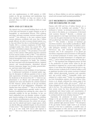
Here, we reviewed recent in vitro and in vivo data on the
effects of both CKD and oral iron on the gut microbiome
and metabolome, and immunity. Collectively, these data
show it is conceivable that oral iron supplementation in
iron deficient predialysis CKD patients may further wors-
en their clinical condition by adversely changing gut
microbiome composition, the gut and systemic metabo-
lome, and host immunity and infection (Figure 1). Future
studies are warranted to confirm these concerns and to
assess whether—compared to IV iron supplementation
and placebo—oral iron supplementation negatively
impacts on the disease progression of CKD patients.
Therefore, until more is known about local gut and sys-
temic adverse effects of oral iron in patients with CKD,
we recommend to carefully weigh the positive effects of
supplementary iron on preventing symptoms of iron defi-
ciency against the possible adverse effects on gut micro-
biome composition and activity, the systemic
metabolome, infection, and host immunity.
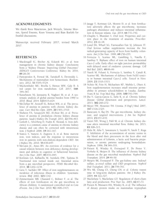
Figure 1 Combined effects of oral iron supplementation
and CKD on gut microbiome composition, metabolome,
and host immunity and infection. These effects add to other
(inborn and environmental) factors, and together will deter-
mine the morbidity (e.g., progression of the disease) of the
patient. [Color figure can be viewed at wileyonlinelibrary.
com]
Kortman et al.
6 Hemodialysis International 2017; 00:00–00
7. ACKNOWLEDGMENTS
We thank Roos Masereeuw, Jack Wetzels, Simone Moo-
ren, Sjoerd Emonts, Koen Venema and Rian Roelofs for
fruitful discussions.
Manuscript received February 2017; revised March
2017.
REFERENCES
1 Macdougall IC, Bircher AJ, Eckardt KU, et al. Iron
management in chronic kidney disease: Conclusions
from a “Kidney Disease: Improving Global Outcomes”
(KDIGO) Controversies Conference. Kidney Int. 2016;
89:28–39.
2 Pantopoulos K, Porwal SK, Tartakoff A, Devireddy L.
Mechanisms of mammalian iron homeostasis. Biochem-
istry. 2012; 51:5705–5724.
3 Muckenthaler MU, Rivella S, Hentze MW, Galy B. A
red carpet for iron metabolism. Cell. 2017; 168:
344–361.
4 Kassebaum NJ, Jasrasaria R, Naghavi M, et al. A sys-
tematic analysis of global anemia burden from 1990 to
2010. Blood. 2014; 123:615–624.
5 McClellan W, Aronoff SL, Bolton WK, et al. The preva-
lence of anemia in patients with chronic kidney dis-
ease. Curr Med Res Opin. 2004; 20:1501–1510.
6 Shaheen FA, Souqiyyeh MZ, Al-Attar BA, et al. Preva-
lence of anemia in predialysis chronic kidney disease
patients. Saudi J Kidney Dis Transpl. 2011; 22:456–463.
7 Gotloib L, Silverberg D, Fudin R, Shostak A. Iron defi-
ciency is a common cause of anemia in chronic kidney
disease and can often be corrected with intravenous
iron. J Nephrol. 2006; 19:161–167.
8 Stancu S, Stanciu A, Zugravu A, et al. Bone marrow
iron, iron indices, and the response to intravenous
iron in patients with non-dialysis-dependent CKD. Am
J Kidney Dis. 2010; 55:639–647.
9 McCann JC, Ames BN. An overview of evidence for a
causal relation between iron deficiency during develop-
ment and deficits in cognitive or behavioral function.
Am J Clin Nutr. 2007; 85:931–945.
10 Kortman GA, Raffatellu M, Swinkels DW, Tjalsma H.
Nutritional iron turned inside out: Intestinal stress
from a gut microbial perspective. FEMS Microbiol Rev.
2014; 38:1202–1234.
11 Gera T, Sachdev HP. Effect of iron supplementation on
incidence of infectious illness in children: Systematic
review. BMJ. 2002; 325:1142.
12 Zimmermann MB, Chassard C, Rohner F, et al. The
effects of iron fortification on the gut microbiota in
African children: A randomized controlled trial in Cote
d’Ivoire. Am J Clin Nutr. 2010; 92:1406–1415.
13 Jaeggi T, Kortman GA, Moretti D, et al. Iron fortifica-
tion adversely affects the gut microbiome, increases
pathogen abundance and induces intestinal inflamma-
tion in Kenyan infants. Gut. 2014; 64:731–742.
14 Chaplin S, Bhandari S. Oral iron: Properties and cur-
rent place in the treatment of anaemia. Prescriber.
2012; 23:12–18.
15 Lund EK, Wharf SG, Fairweather-Tait SJ, Johnson IT.
Oral ferrous sulfate supplements increase the free
radical-generating capacity of feces from healthy volun-
teers. Am J Clin Nutr. 1999; 69:250–255.
16 Ferruzza S, Scarino ML, Gambling L, Natella F,
Sambuy Y. Biphasic effect of iron on human intestinal
Caco-2 cells: Early effect on tight junction permeability
with delayed onset of oxidative cytotoxic damage. Cell
Mol Biol. 2003; 49:89–99.
17 Natoli M, Felsani A, Ferruzza S, Sambuy Y, Canali R,
Scarino ML. Mechanisms of defence from Fe(II) toxici-
ty in human intestinal Caco-2 cells. Toxicol in Vitro.
2009; 23:1510–1515.
18 Nchito M, Friis H, Michaelsen KF, Mubila L, Olsen A.
Iron supplementation increases small intestine perme-
ability in primary schoolchildren in Lusaka, Zambia.
Trans R Soc Trop Med Hyg. 2006; 100:791–794.
19 Jha V, Garcia-Garcia G, Iseki K, et al. Chronic kidney
disease: Global dimension and perspectives. Lancet.
2013; 382:260–272.
20 Meyer TW, Hostetter TH. Uremia. N Engl J Med. 2007;
357:1316–1325.
21 Ramezani A, Raj DS. The gut microbiome, kidney dis-
ease, and targeted interventions. J Am Soc Nephrol.
2014; 25:657–670.
22 Vaziri ND, Wong J, Pahl M, et al. Chronic kidney dis-
ease alters intestinal microbial flora. Kidney Int. 2013;
83:308–315.
23 Hida M, Aiba Y, Sawamura S, Suzuki N, Satoh T, Koga
Y. Inhibition of the accumulation of uremic toxins in
the blood and their precursors in the feces after oral
administration of Lebenin, a lactic acid bacteria prepa-
ration, to uremic patients undergoing hemodialysis.
Nephron. 1996; 74:349–355.
24 Poesen R, Windey K, Evenepoel P, De Preter V,
Verbeke K, Meijers B. The influence of chronic kidney
disease on the gut microbial metabolism. Nephrol Dial
Transplant. 2014; 29:49–50.
25 Meijers BK, Evenepoel P. The gut-kidney axis: Indoxyl
sulfate, p-cresyl sulfate and CKD progression. Nephrol
Dial Transplant. 2011; 26:759–761.
26 Wu MJ, Chang CS, Cheng CH, et al. Colonic transit
time in long-term dialysis patients. Am J Kidney Dis.
2004; 44:322–327.
27 Macfarlane S, Macfarlane GT. Regulation of short-chain
fatty acid production. Proc Nutr Soc. 2003; 62:67–72.
28 Poesen R, Mutsaers HA, Windey K, et al. The influence
of dietary protein intake on mammalian tryptophan
Oral iron and the gut microbiome in CKD
Hemodialysis International 2017; 00:00–00 7
8. and phenolic metabolites. PLoS One. 2015; 10:
e0140820.
29 Poesen R, Claes K, Evenepoel P, et al. Microbiota-
derived phenylacetylglutamine associates with overall
mortality and cardiovascular disease in patients with
CKD. J Am Soc Nephrol. 2016; 27:3479–3487.
30 Aronov PA, Luo FJ, Plummer NS, et al. Colonic contri-
bution to uremic solutes. J Am Soc Nephrol. 2011; 22:
1769–1776.
31 Evenepoel P, Poesen R, Meijers B. The gut-kidney axis.
Pediatr Nephrol. 2016, in press.
32 Gryp T, Vanholder R, Vaneechoutte M, Glorieux G. p-
Cresyl sulfate. Toxins. 2017; 9:52.
33 Vaziri ND, Yuan J, Rahimi A, Ni Z, Said H,
Subramanian VS. Disintegration of colonic epithelial
tight junction in uremia: A likely cause of CKD-
associated inflammation. Nephrol Dial Transplant. 2012;
27:2686–2693.
34 Leemans JC, Kors L, Anders HJ, Florquin S. Pattern
recognition receptors and the inflammasome in kidney
disease. Nat Rev Nephrol. 2014; 10:398–414.
35 Felizardo RJ, Castoldi A, Andrade-Oliveira V,
Camara NO. The microbiota and chronic kidney
diseases: A double-edged sword. Clin Trans Immunol.
2016; 5: e86.
36 Vaziri ND, Zhao YY, Pahl MV. Altered intestinal micro-
bial flora and impaired epithelial barrier structure and
function in CKD: The nature, mechanisms, conse-
quences and potential treatment. Nephrol Dial Trans-
plant. 2016; 31:737–746.
37 Ramezani A, Massy ZA, Meijers B, Evenepoel P,
Vanholder R, Raj DS. Role of the gut microbiome in
uremia: A potential therapeutic target. Am J Kidney Dis.
2016; 67:483–498.
38 Andersen K, Kesper MS, Marschner JA, et al. Intestinal
dysbiosis, barrier dysfunction, and bacterial transloca-
tion account for CKD-related systemic inflammation.
J Am Soc Nephrol. 2017; 28:76–83.
39 Nallu A, Sharma S, Ramezani A, Muralidharan J, Raj
D. Gut microbiome in chronic kidney disease: Chal-
lenges and opportunities. Transl Res. 2017; 179:
24–37.
40 Canfora EE, Jocken JW, Blaak EE. Short-chain fatty
acids in control of body weight and insulin sensitivity.
Nat Rev Endocrinol. 2015; 11:577–591.
41 Poesen R, Windey K, Neven E, et al. The influence of
CKD on colonic microbial metabolism. J Am Soc Neph-
rol. 2016; 27:1389–1399.
42 Soares MP, Weiss G. The iron age of host-microbe
interactions. EMBO Rep. 2015; 16:1482–1500.
43 Weinberg ED. Iron availability and infection. Biochim
Biophys Acta. 2009; 1790:600–605.
44 Nairz M, Schroll A, Sonnweber T, Weiss G. The strug-
gle for iron—A metal at the host-pathogen interface.
Cell Microbiol. 2010; 12:1691–1702.
45 Weiss G, Schett G. Anaemia in inflammatory rheumatic
diseases. Nat Rev Rheumatol. 2013; 9:205–215.
46 Ganz T. Iron in innate immunity: Starve the invaders.
Curr Opin Immunol. 2009; 21:63–67.
47 Macdougall IC, Bock A, Carrera F, et al. The FIND-
CKD study–a randomized controlled trial of intrave-
nous iron versus oral iron in non-dialysis chronic kid-
ney disease patients: Background and rationale.
Nephrol Dial. 2014; 29:843–850.
48 Avni T, Bieber A, Steinmetz T, Leibovici L, Gafter-Gvili
A. Treatment of anemia in inflammatory bowel disease-
systematic review and meta-analysis. PLoS One. 2013;
8: e75540.
49 Lee TW, Kolber MR, Fedorak RN, van Zanten SV. Iron
replacement therapy in inflammatory bowel disease
patients with iron deficiency anemia: A systematic
review and meta-analysis. J Crohns Colitis. 2012; 6:
267–275.
50 Zhu A, Kaneshiro M, Kaunitz JD. Evaluation and treat-
ment of iron deficiency anemia: A gastroenterological
perspective. Dig Dis Sci. 2010; 55:548–559.
51 Powell JJ, Cook WB, Hutchinson C, et al. Dietary forti-
ficant iron intake is negatively associated with quality
of life in patients with mildly active inflammatory bow-
el disease. Nutr Metab (Lond). 2013; 10:9.
52 Tolkien Z, Pereira DI, Prassmayer L, et al. Dietary iron
does not impact the quality of life of patients with qui-
escent ulcerative colitis: An observational study. Nutr J.
2013; 12:152.
53 Kortman GA, Dutilh BE, Maathuis AJ, et al. Microbial
metabolism shifts towards an adverse profile with sup-
plementary iron in the TIM-2 in vitro model of the
human colon. Front Microbiol. 2015; 6:1481.
54 Montemurno E, Cosola C, Dalfino G, et al. What
would you like to eat, Mr CKD Microbiota? A Mediter-
ranean Diet, please!. Kidney Blood Press Res. 2014; 39:
114–123.
55 Evenepoel P, Meijers BK, Bammens BR, Verbeke K.
Uremic toxins originating from colonic microbial
metabolism. Kidney Int. 2009; S12–S19.
56 Vanholder RC, Glorieux GL. An overview of uremic
toxicity. Hemodial Int. 2003; 7:156–161.
57 Low MS, Speedy J, Styles CE, De-Regil LM,
Pasricha SR. Daily iron supplementation for
improving anaemia, iron status and health in men-
struating women. Cochrane Database Syst Rev. 2016;
4:CD009747.
58 Fishbane S, Block GA, Loram L, Neylan J, Pergola PE,
Uhlig K, Chertow GM. Effects of ferric citrate in
patients with nondialysis-dependent CKD and iron
deficiency anemia. J Am Soc Nephrol. 2017, in press.
59 Lee T, Clavel T, Smirnov K, et al. Oral versus intra-
venous iron replacement therapy distinctly alters the
gut microbiota and metabolome in patients with IBD.
Gut. 2016, in press.
Kortman et al.
8 Hemodialysis International 2017; 00:00–00
9. 60 Lewis JB, Sika M, Koury MJ, et al. Ferric citrate con-
trols phosphorus and delivers iron in patients on dialy-
sis. J Am Soc Nephrol. 2015; 26:493–503.
61 Pai AB, Jang SM, Wegrzyn N. Iron-based phosphate
binders–a new element in management of hyperphos-
phatemia. Expert Opin Drug Metab Toxicol. 2016; 12:
115–127.
62 Pizarro F, Amar M, Stekel A. Determination of iron in
stools as a method to monitor consumption of iron-
fortified products in infants. Am J Clin Nutr. 1987; 45:
484–487.
63 Andrews SC, Robinson AK, Rodriguez-Quinones F.
Bacterial iron homeostasis. FEMS Microbiol Rev. 2003;
27:215–237.
64 Li H, Limenitakis JP, Fuhrer T, et al. The outer mucus
layer hosts a distinct intestinal microbial niche. Nat
Comms. 2015; 6:8292.
65 Vieira AT, Teixeira MM, Martins FS. The role of probi-
otics and prebiotics in inducing gut immunity. Front
Immunol. 2013; 4:445.
66 Petry N, Egli I, Chassard C, Lacroix C, Hurrell R. Inu-
lin modifies the bifidobacteria population, fecal lactate
concentration, and fecal pH but does not influence
iron absorption in women with low iron status. Am J
Clin Nutr. 2012; 96:325–331.
67 Burkitt DP, Walker AR, Painter NS. Effect of dietary
fibre on stools and the transit-times, and its role in the
causation of disease. Lancet. 1972; 2:1408–1412.
68 Koppe L, Fouque D. Microbiota and prebiotics modu-
late uremic toxins generation. Panminerva Medi. 2016,
in press.
69 Hilty FM, Arnold M, Hilbe M, et al. Iron from nano-
compounds containing iron and zinc is highly bioavail-
able in rats without tissue accumulation. Nat
Nanotechnol. 2010; 5:374–380.
70 Pereira DI, Mergler BI, Faria N, et al. Caco-2 cell
acquisition of dietary iron(III) invokes a nanoparticu-
late endocytic pathway. PLoS One. 2013; 8: e81250.
71 Powell JJ, Bruggraber SF, Faria N, Poots LK, Hondow N,
Pennycook TJ, etet al. A nano-disperse ferritin-core
mimetic that efficiently corrects anemia without luminal
iron redox activity. Nanomedicine. 2014; 10:1529–1538.
72 Pereira DI, Aslam MF, Frazer DM, et al. Dietary iron
depletion at weaning imprints low microbiome diversi-
ty and this is not recovered with oral Nano Fe(III).
MicrobiologyOpen. 2015; 4:12–27.
73 Lonnerdal B. Nutritional roles of lactoferrin. Curr Opin
Clin Nutr Metab Care. 2009; 12:293–297.
74 La Carpia F, Wojczyk B, Rebbaa A, Tang A, Hod EA.
Chronic transfusion and iron overload modify the
mouse gut microbiome. Blood. 2016; 128:200.
75 Falony G, Joossens M, Vieira-Silva S, et al. Population-
level analysis of gut microbiome variation. Science.
2016; 352:560–564.
76 Zhernakova A, Kurilshikov A, Bonder MJ, et al. Popu-
lation-based metagenomics analysis reveals markers for
gut microbiome composition and diversity. Science.
2016; 352:565–569.
77 KDIGO. Kidney disease: Improving global outcomes
(KDIGO) Anemia Work Group. KDIGO clinical prac-
tice guideline for anemia in chronic kidney disease.
Kidney Int. supplements 2012; 2:279–335.
78 Rostoker G, Griuncelli M, Loridon C, et al. Hemodialy-
sis-associated hemosiderosis in the era of
erythropoiesis-stimulating agents: A MRI study. Am J
Med. 2012; 125:991–999.e1.
79 Kalra PA, Bhandari S, Saxena S, et al. A randomized
trial of iron isomaltoside 1000 versus oral iron in non-
dialysis-dependent chronic kidney disease patients
with anaemia. Nephrol Dial Transplant. 2016; 31:
646–655.
80 Macdougall IC, Bock AH, Carrera F, et al. FIND-CKD:
A randomized trial of intravenous ferric carboxymal-
tose versus oral iron in patients with chronic kidney
disease and iron deficiency anaemia. Nephrol Dial
Transplant. 2014; 29:2075–2084.
81 Agarwal R, Kusek JW, Pappas MK. A randomized trial
of intravenous and oral iron in chronic kidney disease.
Kidney Int. 2015; 88:905–914.
82 Locatelli F, Barany P, Covic A, et al. Kidney disease:
Improving global outcomes guidelines on anaemia
management in chronic kidney disease: a European
Renal Best Practice position statement. Nephrol Dial
Transplant. 2013; 28:1346–1359.
Oral iron and the gut microbiome in CKD
Hemodialysis International 2017; 00:00–00 9