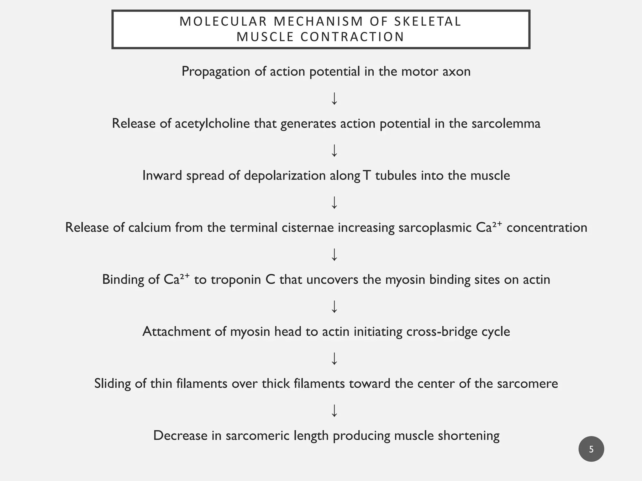
MOLECULAR MECHANISM OFSKELETAL
MUSCLE CONTRACTION
5
Propagation of action potential in the motor axon
↓
Release of acetylcholine that generates action potential in the sarcolemma
↓
Inward spread of depolarization along T tubules into the muscle
↓
Release of calcium from the terminal cisternae increasing sarcoplasmic Ca²⁺ concentration
↓
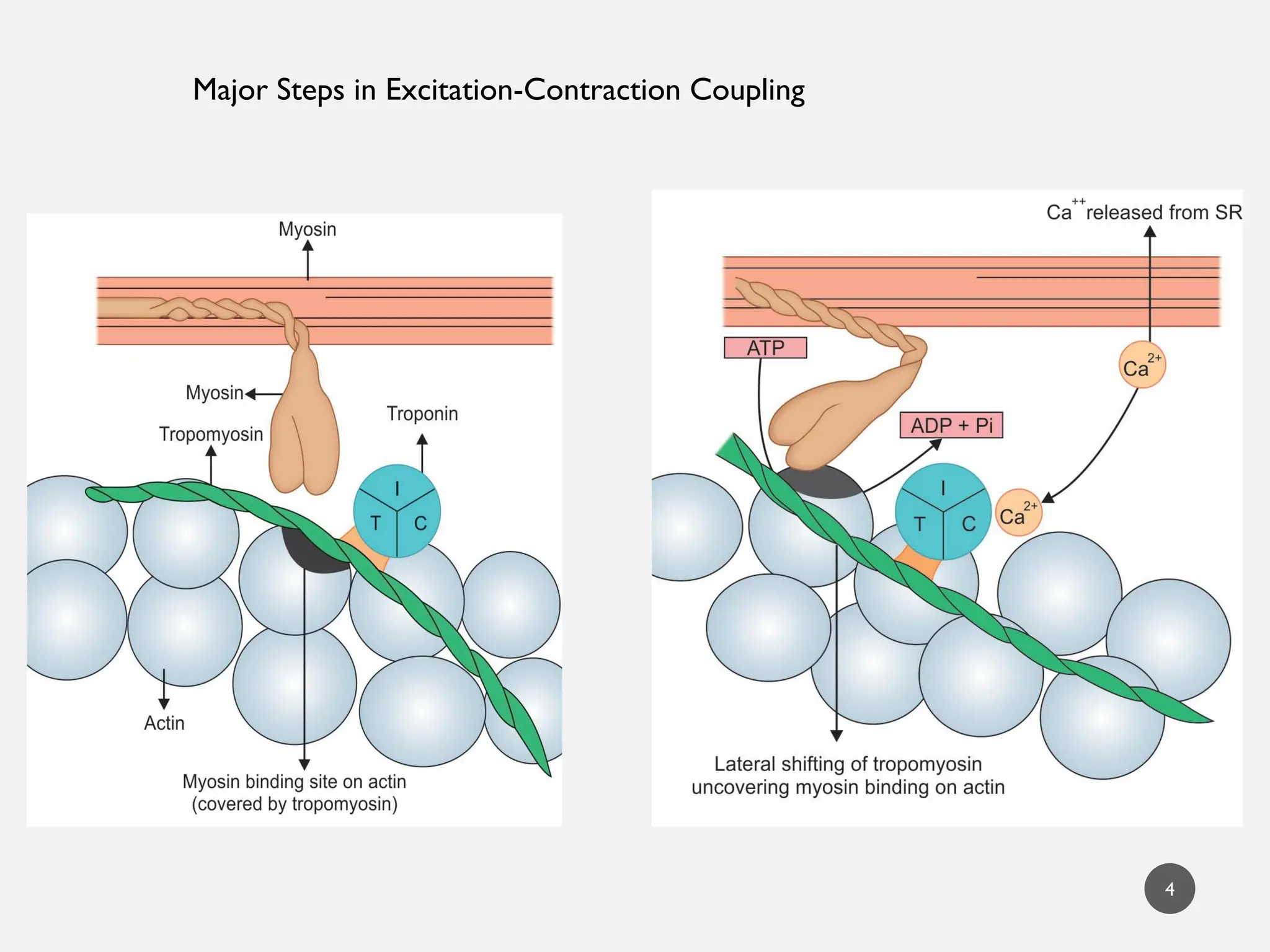
Binding of Ca²⁺ to troponin C that uncovers the myosin binding sites on actin
↓
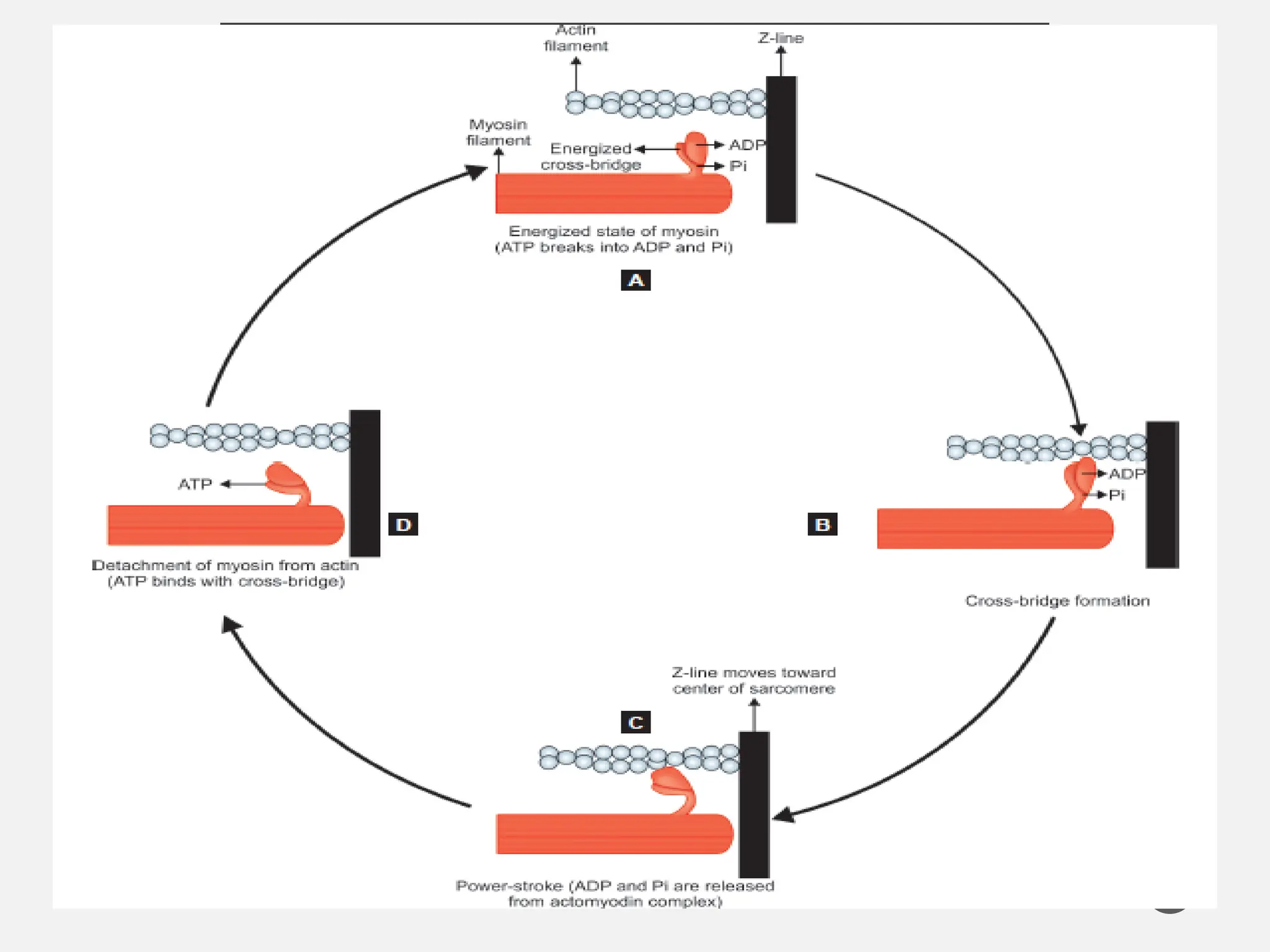
Attachment of myosin head to actin initiating cross-bridge cycle
↓
Sliding of thin filaments over thick filaments toward the center of the sarcomere
↓
Decrease in sarcomeric length producing muscle shortening
6.
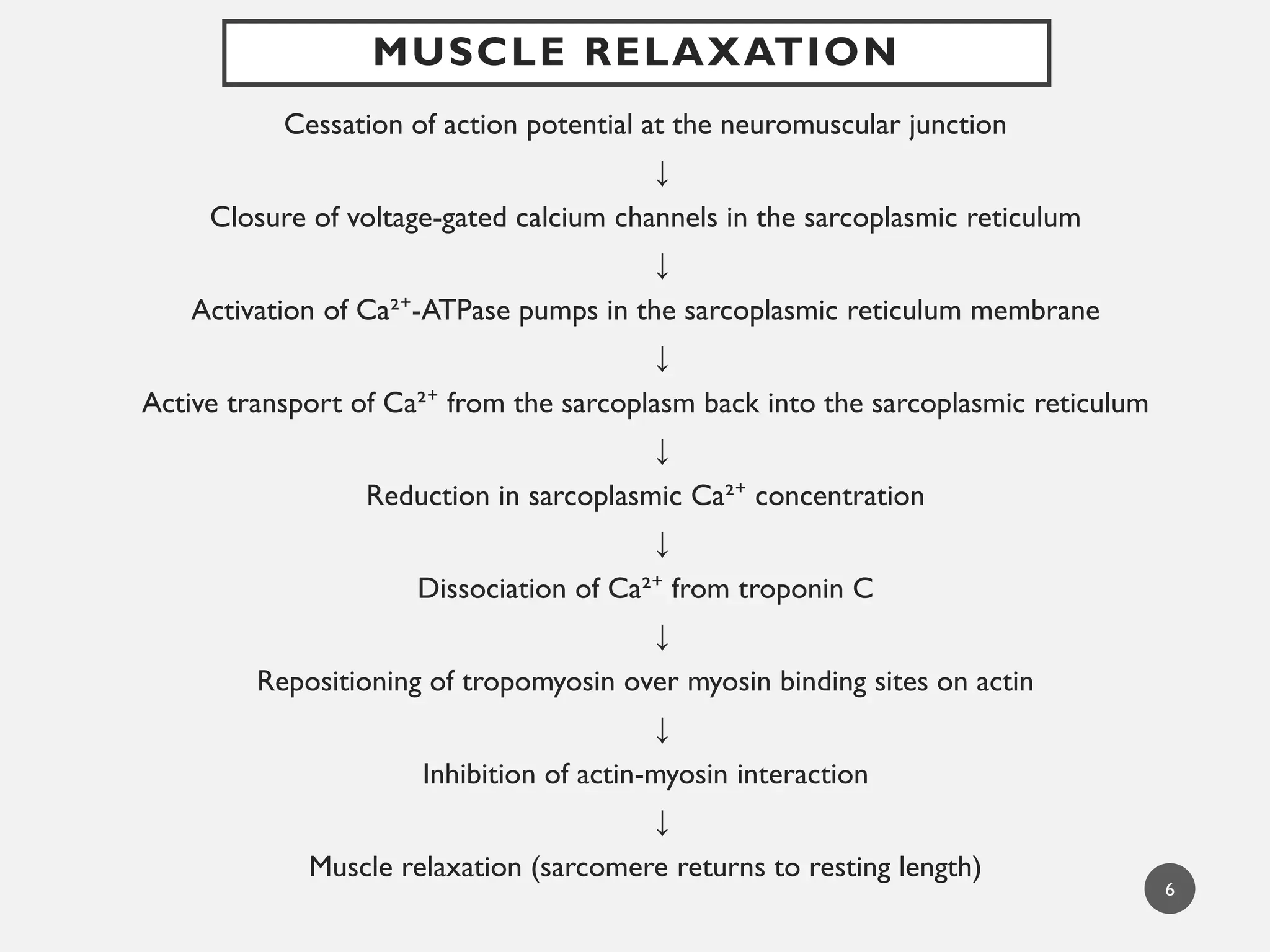
MUSCLE RELAXATION
Cessation ofaction potential at the neuromuscular junction
↓
Closure of voltage-gated calcium channels in the sarcoplasmic reticulum
↓
Activation of Ca²⁺-ATPase pumps in the sarcoplasmic reticulum membrane
↓
Active transport of Ca²⁺ from the sarcoplasm back into the sarcoplasmic reticulum
↓
Reduction in sarcoplasmic Ca²⁺ concentration
↓
Dissociation of Ca²⁺ from troponin C
↓
Repositioning of tropomyosin over myosin binding sites on actin
↓
Inhibition of actin-myosin interaction
↓
Muscle relaxation (sarcomere returns to resting length)
6
7.
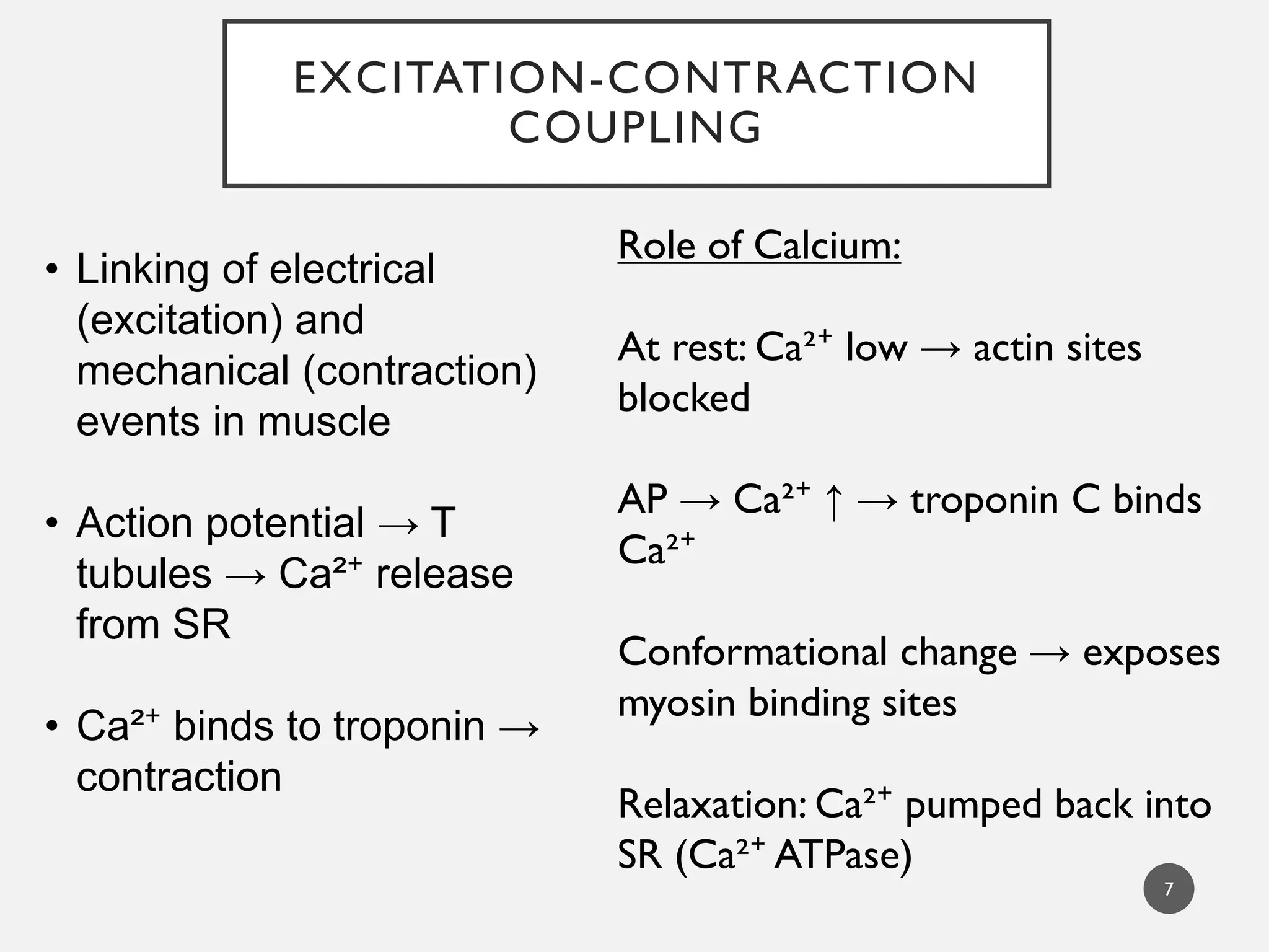
EXCITATION-CONTRACTION
COUPLING
7
• Linking ofelectrical
(excitation) and
mechanical (contraction)
events in muscle
• Action potential → T
tubules → Ca²⁺ release
from SR
• Ca²⁺ binds to troponin →
contraction
Role of Calcium:
At rest: Ca²⁺ low → actin sites
blocked
AP → Ca²⁺ ↑ → troponin C binds
Ca²⁺
Conformational change → exposes
myosin binding sites
Relaxation: Ca²⁺ pumped back into
SR (Ca²⁺ ATPase)
RIGOR MORTIS
• Stiffeningof the muscle after death is called rigor mortis.
• Shortening and rigidity of all the body muscles which occurs some
hours after death.
• Depletion of ATP fails to detach the cross-bridge - muscle remains
in a state of contraction or rigidity.
• Due to rigor mortis, body remains in same position for a longer
time.
• Thus, rigor mortis not only speaks about the time of death, but also
the nature of death, which helps in medicolegal investigations in
case of mysterious death.
9