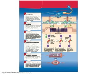
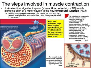
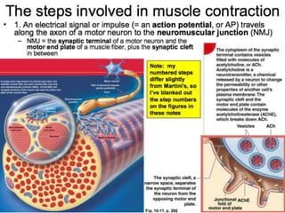
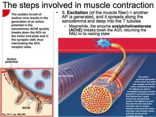
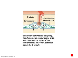
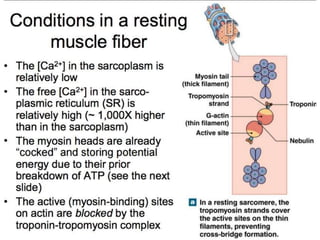
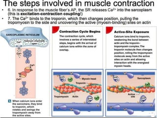
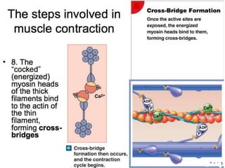
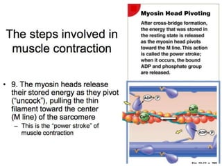
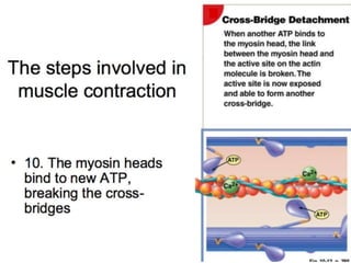
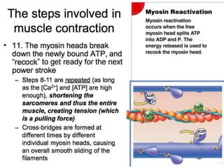
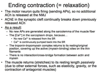
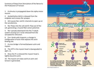
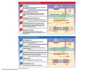
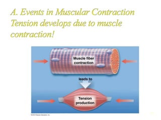
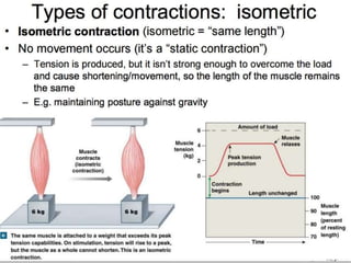
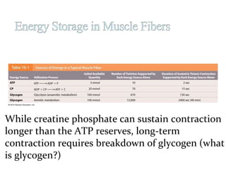
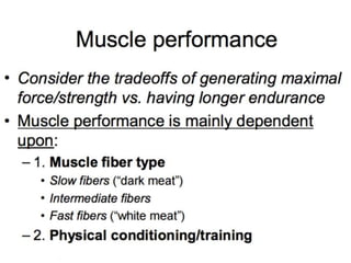
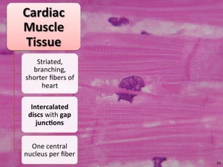
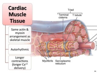
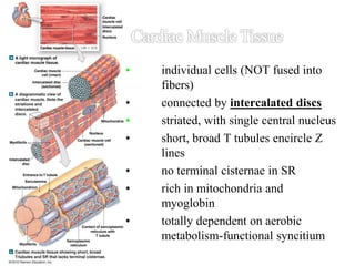
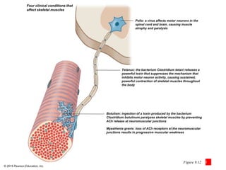
SlideShare a Scribd company logo
© 2015 Pearson Education, Inc.
Chapter 10 part 3
© 2015 Pearson Education, Inc.
Motor end
plate
Synaptic terminal
Sarcoplasmic
reticulum
Myofibril
Motor neuron
Axon
Motor end plate
Path of electrical
impulse (action
potential)
Neuromuscular
junction
Myofibril
The structural relationship between a skeletal
muscle fiber and its lone neuromuscular junction
3
Muscle contraction begins when a motor neuron
impulse stimulates an impulse in a muscle fiber.
Nerve impulse is generated and sent as an electrical
current (action potential) along sarcolemma
The neuromuscular junction is the region where the
motor neuron comes into close proximity to the muscle
fiber.
The neuromuscular junction (NMJ)
Special intercellular connection between the
nervous system and skeletal muscle fiber
Controls calcium ion release into the sarcoplasm
© 2015 Pearson Education, Inc.
Excitation-contraction coupling,
the dumping of calcium ions onto
sarcomeres as a result of the
movement of an action potential
down the T tubule
T tubule
Sarcoplasm
Sarcoplasmic
reticulum (SR)
Ca2+ Ca2+
© 2015 Pearson Education, Inc.
18
Summary of Steps from Stimulation of the Nerve to
the Production of Tension
1. A stimulus is propagated down the alpha motor
neuron.
2. Acetylcholine (Ach) is released from the
endplate and crosses the synapse.
3. Ach causes Na+ and K+ channels to open up on
the sarcolemma.
4. Na+ flows into the cell and K+ flow out of the
cell, generating a muscle fiber action potential.
5. Na+ spreads downward into the T-tubule
system causing Ca++ to be released from the
sarcoplasmic reticulum.
6. Ca++ binds with troponin, a change in
configuration of actin that exposes the actin
binding site.
7. A cross-bridge is formed between actin and
myosin.
8. The ATP in the myosin head is downgraded to
ADP + Pi.
9. Once the Pi is released the myosin head is
tightly bound to actin.
10. The myosin arm does work on actin and
tension is generated.
© 2015 Pearson Education, Inc.
20
 Tension = pulling force
• Tension Production by Muscles Fibers
• As a whole, a muscle fiber is either contracted or relaxed
 The all–or–none principal: as a whole, a muscle fiber is
either contracted or relaxed
 Amount of tension in a muscle is determined by the
following:
 Resting length of the sarcomere at the time of
stimulation (how much overlap)
 Frequency of stimulation….which impacts the
concentration of calcium ions
© 2015 Pearson Education, Inc.
25
Myogram -
records
muscle
contraction
(force of
contraction
in
relationship
to time
© 2015 Pearson Education, Inc.
The production of lactate during peak activity, its conversion to glucose in the liver, and the rebuilding of
glycogen reserves in the muscles during recovery
Lactate
Pyruvate Glucose
Glucose
Pyruvate
Lactate
Glucose
70–80%
20–30%
LIVER
MUSCLE
Glycogen reserves in muscle
Peak Activity Recovery
Much of the large amounts of lactate
produced during peak exertion diffuses
out of the muscle fibers and into the
bloodstream. The liver absorbs this lactate
and begins converting it into pyruvate.
This process continues after exertion has ended, because lactate
levels within muscle fibers remain relatively high, and lactate
continues to diffuse into the bloodstream. After the absorbed
lactate is converted to pyruvate in the liver, roughly 30 percent of
the new pyruvate molecules are broken down in the
mitochondria, providing the ATP needed to convert the remaining
70 percent of pyruvate
molecules into
glucose. The
glucose molecules
are then released
into the circulation,
where they are
absorbed by skeletal
muscle fibers and used
to rebuild their glycogen
reserves.
While creatine phosphate can sustain contraction
longer than the ATP reserves, long-term
contraction requires breakdown of glycogen (what
is glycogen?)
© 2015 Pearson Education, Inc.
Slow (R)—more mitochondria (M) and
capillaries (cap) than fast (W)
Table 10-2
Intermediate
Intermediate
Intermediate
© 2015 Pearson Education, Inc.
Figure 10-22
• individual cells (NOT fused into
fibers)
• connected by intercalated discs
• striated, with single central nucleus
• short, broad T tubules encircle Z
lines
• no terminal cisternae in SR
• rich in mitochondria and
myoglobin
• totally dependent on aerobic
metabolism-functional syncitium
56
© 2015 Pearson Education, Inc.
Factors and clinical conditions affecting muscles
• Hypertrophy
– Increase in muscle size due to:
• Increase in myofilaments
• Increase in myofibril size
• Increase in mitochondria
• More glycogen and glycolytic enzymes
– As a result of repeated exhaustive stimulation
• Can be promoted by administration of steroid hormones
© 2015 Pearson Education, Inc.
Factors and clinical conditions affecting muscles
• Atrophy
– Decrease in muscle size, tone, and power
– As a result of decreased stimulation such as during:
• Paralysis by spinal injury
• Damage to nervous system
• Having body part in cast after bone fracture
– Initially reversible, but after prolonged disuse,
muscle fibers can die and not be replaced
© 2015 Pearson Education, Inc.
Factors and clinical conditions affecting muscles
• Clinical conditions
– Polio
• Virus attacks motor neurons of brain and spinal cord causing
paralysis (lost of voluntary movement)
– Tetanus
• Toxin from bacteria (Clostridium tetani) that suppresses the
mechanism inhibiting motor neuron activity
• Thrives in low-oxygen areas like deep punctured tissues
• Results in sustained, powerful contractions of affected muscles
• Severe tetanus can have 40%–60% mortality
– Deaths rare due to immunization in U.S.
© 2015 Pearson Education, Inc.
Factors and clinical conditions affecting
muscles
• Clinical conditions
• Botulism
• Toxin from bacteria (Clostridium botulinum) that blocks
ACh release at neuromuscular junctions
• Acquired through bacteria-contaminated food
– Myasthenia gravis
• Loss of ACh receptors at neuromuscular junctions
• Results in progressive weakness
© 2015 Pearson Education, Inc.
Factors and clinical conditions affecting muscles
• Clinical conditions Rigor mortis
• Generalized muscle contraction shortly after death
(2–7 hours)
• Begins with small muscles of face, neck, and arms
• Due to depletion of ATP, leaving myosin cross-bridges
attached to actin
• Ends 1–6 days later as muscular tissue decomposes
© 2015 Pearson Education, Inc.
Figure 9.12 3
Four clinical conditions that
affect skeletal muscles
Polio: a virus affects motor neurons in the
spinal cord and brain, causing muscle
atrophy and paralysis
Tetanus: the bacterium Clostridium tetani releases a
powerful toxin that suppresses the mechanism that
inhibits motor neuron activity, causing sustained,
powerful contraction of skeletal muscles throughout
the body
Botulism: ingestion of a toxin produced by the bacterium
Clostridium botulinum paralyzes skeletal muscles by preventing
ACh release at neuromuscular junctions
Myasthenia gravis: loss of ACh receptors at the neuromuscular
junctions results in progressive muscular weakness

More Related Content

What's hot

Mechanism of muscle contraction (saran.s)
Mechanism of muscle contraction (saran.s)Mechanism of muscle contraction (saran.s)
Mechanism of muscle contraction (saran.s)
Saran S
 
Fiber types, energy sources
Fiber types, energy sourcesFiber types, energy sources
Fiber types, energy sources
Pradeep Singh Narwat
 
Muscle contraction
Muscle contractionMuscle contraction
Muscle contractionRajesh Goit
 
7. muscle contraction lecture 2
7. muscle contraction lecture 27. muscle contraction lecture 2
7. muscle contraction lecture 2
Sam Phiri
 
Muscles Contraction
Muscles Contraction Muscles Contraction
Muscles Contraction
Manish Kuthyal
 
Mechanism Of Muscle Contraction&Neural Control
Mechanism Of Muscle Contraction&Neural ControlMechanism Of Muscle Contraction&Neural Control
Mechanism Of Muscle Contraction&Neural Controlraj kumar
 
APU6.4 Skeletal Muscle Contractions
APU6.4 Skeletal Muscle ContractionsAPU6.4 Skeletal Muscle Contractions
APU6.4 Skeletal Muscle Contractions
NeQuelle DeFord
 
Muscle Contraction
Muscle ContractionMuscle Contraction
Muscle Contractionguest12e21
 
11.2 muscle contraction
11.2  muscle contraction11.2  muscle contraction
11.2 muscle contraction
cartlidge
 
Controlling muscle contraction
Controlling muscle contractionControlling muscle contraction
Controlling muscle contractionPersonal
 
Ppt notes muscle contraction
Ppt notes   muscle contractionPpt notes   muscle contraction
Ppt notes muscle contractionBilly Joe Garma
 
Muscle contraction with medecine
Muscle contraction with medecineMuscle contraction with medecine
Muscle contraction with medecineHussy Dimaangay
 
Sliding filament theory muscle contraction
Sliding filament theory   muscle contractionSliding filament theory   muscle contraction
Sliding filament theory muscle contractionLloyd Dean
 
Skeletal muscle contraction
Skeletal muscle contractionSkeletal muscle contraction
Skeletal muscle contraction
muhammad aleem ijaz
 
muscle fatigue
 muscle fatigue muscle fatigue
muscle fatigue
Ali Faris
 
section 4, chapter 9: energy for muscles
section 4, chapter 9: energy for musclessection 4, chapter 9: energy for muscles
section 4, chapter 9: energy for muscles
Michael Walls
 
SARCOTUBULAR SYSTEM
SARCOTUBULAR SYSTEMSARCOTUBULAR SYSTEM
SARCOTUBULAR SYSTEM
Dr Nilesh Kate
 
Muscle contraction
Muscle contractionMuscle contraction
Muscle contraction
Suprabha Panda
 

What's hot (20)

Mechanism of muscle contraction (saran.s)
Mechanism of muscle contraction (saran.s)Mechanism of muscle contraction (saran.s)
Mechanism of muscle contraction (saran.s)
 
Fiber types, energy sources
Fiber types, energy sourcesFiber types, energy sources
Fiber types, energy sources
 
Muscle contraction
Muscle contractionMuscle contraction
Muscle contraction
 
Muscle
MuscleMuscle
Muscle
 
7. muscle contraction lecture 2
7. muscle contraction lecture 27. muscle contraction lecture 2
7. muscle contraction lecture 2
 
Muscles Contraction
Muscles Contraction Muscles Contraction
Muscles Contraction
 
Mechanism Of Muscle Contraction&Neural Control
Mechanism Of Muscle Contraction&Neural ControlMechanism Of Muscle Contraction&Neural Control
Mechanism Of Muscle Contraction&Neural Control
 
APU6.4 Skeletal Muscle Contractions
APU6.4 Skeletal Muscle ContractionsAPU6.4 Skeletal Muscle Contractions
APU6.4 Skeletal Muscle Contractions
 
Bio
BioBio
Bio
 
Muscle Contraction
Muscle ContractionMuscle Contraction
Muscle Contraction
 
11.2 muscle contraction
11.2  muscle contraction11.2  muscle contraction
11.2 muscle contraction
 
Controlling muscle contraction
Controlling muscle contractionControlling muscle contraction
Controlling muscle contraction
 
Ppt notes muscle contraction
Ppt notes   muscle contractionPpt notes   muscle contraction
Ppt notes muscle contraction
 
Muscle contraction with medecine
Muscle contraction with medecineMuscle contraction with medecine
Muscle contraction with medecine
 
Sliding filament theory muscle contraction
Sliding filament theory   muscle contractionSliding filament theory   muscle contraction
Sliding filament theory muscle contraction
 
Skeletal muscle contraction
Skeletal muscle contractionSkeletal muscle contraction
Skeletal muscle contraction
 
muscle fatigue
 muscle fatigue muscle fatigue
muscle fatigue
 
section 4, chapter 9: energy for muscles
section 4, chapter 9: energy for musclessection 4, chapter 9: energy for muscles
section 4, chapter 9: energy for muscles
 
SARCOTUBULAR SYSTEM
SARCOTUBULAR SYSTEMSARCOTUBULAR SYSTEM
SARCOTUBULAR SYSTEM
 
Muscle contraction
Muscle contractionMuscle contraction
Muscle contraction
 

Similar to Chapter10 part 3

Presented by yasmeen zartash
Presented by yasmeen zartashPresented by yasmeen zartash
Presented by yasmeen zartash
Ayannoor1
 
muscle7-smoothmuscle-180414110808.pptx
muscle7-smoothmuscle-180414110808.pptxmuscle7-smoothmuscle-180414110808.pptx
muscle7-smoothmuscle-180414110808.pptx
LaviBharti1
 
EXCITATION CONTRACTION COUPLING physiology
EXCITATION CONTRACTION COUPLING physiologyEXCITATION CONTRACTION COUPLING physiology
EXCITATION CONTRACTION COUPLING physiology
RudrapratapSinghJodh
 
Smooth muscle contraction (updated).pptx
Smooth muscle contraction (updated).pptxSmooth muscle contraction (updated).pptx
Smooth muscle contraction (updated).pptx
asdfg953797
 
Contraction of skeletal and smooth muscles
Contraction of skeletal and smooth musclesContraction of skeletal and smooth muscles
Contraction of skeletal and smooth muscles
Niyamat Chimthanawala
 
Anatomy & Physiology 2e Chapter 10 Muscle Tissue
Anatomy & Physiology 2e Chapter 10 Muscle TissueAnatomy & Physiology 2e Chapter 10 Muscle Tissue
Anatomy & Physiology 2e Chapter 10 Muscle Tissue
dmarantz81
 
General physiology lecture 3
General physiology lecture 3General physiology lecture 3
General physiology lecture 3
Huang Yu-Wen
 
NeuroTrac Sports XL electrode placement manual (English)
NeuroTrac Sports XL electrode placement manual (English)NeuroTrac Sports XL electrode placement manual (English)
NeuroTrac Sports XL electrode placement manual (English)
mareden01
 
Muscular system
Muscular systemMuscular system
Muscular systemkimcoover
 
1587124408-biomechanics-of-skeletal-muscles.pdf
1587124408-biomechanics-of-skeletal-muscles.pdf1587124408-biomechanics-of-skeletal-muscles.pdf
1587124408-biomechanics-of-skeletal-muscles.pdf
zaiqar1
 
Corrected version of smooth muscle by prof.Samia.pptx
Corrected version of smooth muscle by prof.Samia.pptxCorrected version of smooth muscle by prof.Samia.pptx
Corrected version of smooth muscle by prof.Samia.pptx
bobehe4189
 
Chapter 39 - Motor Mechanisms and Behavior.pdf
Chapter 39 - Motor Mechanisms and Behavior.pdfChapter 39 - Motor Mechanisms and Behavior.pdf
Chapter 39 - Motor Mechanisms and Behavior.pdf
De La Salle University
 
Biology in Focus - Chapter 39
Biology in Focus - Chapter 39Biology in Focus - Chapter 39
Biology in Focus - Chapter 39
mpattani
 
Biochemistry of Muscles.pptx
Biochemistry of Muscles.pptxBiochemistry of Muscles.pptx
Biochemistry of Muscles.pptx
DamiJunior
 
Chapter 8 the muscular system
Chapter 8  the muscular systemChapter 8  the muscular system
Chapter 8 the muscular system
Nada G.Youssef
 
MUSCLE TISSUE.pptx
MUSCLE TISSUE.pptxMUSCLE TISSUE.pptx
MUSCLE TISSUE.pptx
DrRashidKaziMahbubur
 
lecture 6a physio.docx
lecture 6a physio.docxlecture 6a physio.docx
lecture 6a physio.docx
Superior college
 
1587411978-skeletal-muscle.pptx
1587411978-skeletal-muscle.pptx1587411978-skeletal-muscle.pptx
1587411978-skeletal-muscle.pptx
ym782amnashaheen
 

Similar to Chapter10 part 3 (20)

Presented by yasmeen zartash
Presented by yasmeen zartashPresented by yasmeen zartash
Presented by yasmeen zartash
 
muscle7-smoothmuscle-180414110808.pptx
muscle7-smoothmuscle-180414110808.pptxmuscle7-smoothmuscle-180414110808.pptx
muscle7-smoothmuscle-180414110808.pptx
 
EXCITATION CONTRACTION COUPLING physiology
EXCITATION CONTRACTION COUPLING physiologyEXCITATION CONTRACTION COUPLING physiology
EXCITATION CONTRACTION COUPLING physiology
 
Smooth muscle contraction (updated).pptx
Smooth muscle contraction (updated).pptxSmooth muscle contraction (updated).pptx
Smooth muscle contraction (updated).pptx
 
Contraction of skeletal and smooth muscles
Contraction of skeletal and smooth musclesContraction of skeletal and smooth muscles
Contraction of skeletal and smooth muscles
 
Anatomy & Physiology 2e Chapter 10 Muscle Tissue
Anatomy & Physiology 2e Chapter 10 Muscle TissueAnatomy & Physiology 2e Chapter 10 Muscle Tissue
Anatomy & Physiology 2e Chapter 10 Muscle Tissue
 
General physiology lecture 3
General physiology lecture 3General physiology lecture 3
General physiology lecture 3
 
NeuroTrac Sports XL electrode placement manual (English)
NeuroTrac Sports XL electrode placement manual (English)NeuroTrac Sports XL electrode placement manual (English)
NeuroTrac Sports XL electrode placement manual (English)
 
Anatomy
AnatomyAnatomy
Anatomy
 
Muscular system
Muscular systemMuscular system
Muscular system
 
1587124408-biomechanics-of-skeletal-muscles.pdf
1587124408-biomechanics-of-skeletal-muscles.pdf1587124408-biomechanics-of-skeletal-muscles.pdf
1587124408-biomechanics-of-skeletal-muscles.pdf
 
Corrected version of smooth muscle by prof.Samia.pptx
Corrected version of smooth muscle by prof.Samia.pptxCorrected version of smooth muscle by prof.Samia.pptx
Corrected version of smooth muscle by prof.Samia.pptx
 
Chapter 39 - Motor Mechanisms and Behavior.pdf
Chapter 39 - Motor Mechanisms and Behavior.pdfChapter 39 - Motor Mechanisms and Behavior.pdf
Chapter 39 - Motor Mechanisms and Behavior.pdf
 
Muscle tissue
Muscle tissueMuscle tissue
Muscle tissue
 
Biology in Focus - Chapter 39
Biology in Focus - Chapter 39Biology in Focus - Chapter 39
Biology in Focus - Chapter 39
 
Biochemistry of Muscles.pptx
Biochemistry of Muscles.pptxBiochemistry of Muscles.pptx
Biochemistry of Muscles.pptx
 
Chapter 8 the muscular system
Chapter 8  the muscular systemChapter 8  the muscular system
Chapter 8 the muscular system
 
MUSCLE TISSUE.pptx
MUSCLE TISSUE.pptxMUSCLE TISSUE.pptx
MUSCLE TISSUE.pptx
 
lecture 6a physio.docx
lecture 6a physio.docxlecture 6a physio.docx
lecture 6a physio.docx
 
1587411978-skeletal-muscle.pptx
1587411978-skeletal-muscle.pptx1587411978-skeletal-muscle.pptx
1587411978-skeletal-muscle.pptx
 

Recently uploaded

Biological Screening of Herbal Drugs in detailed.
Biological Screening of Herbal Drugs in detailed.Biological Screening of Herbal Drugs in detailed.
Biological Screening of Herbal Drugs in detailed.
Ashokrao Mane college of Pharmacy Peth-Vadgaon
 
A Strategic Approach: GenAI in Education
A Strategic Approach: GenAI in EducationA Strategic Approach: GenAI in Education
A Strategic Approach: GenAI in Education
Peter Windle
 
A Survey of Techniques for Maximizing LLM Performance.pptx
A Survey of Techniques for Maximizing LLM Performance.pptxA Survey of Techniques for Maximizing LLM Performance.pptx
A Survey of Techniques for Maximizing LLM Performance.pptx
thanhdowork
 
Best Digital Marketing Institute In NOIDA
Best Digital Marketing Institute In NOIDABest Digital Marketing Institute In NOIDA
Best Digital Marketing Institute In NOIDA
deeptiverma2406
 
special B.ed 2nd year old paper_20240531.pdf
special B.ed 2nd year old paper_20240531.pdfspecial B.ed 2nd year old paper_20240531.pdf
special B.ed 2nd year old paper_20240531.pdf
Special education needs
 
How libraries can support authors with open access requirements for UKRI fund...
How libraries can support authors with open access requirements for UKRI fund...How libraries can support authors with open access requirements for UKRI fund...
How libraries can support authors with open access requirements for UKRI fund...
Jisc
 
Honest Reviews of Tim Han LMA Course Program.pptx
Honest Reviews of Tim Han LMA Course Program.pptxHonest Reviews of Tim Han LMA Course Program.pptx
Honest Reviews of Tim Han LMA Course Program.pptx
timhan337
 
Unit 8 - Information and Communication Technology (Paper I).pdf
Unit 8 - Information and Communication Technology (Paper I).pdfUnit 8 - Information and Communication Technology (Paper I).pdf
Unit 8 - Information and Communication Technology (Paper I).pdf
Thiyagu K
 
"Protectable subject matters, Protection in biotechnology, Protection of othe...
"Protectable subject matters, Protection in biotechnology, Protection of othe..."Protectable subject matters, Protection in biotechnology, Protection of othe...
"Protectable subject matters, Protection in biotechnology, Protection of othe...
SACHIN R KONDAGURI
 
Synthetic Fiber Construction in lab .pptx
Synthetic Fiber Construction in lab .pptxSynthetic Fiber Construction in lab .pptx
Synthetic Fiber Construction in lab .pptx
Pavel ( NSTU)
 
aaaaaaaaaaaaaaaaaaaaaaaaaaaaaaaaaaaaaaaaa
aaaaaaaaaaaaaaaaaaaaaaaaaaaaaaaaaaaaaaaaaaaaaaaaaaaaaaaaaaaaaaaaaaaaaaaaaaaaaaaaaa
aaaaaaaaaaaaaaaaaaaaaaaaaaaaaaaaaaaaaaaaa
siemaillard
 
TESDA TM1 REVIEWER FOR NATIONAL ASSESSMENT WRITTEN AND ORAL QUESTIONS WITH A...
TESDA TM1 REVIEWER  FOR NATIONAL ASSESSMENT WRITTEN AND ORAL QUESTIONS WITH A...TESDA TM1 REVIEWER  FOR NATIONAL ASSESSMENT WRITTEN AND ORAL QUESTIONS WITH A...
TESDA TM1 REVIEWER FOR NATIONAL ASSESSMENT WRITTEN AND ORAL QUESTIONS WITH A...
EugeneSaldivar
 
Marketing internship report file for MBA
Marketing internship report file for MBAMarketing internship report file for MBA
Marketing internship report file for MBA
gb193092
 
STRAND 3 HYGIENIC PRACTICES.pptx GRADE 7 CBC
STRAND 3 HYGIENIC PRACTICES.pptx GRADE 7 CBCSTRAND 3 HYGIENIC PRACTICES.pptx GRADE 7 CBC
STRAND 3 HYGIENIC PRACTICES.pptx GRADE 7 CBC
kimdan468
 
Home assignment II on Spectroscopy 2024 Answers.pdf
Home assignment II on Spectroscopy 2024 Answers.pdfHome assignment II on Spectroscopy 2024 Answers.pdf
Home assignment II on Spectroscopy 2024 Answers.pdf
Tamralipta Mahavidyalaya
 
Normal Labour/ Stages of Labour/ Mechanism of Labour
Normal Labour/ Stages of Labour/ Mechanism of LabourNormal Labour/ Stages of Labour/ Mechanism of Labour
Normal Labour/ Stages of Labour/ Mechanism of Labour
Wasim Ak
 
Chapter 3 - Islamic Banking Products and Services.pptx
Chapter 3 - Islamic Banking Products and Services.pptxChapter 3 - Islamic Banking Products and Services.pptx
Chapter 3 - Islamic Banking Products and Services.pptx
Mohd Adib Abd Muin, Senior Lecturer at Universiti Utara Malaysia
 
1.4 modern child centered education - mahatma gandhi-2.pptx
1.4 modern child centered education - mahatma gandhi-2.pptx1.4 modern child centered education - mahatma gandhi-2.pptx
1.4 modern child centered education - mahatma gandhi-2.pptx
JosvitaDsouza2
 
2024.06.01 Introducing a competency framework for languag learning materials ...
2024.06.01 Introducing a competency framework for languag learning materials ...2024.06.01 Introducing a competency framework for languag learning materials ...
2024.06.01 Introducing a competency framework for languag learning materials ...
Sandy Millin
 
June 3, 2024 Anti-Semitism Letter Sent to MIT President Kornbluth and MIT Cor...
June 3, 2024 Anti-Semitism Letter Sent to MIT President Kornbluth and MIT Cor...June 3, 2024 Anti-Semitism Letter Sent to MIT President Kornbluth and MIT Cor...
June 3, 2024 Anti-Semitism Letter Sent to MIT President Kornbluth and MIT Cor...
Levi Shapiro
 

Recently uploaded (20)

Biological Screening of Herbal Drugs in detailed.
Biological Screening of Herbal Drugs in detailed.Biological Screening of Herbal Drugs in detailed.
Biological Screening of Herbal Drugs in detailed.
 
A Strategic Approach: GenAI in Education
A Strategic Approach: GenAI in EducationA Strategic Approach: GenAI in Education
A Strategic Approach: GenAI in Education
 
A Survey of Techniques for Maximizing LLM Performance.pptx
A Survey of Techniques for Maximizing LLM Performance.pptxA Survey of Techniques for Maximizing LLM Performance.pptx
A Survey of Techniques for Maximizing LLM Performance.pptx
 
Best Digital Marketing Institute In NOIDA
Best Digital Marketing Institute In NOIDABest Digital Marketing Institute In NOIDA
Best Digital Marketing Institute In NOIDA
 
special B.ed 2nd year old paper_20240531.pdf
special B.ed 2nd year old paper_20240531.pdfspecial B.ed 2nd year old paper_20240531.pdf
special B.ed 2nd year old paper_20240531.pdf
 
How libraries can support authors with open access requirements for UKRI fund...
How libraries can support authors with open access requirements for UKRI fund...How libraries can support authors with open access requirements for UKRI fund...
How libraries can support authors with open access requirements for UKRI fund...
 
Honest Reviews of Tim Han LMA Course Program.pptx
Honest Reviews of Tim Han LMA Course Program.pptxHonest Reviews of Tim Han LMA Course Program.pptx
Honest Reviews of Tim Han LMA Course Program.pptx
 
Unit 8 - Information and Communication Technology (Paper I).pdf
Unit 8 - Information and Communication Technology (Paper I).pdfUnit 8 - Information and Communication Technology (Paper I).pdf
Unit 8 - Information and Communication Technology (Paper I).pdf
 
"Protectable subject matters, Protection in biotechnology, Protection of othe...
"Protectable subject matters, Protection in biotechnology, Protection of othe..."Protectable subject matters, Protection in biotechnology, Protection of othe...
"Protectable subject matters, Protection in biotechnology, Protection of othe...
 
Synthetic Fiber Construction in lab .pptx
Synthetic Fiber Construction in lab .pptxSynthetic Fiber Construction in lab .pptx
Synthetic Fiber Construction in lab .pptx
 
aaaaaaaaaaaaaaaaaaaaaaaaaaaaaaaaaaaaaaaaa
aaaaaaaaaaaaaaaaaaaaaaaaaaaaaaaaaaaaaaaaaaaaaaaaaaaaaaaaaaaaaaaaaaaaaaaaaaaaaaaaaa
aaaaaaaaaaaaaaaaaaaaaaaaaaaaaaaaaaaaaaaaa
 
TESDA TM1 REVIEWER FOR NATIONAL ASSESSMENT WRITTEN AND ORAL QUESTIONS WITH A...
TESDA TM1 REVIEWER  FOR NATIONAL ASSESSMENT WRITTEN AND ORAL QUESTIONS WITH A...TESDA TM1 REVIEWER  FOR NATIONAL ASSESSMENT WRITTEN AND ORAL QUESTIONS WITH A...
TESDA TM1 REVIEWER FOR NATIONAL ASSESSMENT WRITTEN AND ORAL QUESTIONS WITH A...
 
Marketing internship report file for MBA
Marketing internship report file for MBAMarketing internship report file for MBA
Marketing internship report file for MBA
 
STRAND 3 HYGIENIC PRACTICES.pptx GRADE 7 CBC
STRAND 3 HYGIENIC PRACTICES.pptx GRADE 7 CBCSTRAND 3 HYGIENIC PRACTICES.pptx GRADE 7 CBC
STRAND 3 HYGIENIC PRACTICES.pptx GRADE 7 CBC
 
Home assignment II on Spectroscopy 2024 Answers.pdf
Home assignment II on Spectroscopy 2024 Answers.pdfHome assignment II on Spectroscopy 2024 Answers.pdf
Home assignment II on Spectroscopy 2024 Answers.pdf
 
Normal Labour/ Stages of Labour/ Mechanism of Labour
Normal Labour/ Stages of Labour/ Mechanism of LabourNormal Labour/ Stages of Labour/ Mechanism of Labour
Normal Labour/ Stages of Labour/ Mechanism of Labour
 
Chapter 3 - Islamic Banking Products and Services.pptx
Chapter 3 - Islamic Banking Products and Services.pptxChapter 3 - Islamic Banking Products and Services.pptx
Chapter 3 - Islamic Banking Products and Services.pptx
 
1.4 modern child centered education - mahatma gandhi-2.pptx
1.4 modern child centered education - mahatma gandhi-2.pptx1.4 modern child centered education - mahatma gandhi-2.pptx
1.4 modern child centered education - mahatma gandhi-2.pptx
 
2024.06.01 Introducing a competency framework for languag learning materials ...
2024.06.01 Introducing a competency framework for languag learning materials ...2024.06.01 Introducing a competency framework for languag learning materials ...
2024.06.01 Introducing a competency framework for languag learning materials ...
 
June 3, 2024 Anti-Semitism Letter Sent to MIT President Kornbluth and MIT Cor...
June 3, 2024 Anti-Semitism Letter Sent to MIT President Kornbluth and MIT Cor...June 3, 2024 Anti-Semitism Letter Sent to MIT President Kornbluth and MIT Cor...
June 3, 2024 Anti-Semitism Letter Sent to MIT President Kornbluth and MIT Cor...
 

Chapter10 part 3

  • 1. © 2015 Pearson Education, Inc. Chapter 10 part 3
  • 2. © 2015 Pearson Education, Inc. Motor end plate Synaptic terminal Sarcoplasmic reticulum Myofibril Motor neuron Axon Motor end plate Path of electrical impulse (action potential) Neuromuscular junction Myofibril The structural relationship between a skeletal muscle fiber and its lone neuromuscular junction
  • 3. 3 Muscle contraction begins when a motor neuron impulse stimulates an impulse in a muscle fiber. Nerve impulse is generated and sent as an electrical current (action potential) along sarcolemma The neuromuscular junction is the region where the motor neuron comes into close proximity to the muscle fiber. The neuromuscular junction (NMJ) Special intercellular connection between the nervous system and skeletal muscle fiber Controls calcium ion release into the sarcoplasm
  • 4. © 2015 Pearson Education, Inc.
  • 5. © 2015 Pearson Education, Inc.
  • 6. © 2015 Pearson Education, Inc.
  • 7. © 2015 Pearson Education, Inc.
  • 8. © 2015 Pearson Education, Inc.
  • 9. © 2015 Pearson Education, Inc.
  • 10. © 2015 Pearson Education, Inc. Excitation-contraction coupling, the dumping of calcium ions onto sarcomeres as a result of the movement of an action potential down the T tubule T tubule Sarcoplasm Sarcoplasmic reticulum (SR) Ca2+ Ca2+
  • 11. © 2015 Pearson Education, Inc.
  • 12. © 2015 Pearson Education, Inc.
  • 13. © 2015 Pearson Education, Inc.
  • 14. © 2015 Pearson Education, Inc.
  • 15. © 2015 Pearson Education, Inc.
  • 16. © 2015 Pearson Education, Inc.
  • 17. © 2015 Pearson Education, Inc.
  • 18. 18 Summary of Steps from Stimulation of the Nerve to the Production of Tension 1. A stimulus is propagated down the alpha motor neuron. 2. Acetylcholine (Ach) is released from the endplate and crosses the synapse. 3. Ach causes Na+ and K+ channels to open up on the sarcolemma. 4. Na+ flows into the cell and K+ flow out of the cell, generating a muscle fiber action potential. 5. Na+ spreads downward into the T-tubule system causing Ca++ to be released from the sarcoplasmic reticulum. 6. Ca++ binds with troponin, a change in configuration of actin that exposes the actin binding site. 7. A cross-bridge is formed between actin and myosin. 8. The ATP in the myosin head is downgraded to ADP + Pi. 9. Once the Pi is released the myosin head is tightly bound to actin. 10. The myosin arm does work on actin and tension is generated.
  • 19. © 2015 Pearson Education, Inc.
  • 20. 20
  • 21.  Tension = pulling force • Tension Production by Muscles Fibers • As a whole, a muscle fiber is either contracted or relaxed  The all–or–none principal: as a whole, a muscle fiber is either contracted or relaxed  Amount of tension in a muscle is determined by the following:  Resting length of the sarcomere at the time of stimulation (how much overlap)  Frequency of stimulation….which impacts the concentration of calcium ions
  • 22. © 2015 Pearson Education, Inc.
  • 23. © 2015 Pearson Education, Inc.
  • 24. © 2015 Pearson Education, Inc.
  • 26. © 2015 Pearson Education, Inc.
  • 27. © 2015 Pearson Education, Inc.
  • 28. © 2015 Pearson Education, Inc.
  • 29. © 2015 Pearson Education, Inc.
  • 30. © 2015 Pearson Education, Inc.
  • 31. © 2015 Pearson Education, Inc.
  • 32. © 2015 Pearson Education, Inc.
  • 33. © 2015 Pearson Education, Inc.
  • 34. © 2015 Pearson Education, Inc.
  • 35. © 2015 Pearson Education, Inc.
  • 36.
  • 37. © 2015 Pearson Education, Inc.
  • 38. © 2015 Pearson Education, Inc.
  • 39. © 2015 Pearson Education, Inc.
  • 40.
  • 41. © 2015 Pearson Education, Inc.
  • 42. © 2015 Pearson Education, Inc.
  • 43. © 2015 Pearson Education, Inc.
  • 44. © 2015 Pearson Education, Inc. The production of lactate during peak activity, its conversion to glucose in the liver, and the rebuilding of glycogen reserves in the muscles during recovery Lactate Pyruvate Glucose Glucose Pyruvate Lactate Glucose 70–80% 20–30% LIVER MUSCLE Glycogen reserves in muscle Peak Activity Recovery Much of the large amounts of lactate produced during peak exertion diffuses out of the muscle fibers and into the bloodstream. The liver absorbs this lactate and begins converting it into pyruvate. This process continues after exertion has ended, because lactate levels within muscle fibers remain relatively high, and lactate continues to diffuse into the bloodstream. After the absorbed lactate is converted to pyruvate in the liver, roughly 30 percent of the new pyruvate molecules are broken down in the mitochondria, providing the ATP needed to convert the remaining 70 percent of pyruvate molecules into glucose. The glucose molecules are then released into the circulation, where they are absorbed by skeletal muscle fibers and used to rebuild their glycogen reserves.
  • 45. While creatine phosphate can sustain contraction longer than the ATP reserves, long-term contraction requires breakdown of glycogen (what is glycogen?)
  • 46. © 2015 Pearson Education, Inc.
  • 47. © 2015 Pearson Education, Inc.
  • 48.
  • 49. © 2015 Pearson Education, Inc.
  • 50. Slow (R)—more mitochondria (M) and capillaries (cap) than fast (W)
  • 52. © 2015 Pearson Education, Inc.
  • 53.
  • 54.
  • 55. Figure 10-22 • individual cells (NOT fused into fibers) • connected by intercalated discs • striated, with single central nucleus • short, broad T tubules encircle Z lines • no terminal cisternae in SR • rich in mitochondria and myoglobin • totally dependent on aerobic metabolism-functional syncitium
  • 56. 56
  • 57.
  • 58. © 2015 Pearson Education, Inc.
  • 59. © 2015 Pearson Education, Inc.
  • 60.
  • 61. © 2015 Pearson Education, Inc. Factors and clinical conditions affecting muscles • Hypertrophy – Increase in muscle size due to: • Increase in myofilaments • Increase in myofibril size • Increase in mitochondria • More glycogen and glycolytic enzymes – As a result of repeated exhaustive stimulation • Can be promoted by administration of steroid hormones
  • 62. © 2015 Pearson Education, Inc. Factors and clinical conditions affecting muscles • Atrophy – Decrease in muscle size, tone, and power – As a result of decreased stimulation such as during: • Paralysis by spinal injury • Damage to nervous system • Having body part in cast after bone fracture – Initially reversible, but after prolonged disuse, muscle fibers can die and not be replaced
  • 63. © 2015 Pearson Education, Inc. Factors and clinical conditions affecting muscles • Clinical conditions – Polio • Virus attacks motor neurons of brain and spinal cord causing paralysis (lost of voluntary movement) – Tetanus • Toxin from bacteria (Clostridium tetani) that suppresses the mechanism inhibiting motor neuron activity • Thrives in low-oxygen areas like deep punctured tissues • Results in sustained, powerful contractions of affected muscles • Severe tetanus can have 40%–60% mortality – Deaths rare due to immunization in U.S.
  • 64. © 2015 Pearson Education, Inc. Factors and clinical conditions affecting muscles • Clinical conditions • Botulism • Toxin from bacteria (Clostridium botulinum) that blocks ACh release at neuromuscular junctions • Acquired through bacteria-contaminated food – Myasthenia gravis • Loss of ACh receptors at neuromuscular junctions • Results in progressive weakness
  • 65. © 2015 Pearson Education, Inc. Factors and clinical conditions affecting muscles • Clinical conditions Rigor mortis • Generalized muscle contraction shortly after death (2–7 hours) • Begins with small muscles of face, neck, and arms • Due to depletion of ATP, leaving myosin cross-bridges attached to actin • Ends 1–6 days later as muscular tissue decomposes
  • 66. © 2015 Pearson Education, Inc. Figure 9.12 3 Four clinical conditions that affect skeletal muscles Polio: a virus affects motor neurons in the spinal cord and brain, causing muscle atrophy and paralysis Tetanus: the bacterium Clostridium tetani releases a powerful toxin that suppresses the mechanism that inhibits motor neuron activity, causing sustained, powerful contraction of skeletal muscles throughout the body Botulism: ingestion of a toxin produced by the bacterium Clostridium botulinum paralyzes skeletal muscles by preventing ACh release at neuromuscular junctions Myasthenia gravis: loss of ACh receptors at the neuromuscular junctions results in progressive muscular weakness

Editor's Notes

  1. Figure 9.4.1 A skeletal muscle fiber contracts when stimulated by a motor neuron
  2. Figure 9.4.8 A skeletal muscle fiber contracts when stimulated by a motor neuron
  3. ATP Provides Energy for Muscle Contraction Sustained muscle contraction uses a lot of ATP energy Muscles store enough energy to start contraction Muscle fibers must manufacture more ATP as needed ATP Generation Cells produce ATP in two ways Aerobic metabolism of fatty acids in the mitochondria Anaerobic glycolysis in the cytoplasm Energy demands: ATP consumption: trillions of molecules per second per fiber sustained contraction requires continuous regeneration of ATP Production of ATP for muscle contraction. (a) Creatine phosphate, formed from ATP while the muscle is relaxed, transfers a high-energy phosphate group to ADP, forming ATP during muscle contraction. (b) Breakdown of muscle glycogen into glucose and production of pyruvic acid from glucose via glycolysis produce both ATP and lactic acid. Because no oxygen is needed, this is an anaerobic pathway. (c) Within mitochondria, pyruvic acid, fatty acids, and amino acids are used to produce ATP via aerobic respiration, an oxygen-requiring set of reactions.
  4. Figure 9.10.3 Muscles are subject to fatigue and may require an extended recovery period
  5. Skeletal muscle fibers are not all alike in composition and function. For example, muscle fibers vary in their content of myoglobin, the red-colored protein that binds oxygen in muscle fibers. Skeletal muscle fibers that have a high myoglobin content are termed red muscle fibers and appear darker (the dark meat in chicken legs and thighs); those that have a low content of myoglobin are called white muscle fibers and appear lighter (the white meat in chicken breasts). Red muscle fibers also contain more mitochondria and are supplied by more blood capillaries. Skeletal muscle fibers also contract and relax at different speeds, and vary in which metabolic reactions they use to generate ATP and in how quickly they fatigue. For example, a fiber is categorized as either slow or fast depending on how rapidly the ATPase in its myosin heads hydrolyzes ATP. Based on all these structural and functional characteristics, skeletal muscle fibers are classified into three main types: (1) slow oxidative fibers, (2) fast oxidative–glycolytic fibers, and (3) fast glycolytic fibers.
  6. Within a particular motor unit, all of the skeletal muscle fibers are of the same type. The different motor units in a muscle are recruited in a specific order, depending on need. For example, if weak contractions suffice to perform a task, only SO motor units are activated. If more force is needed, the motor units of FOG fibers are also recruited. Finally, if maximal force is required, motor units of FG fibers are also called into action with the other two types. Activation of various motor units is controlled by the brain and spinal cord. The relative ratio of fast glycolytic (FG) and slow oxidative (SO) fibers in each muscle is genetically determined and helps account for individual differences in physical performance. For example, people with a higher proportion of FG fibers (see Table 10.4) often excel in activities that require periods of intense activity, such as weight lifting or sprinting. People with higher percentages of SO fibers are better at activities that require endurance, such as long-distance running.
  7. Although the total number of skeletal muscle fibers usually does not increase with exercise, the characteristics of those present can change to some extent. Various types of exercises can induce changes in the fibers in a skeletal muscle. Endurance-type (aerobic) exercises, such as running or swimming, cause a gradual transformation of some FG fibers into fast oxidative–glycolytic (FOG) fibers. The transformed muscle fibers show slight increases in diameter, number of mitochondria, blood supply, and strength. Endurance exercises also result in cardiovascular and respiratory changes that cause skeletal muscles to receive better supplies of oxygen and nutrients but do not increase muscle mass. By contrast, exercises that require great strength for short periods produce an increase in the size and strength of FG fibers. The increase in size is due to increased synthesis of thick and thin filaments. The overall result is muscle enlargement (hypertrophy), as evidenced by the bulging muscles of body builders. A certain degree of elasticity is an important attribute of skeletal muscles and their connective tissue attachments. Greater elasticity contributes to a greater degree of flexibility, increasing the range of motion of a joint. When a relaxed muscle is physically stretched, its ability to lengthen is limited by connective tissue structures, such as fasciae. Regular stretching gradually lengthens these structures, but the process occurs very slowly. To see an improvement in flexibility, stretching exercises must be performed regularly—daily, if possible—for many weeks.
  8. Between the layers of cardiac muscle fibers, the contractile cells of the heart, are sheets of connective tissue that contain blood vessels, nerves, and the conduction system of the heart. Cardiac muscle fibers have the same arrangement of actin and myosin and the same bands, zones, and Z discs as skeletal muscle fibers. However, intercalated discs (in-TER-ka-lāt-ed; intercal- = to insert between) are unique to cardiac muscle fibers. These microscopic structures are irregular transverse thickenings of the sarcolemma that connect the ends of cardiac muscle fibers to one another. The discs contain desmosomes, which hold the fibers together, and gap junctions, which allow muscle action potentials to spread from one cardiac muscle fiber to another. Cardiac muscle tissue has an endomysium and perimysium, but lacks an epimysium.
  9. Cardiac muscle cells (cardiocytes): individual cells (NOT fused into fibers) connected by intercalated discs striated, with single central nucleus short, broad T tubules encircle Z lines no terminal cisternae in SR rich in mitochondria and myoglobin totally dependent on aerobic metabolism In response to a single action potential, cardiac muscle tissue remains contracted 10 to 15 times longer than skeletal muscle tissue. The long contraction is due to prolonged delivery of Ca2+ into the sarcoplasm. In cardiac muscle fibers, Ca2+ enters the sarcoplasm both from the sarcoplasmic reticulum (as in skeletal muscle fibers) and from the interstitial fluid that bathes the fibers. Because the channels that allow inflow of Ca2+ from interstitial fluid stay open for a relatively long time, a cardiac muscle contraction lasts much longer than a skeletal muscle twitch.
  10. Intercalated Discs Are specialized contact points between cardiocytes Join cell membranes of adjacent cardiocytes (gap junctions, desmosomes) Functions of intercalated discs Maintain structure Enhance molecular and electrical connections Conduct action potentials Coordination of cardiocytes Because intercalated discs link heart cells mechanically, chemically, and electrically, the heart functions like a single, fused mass of cells We have seen that skeletal muscle tissue contracts only when stimulated by acetylcholine released by a nerve impulse in a motor neuron. In contrast, cardiac muscle tissue contracts when stimulated by its own autorhythmic muscle fibers. Under normal resting conditions, cardiac muscle tissue contracts and relaxes about 75 times a minute. This continuous, rhythmic activity is a major physiological difference between cardiac and skeletal muscle tissue. The mitochondria in cardiac muscle fibers are larger and more numerous than in skeletal muscle fibers. This structural feature correctly suggests that cardiac muscle depends largely on aerobic respiration to generate ATP, and thus requires a constant supply of oxygen. Cardiac muscle fibers can also use lactic acid produced by skeletal muscle fibers to make ATP, a benefit during exercise. Like skeletal muscle, cardiac muscle fibers can undergo hypertrophy in response to an increased workload. This is called a physiological enlarged heart and it is why many athletes have enlarged hearts. By contrast, a pathological enlarged heart is related to significant heart disease.
  11. Action potentials: long duration compared to skeletal muscle slow calcium channels delay repolarization (return to a relaxed state) long refractory period during which these cells cannot be stimulated (NO wave summation or tetany)
  12. spindle-shaped, non-striated single, central nucleus no T tubules; SR forms a loose network filaments NOT arranged in sarcomeres thin filaments attach to dense bodies Like cardiac muscle tissue, smooth muscle tissue is usually activated involuntarily. Of the two types of smooth muscle tissue, the more common type is visceral (single-unit) smooth muscle tissue (Figure 10.16a). It is found in the skin and in tubular arrangements that form part of the walls of small arteries and veins and of hollow organs such as the stomach, intestines, uterus, and urinary bladder. Like cardiac muscle, visceral smooth muscle is autorhythmic. The fibers connect to one another by gap junctions, forming a network through which muscle action potentials can spread. When a neurotransmitter, hormone, or autorhythmic signal stimulates one fiber, the muscle action potential is transmitted to neighboring fibers, which then contract in unison, as a single unit.
  13. Figure 9.12.3 Many factors can result in muscle hypertrophy, atrophy, or paralysis