Download to read offline

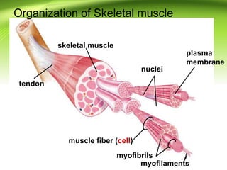
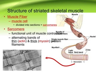
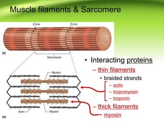
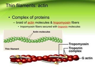
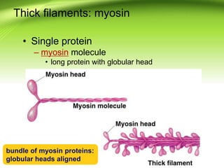
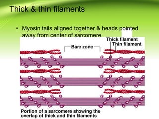
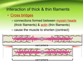
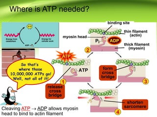


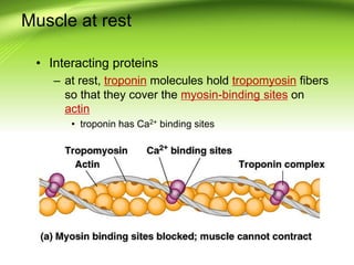
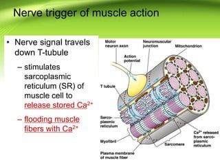
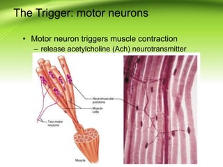
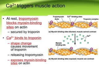
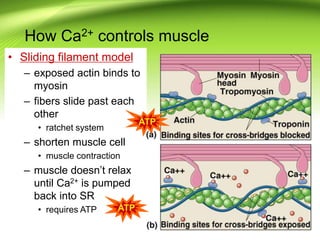
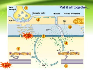
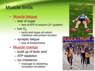
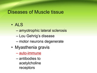
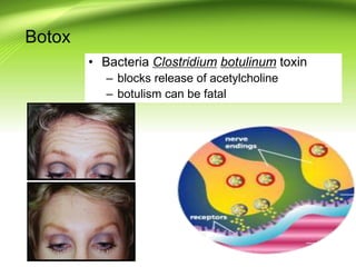


The document discusses the structure and function of skeletal muscle cells, including details on muscle fibers, sarcomeres, and the role of proteins like actin and myosin in muscle contraction. It explains the importance of ATP and calcium ions in muscle action, detailing the process of muscle contraction and relaxation, as well as differentiating between slow and fast twitch muscle fibers. Additionally, it addresses muscle fatigue, diseases affecting muscle tissue, and the effects of conditions like botulism.


![2022 bio20-8a - MuscularSystemreview [ST].pdf](https://cdn.slidesharecdn.com/ss_thumbnails/2022bio20-8a-muscularsystemst-250521201551-86d3a57c-thumbnail.jpg?width=640&height=640&fit=bounds)
![Chapt09 Holes Lecture Animation[1]](https://cdn.slidesharecdn.com/ss_thumbnails/chapt09holeslectureanimation1-091122122851-phpapp02-thumbnail.jpg?width=640&height=640&fit=bounds)


