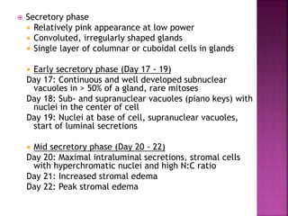
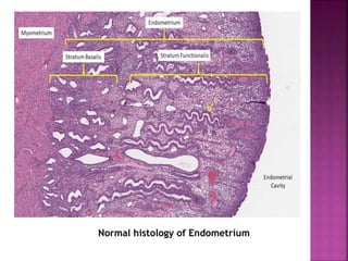
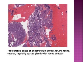
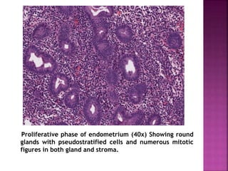
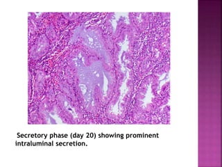
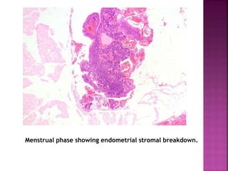
- by Dr. Sampada Jaware
 Mesodermal origin, formed secondary to fusion of
mullerian ducts between 8th and 9th weeks.
 Upto 20th week of gestation, single layer of columnar
epithelium supported by thick layer of fibroblastic
stroma
 After 20th week, surface epithelium invaginates in to
underlying stroma forming glandular structures that
extend toward the underlying myometrium.
 At birth, surface and glands are lined by low
columnar to cuboidal epithelium.
 During reproductive period undergoes cyclic
morphological changes in response to hormones.
 In post menopausal life it is thin with relatively few
glands in lined by cuboidal epithelium that is devoid
of proliferative or secretory activity (inactive and
atrophic endometrium).
 Radial arteries of the myometrium penetrate the
endometrium at regular intervals and give rise to
basal arteries.
 These divide into horizontal branches providing
the blood supply to basal layer and vertical
branches supplying the functionalis layer which
are known as spiral arterioles.
 Sub endothelial elastica is present in the
myometrial arteries but not in endometrial
arteries, except for the basal portion.
 Veins and lymphatics are closely associated with
the endometrial arteries and glands,
respectively. Uterine lymphatics drain from
subserosal uterine plexuses to the pelvic and
periaortic lymph nodes.
 Proliferative phase
 Cellular blue appearance at low power
 Round to tubular glands
 Even, regular spacing between glands
 Pseudostratified columnar cells in glands
 Numerous mitotic figures in glands and stroma
 Interval phase (Day 16)
 Mitoses present, but not as numerous as in
proliferative phase
 Less than 50% of cells in a gland with continuous and
well developed subnuclear vacuoles
 Secretory phase
 Relatively pink appearance at low power
 Convoluted, irregularly shaped glands
 Single layer of columnar or cuboidal cells in glands
 Early secretory phase (Day 17 - 19)
Day 17: Continuous and well developed subnuclear
vacuoles in > 50% of a gland, rare mitoses
Day 18: Sub- and supranuclear vacuoles (piano keys) with
nuclei in the center of cell
Day 19: Nuclei at base of cell, supranuclear vacuoles,
start of luminal secretions
 Mid secretory phase (Day 20 - 22)
Day 20: Maximal intraluminal secretions, stromal cells
with hyperchromatic nuclei and high N:C ratio
Day 21: Increased stromal edema
Day 22: Peak stromal edema
 Late secretory phase (Day 23 - 27)
Day 23: Predecidua surrounds spiral arterioles.
Day 24: Predecidua bridges multiple vessels
Day 25: Thin band of predecidua beneath endometrial
surface
Day 26: Thick band of predecidua beneath surface
Day 27: Abundant predecidua expanding downward
from endometrial surface, increased number of
stromal granulocytes
 Menstrual phase - Endometrial stromal breakdown:
dense round aggregates of stromal cells admixed with
inflammatory cells and blood
 Gestational changes
 Decidual change: stroma gains abundant
eosinophilic cytoplasm, appears polygonal with
distinct cell borders
 Arias-Stella reaction in glandular cells
 Nuclear enlargement and hyperchromasia
 Abundant eosinophilic vacuolated cytoplasm
 Hobnail appearance with cells protruding into
glandular lumen
Morphobiochemical changes leading to menstrual
breakdown
Normal histology of Endometrium
Proliferative phase of endometrium (10x) Showing round,
tubular, regularly spaced glands with round contour
Proliferative phase of endometrium (40x) Showing round
glands with pseudostratified cells and numerous mitotic
figures in both gland and stroma.
Endometrium day 17 (Secretory phase) showing stromal
edema. Some glands are tortuous.
Secretory phase (day 20) showing irregularly shaped
glands with intraluminal secretions.
Endometrium day 17 (secretory phase) showing
subnuclear vacuoles which gives a piano key appearance
Secretory phase (day 20) showing prominent
intraluminal secretion.
Menstrual phase showing endometrial stromal breakdown.
Arias stella reaction showing enlarged nuclei, sub and
supra nuclear vacuoles, intraglandular papillary epithelial
tufts, stromal decidualisation.
Contraceptives –
1. Progestogen only – depending upon dose and
duration 3 effects can be seen
• Decidual pattern – decidualised stroma (large cells
with large round nucleus, abundant eosinophillic
cytoplasm).
• Secretory pattern – mild tortuous secretory glands
lined by columnar cells with basal nuclei and scant
eosinophilic intraluminal secretions.
• Inactive pattern – widely spaced small inactive
tubular glands with scant cytoplasm with minimal to
absent luminal secretions.
2. Combined – widely spaced glands which lack mitosis
or pseudostratification, sparsely cellular stroma.
Tamoxifen (selective estrogen receptor modulator)
1. Diffuse endometrial thickening with cysticaly dilated
glands
2. Polyp are common after prolonged treatment
3. Proliferation of gland and stroma is common.
4. Can cause hyperplasia and carcinoma.
Hormone replacement therapy –
1. Estrogen only – proliferative endometrium
2. Progestogen only - mild tortuous secretory glands
lined by columnar cells with basal nuclei and scant
eosinophilic intraluminal secretions.
3. Combined – mostly inactive/atrophic endometrium,
less common secretory or proliferative pattern
 cervix acts as a barrier to the entry of microorganisms into the
endometrial cavity
 most type of endometritis results from ascending infection
 During menses, abortion, parturition and instrumentation, the
cervical barrier is breached allowing normal vaginal flora access
to the endometrial cavity.
 Based on the type of inflammatory infiltrate endometritis is
classified as acute or chronic.
 Most acute endometritis are caused by hemolytic streptococci,
Staphylococcus, Neisseria gonorrhea, Clostridium welchii.
 Criteria for histologic diagnosis of acute endometritis - moderate
to large number of neutrophils in a non bleeding endometrium,
aggregates of neutrophils in the stroma (micro abscess).
 Diagnosis of chronic endometritis - plasma cells but lymphocytes,
macrophages may be present. plasma cell contain an eccentric
nucleus with a characteristic clumped chromatin pattern and a
paranuclear pale staining area representing the golgi apparatus
with amphophilic cytoplasm.
 Presence of eosinophils associated with plasma cells.
 common causes of chronic endometritis are PID, TB, IUD.
Chronic endometritis showing plasma cells.
Acute endometritis showing prominent intraluminal
neutrophils and neutrophils infiltrating glandular
epithelium.
 Granulomatous endometritis
 Clusters of epithelioid histiocytes rimmed by
lymphocytes
 Cytopathic effects if viral etiology (herpes simplex
virus, cytomegalovirus)
 Tuberculosis: necrotizing granulomas with
multinucleated giant cells, usually in superficial
functional endometrium
 Sarcoidosis: nonnecrotizing granulomatous
inflammation, more commonly in myometrium
 Xanthogranulomatous endometritis
 Abundant foamy histiocytes, neutrophils, plasma
cells, lymphocytes
Xanthogranulomatous endometritis showing endometrial
stroma with prominent inflammation including foamy
histiocytes and lymphocytes.
 Clinical term for uterine bleeding that lacks an underlying
structural abnormality.
 Normal cyclical proliferation, differentiation and shedding of
endometrium requires that all involved pituitary and ovarian
hormones be released at proper time in right amounts.
 Any disturbance of this finely tuned system may result in
dysfunctional uterine bleeding.
 Causes of DUB are
Age group Causes
Prepuberty Precocious puberty
Adolescence Anovulatory cycle, coagulation disorder
reproductive age
 complications of pregnancy (abortion,
trophoblastic disease, ectopic pregnancy)
 Anatomic lesions (Leo myoma, adenomyosis,
polyps, endometrial hyperplasia, carcinoma)
 Anovulatory cycle
 ovulatory dysfunctional bleeding. example
inadequate luteal phase
Perimenopausal
 anovulatory cycle
 anatomic lesions (carcinoma, hyperplasia,
polyps)
Post menopausal
 endometrial atrophy
 Anatomic lesions (carcinoma, hyperplasia,
polyp)
Anovulatory cycle
 It’s a common cause of DUB.
 Failure of ovulation results in excessive endometrial
stimulation by estrogens that is unopposed by
progesterone.
 Under these circumstances, the endometrial glands
undergo mild architectural changes that usually
resolve due to a subsequent ovulatory cycle.
 Repeated anovulation may result in bleeding.
 Anovulation can also be the result of thyroid disease,
a functioning ovarian tumor, polycystic ovaries,
generalized metabolic disturbances like obesity,
malnutrition.
 defined by the presence of ectopic endometrial tissue at a site
outside the uterus. It occurs in the following sites in descending
order - Ovaries, uterine ligaments, recto-vaginal septum, pelvic
peritoneum, serosa of large and small bowel and appendix,
mucosa of cervix, vagina and fallopian tubes, laparotomy scars.
 It can cause infertility, painful menstruation, pelvic pain. It
principally is a disease of women in active reproductive life most
often in the 3rd and 4th decades.
 Pathogenesis of endometriosis is explained by many theories such
as –
 The regurgitation theory proposes that endometrial tissue
implants at ectopic sites via retrograde flow of menstrual
endometrium.
 The benign metastasis theory holds that endometrial tissue from
the uterus can spread to distant sites via blood vessels and
lymphatic channels.
 the metaplastic theory suggests that endometrium arises directly
from coelomic epithelium from which the mullerian ducts and
ultimately the endometrium itself originate during embryonic
development.
 The extrauterine stem or progenitor cell theory proposes that
progenitor cells from the bone marrow differentiate into
endometrial tissue.
 When endometriotic lesions are widespread,
organizing hemorrhage causes extensive fibrous
adhesions between tubes, ovaries and other
structures and obliterates the pouch of Douglas.
 Ovaries may become markedly distorted by large
cystic masses filled with brown fluid resulting from
previous hemorrhage, these are referred to clinically
as chocolate cysts or endometriomas.
 The diagnosis of endometriosis is readily made when
both endometrial glands and stroma are present.
 When endometrial tissue is present in the
myometrium, it is called as Adenomyosis.
Image showing cut section of ovary containing red-
brown fluid in a cystic area.
Endometriotic cyst of the ovary showing abundant
hemosiderin-laden macrophages and fresh hemorrhage in
endometrial stroma.
 Atrophy is most commonly seen in postmenopausal
women due to estrogen withdrawal.
 Microscopically endometrium is composed of a thin
layer of endometrial glands lined by attenuated layer
of inactive epithelial cells surrounded by thin stroma.
No mitotic activity is present.
Atrophic endometrium with detached glands composed of low
columnar to cuboidal cells. At places thin stroma is seen.
 Metaplasia is the presence of any type of glandular
epithelium other than the normal columnar type. It is a
common finding in peri-menopausal and post-menopausal
women and is often associated with abnormal uterine
bleeding or recent use of exogenous hormonal therapy.
 Tubal metaplasia: Consists of foci of normal tubal
epithelium within the endometrial glands, including
ciliated, non ciliated secretary and intercalated cells.
Ratio of the ciliated to non ciliated cell is cyclical and
depends on hormonal influences.
 Ciliated cell metaplasia: Is the most common form of
metaplasia. It is composed of layer of ciliated columnar
cells with round to oval nuclei and abundant pale
eosinophilic cytoplasm. Ciliated cell metaplasia is a normal
response of endometrial epithelium to various hormonal
exposures. It is most commonly found in perimenopausal
endometrium and is associated with endometrial polyps,
anovulatory cycles and exogenous hormonal therapy.
 Squamous metaplasia: Often caused by chronic
irritation and often takes the form of squamous
morules or rounded, swirling nest of squamous
cells.
 Eosinophilic metaplasia: Glandular epithelium with
abundant eosinophilic cytoplasm and central round
to Oval nucleus. It is believed to be a degenerative
change.
 Mucinous metaplasia: It is morphologically similar
to endocervical mucinous epithelium in that it
consists of columnar epithelium with basally
located Oval nuclei and abundant apical mucin.
Tubal (ciliated) metaplasia showing pseudostratified ciliated
cells.
Squamous morular metaplasia showing nests of bland,
immature squamous cells without characteristics features of
squamous differentiation (intercellular bridges,
keratinization)
Eosinophilic metaplasia showing eosinophilic cells with
granular cytoplasm found singly and in small clusters within
glandular lumen.
 These are local overgrowths of endometrial glands
and stroma that are covered by epithelium and
protrude into the endometrial cavity. Polyps are
present in about 20% to 25% of women and are
frequently found in peri and postmenopausal periods.
 Grossly polyps appear as broad based to
pedunculated lesions; some pedunculated polyps can
extend into the endocervical canal, and even through
the os.
 Microscopically polyps are composed of endometrial
glands within a spindled or fibrous stroma; the
presence of thick walled blood vessels within the
fibrous stroma is the most common key to the
diagnosis.
 Adenomyomatous polyp- foci of adenomyosis may
become enlarged into polypoid projections.
 Atypical polypoid adenomyoma- Characterized
histologically by crowded, irregular endometrial glands
with a complex architecture and cytologic atypia, in
stroma that is predominantly composed of smooth
muscle. This lesion has high rate of recurrence after
incomplete surgical removal. If left untreated there is
risk of progression to endometrioid adenocarcinoma.
Uterus has been opened anteriorly through cervix and
into the endometrial cavity. A small polyp is seen high in
fundus projecting into lumen.
Endometrial polyp showing vascular pedicle and few cystic
glands.
Endometrial polyp showing cystically dilated glands in a fibrotic
stroma.
 Defined as abnormal proliferation of endometrial
glands relative to stroma, resulting in increased
gland to stroma ratio when compared to normal
proliferative endometrium.
 Hyperplasia associted with prolonged estrogenic
stimulation of the endometrium can be due to
the following conditions –obesity, pcos,
functioning granulosa cell tumor of ovary,
estrogen replacement therapy.
 Inactivation of the PTEN tumor suppressor gene
is common genetic alteration in both
endometrial hyperplasia and carcinoma.
Hyperplasia without atypia:
 increased gland to stroma ratio with variable size of
glands, cystic dilation. Focal back to back glands may
be seen.
 Caused by persistent eostrogen stimulation.
Atypical Hyperplasia:
 Complex pattern of proliferating glands displaying
nuclear atypia.
 Glands are commonly back to back.
 Cells are rounded and lose normal perpendicular
orientation to basement membrane.
 Nuclei have open vesicular chromatin and
prominent nucleoli.
Hyperplasia without atypia showing increased number of
glands which are closely apposed and have minimal
complexity
Atypical hyperplasia showing packed glands (back to
back arrangement) showing minimal stroma, complex
contours and atypical epithelial lining
Endometrial Intraepithelial Neoplasia (EIN)
 It is characterized by markedly atypical nuclei lining the
surfaces and glands of atrophic endometrium. Nuclear enlarged
with granular or vesicular chromatin, frequently display
enlarged eosinophilic nucleoli. Numerous mitotic figures are
seen. This lesion is also referred as Carcinoma in situ.
 EIN Criterion
 Architecture - Gland area exceeds that of stroma, usually in a
localized region.
 Cytological Alterations - Cytology differs between
architecturally crowded focus and background.
 Size greater than 1mm - Minimum linear dimension should
exceed 1mm.
 Exclude mimics - Basalis, normal secretory, polyps, repair,
lower uterine segment, cystic atrophy, menstrual collapse etc.
 Exclude Cancer - Carcinoma should be diagnosed if: glands are
mazelike and rambling, there are solid areas of epithelial
growth, or there are significant bridges or cribriform areas.
EIN showing closely apposed glands with tufting
intraluminal projections with minimal intervening
stroma
 Most common invasive cancer of the female genital
tract.
 Typically occurs in elderly females, 80% being
postmenopausal during diagnosis.
 Risk factors – family history, nulliparity, early
menarche late menopause, obesity, HRT
 It is divided in 2 types – type 1 and type 2 each with
distinct risk factors, precursor lesions, genetics and
clinical behaviours.
Characteristics TYPE 1 TYPE 2
AGE (Years) 55-65 65-75
Clinical setting
Unopposed estrogen, Obesity,
Hypertension, Diabetes
Atrophy, Thin
physique
Morphology Endometroid
Serous, Clear cell,
Undifferentiated
carcinoma,
Carcinosarcoma
Genetic Abnormalities PTEN, PIK3CA, KRAS, MSI, TP53 TP53, PIK3CA, PP2A
Behaviour
Indolent
Spread via Lymphatics
Aggressive
Intraperitoneal,
Lymphatic spread
Pathogenesis of endometrial carcinoma
 Most common type of endometrial carcinoma accounting for 80-
85% cases.
 Most tumors are well differentiated and mimic proliferative
endometrial glands.
 Most common mutation act to increase signaling through
PI3K/AKT pathway.
 It can take the form of a localized polypoid mass or diffusely
involve endometrial lining. Spread generally occurs by
myometrial invasion followed by direct extension to adjacent
structures/organs.
 Clinical Features
Abnormal, dysfunctional or postmenopausal uterine bleeding
Pelvic pain or mass / compression effect on adjacent structures
Abdominal bloating
Dyspareunia, dysuria
General stigmata of malignancy, i.e. weight and appetite loss,
malaise, fatigue
 Gross description - Mass arising from endometrial surface with
varied appearances / sizes but usually exophytic and friable in
texture.
 Microscopic (histologic) description
 Architecture
 Key feature is confluent or back to back glands lacking
intervening stroma
 Cribriform or microacinar configurations
 Complex papillary, micropapillary or villoglandular structures
 Cytologic features
 Resembles proliferative type endometrium with varying features
/ degrees of atypia but cytology must differ from that of
surrounding non-neoplastic glands
 Cellular / nuclear enlargement
 Nuclear rounding (rather than elongation) with large nucleoli
 Loss of polarity
 Cytoplasmic eosinophilia
 Sharp glandular luminal borders
 Foamy histiocytes in residual stroma
 Its divided into 3 histological grades
 Well differentiated : composed almost entirely of well
formed glands.
 Moderately differentiated : well formed glands mixed with
areas composed of soild sheets of cells which make up <50%
of tumor.
 Poorly differentiated : > 50% of tumor shows solid growth
pattern.
 Myometrial invasion
 Traverses beyond confines of typically irregular endo-
myometrial junction without intervening rim of benign
marker glands or endometrial stroma
 Rounded, smooth pushing invasive front, or
 Infiltrative extension of neoplastic glands
 Stromal response at invasive front variably consists of
fibroblastic proliferation, edema and inflammatory cells
 Ratio of myo-invasion is crucial to staging:
 Numerator: depth of furthest invasion
 Denominator: myometrial thickness
 Following myometrial invasion, direct extension to adjacent
organs and eventually dissemination to regional lymph nodes
occurs.
 FIGO grading system (based primarily on architecture)
 Grade 1: 5% or less nonsquamous solid growth pattern
 Grade 2: 6 - 50% nonsquamous solid growth pattern
 Grade 3: > 50% nonsquamous solid growth pattern
 Nuclear atypia exceeding that expected for the architectural
grade increases FIGO grade by 1.
(atypia here is defined as grade 3 nuclei in >50% of tumor)
 Morphologic variants
 Altered differentiation / metaplasia:
 Squamous or "squamous" morular: usually bland but
occasionally cytologically malignant; former can be
glycogenated which imparts appearance of clear cytoplasm
 Mucinous: intracytoplasmic mucin (intraluminal mucin
pooling does not qualify). >50% cells should show mucinous
differentiation.
 Secretory: sub / supranuclear vacuolization
 Ciliated / tubal: resembles fallopian tube lining; scattered
cells with apical terminal bars and ciliation.
 Papillary type variants:
 Villoglandular
 Small nonvillous papillae
 Micropapillae
 Microglandular hyperplasia-like: microcystic, microacinar
glands with intraluminal neutrophils
 Corded and Hyalinized Endometrial Carcinoma (CHEC):
linear cords of carcinoma cells molded by an abundant
myxohyaline background
Endometrial carcinoma: Gross specimen of anteriorly
opened uterine cavity showing multiple hemorrhagic
polypoidal growths extending into cavity.
Endometroid adenocarcinoma FIGO grade 1: The glands are
well-differentiated and show angulation or branching.
Cytologic atypia is minimal. <5% of solid component. (Low
power)
Endometroid adenocarcinoma FIGO grade 1: There is crowding of
well-differentiated glands showing minimal cytologic atypia and
minimal stroma.
Endometroid adenocarcinoma FIGO grade 2: 6% to 50%
of solid, non-morular component.
Endometroid adenocarcinoma FIGO grade 3: tumors
contain solid areas comprising more than 50% of the
tumor.
Endometroid adenocarcinoma – villoglandular type:
consists of finger-like projections lined by tall columnar
cells with bland nuclei.
 Highly agggressive form of endometrial carcinoma.
 Usually arise in setting of atrophy.
 Age group is 10 years older than that of endometroid
carcinoma.
 These tumors are considered by definition as poorly
differentiated (Grade 3).
 Tp53 mutations are seen in >90% of these tumors
 Their general poorer prognosis is because of propensity to
exfoliate, travel through fallopian tubes and implant on
peritoneal surface. As a result, they have often spread
outside of uterus at the time of diagnosis.
 GROSS : generally, small atrophic uterus with large bulky
tumor mass or invading myometrium.
 MICROSCOPY :
- precursor lesion is serous intraepithelial carcinoma
consists of malignant cells identical to serous carcinoma
confined to epithelial surface.
- invasive lesions have complex papillary growth pattern
composed of cells with marked cytological atypia including
high N/C ratio, atypical mitotic figures, hyperchromasia
and prominent nucleoli.
- at times, can have glandular pattern which is
differentiated from endometroid carcinoma by marked
cytological atypia.
Serous carcinoma: shows complex papillary clusters of high-
grade neoplastic cells (pleomorphic, hyperchromatic nuclei).
Serous carcinoma: image showing complex papillary
pattern of growth.
 Staging system for endometrial carcinoma:
Stage 1 - confined to corpus uteri
Stage 2 - involves corpus and cervix
Stage 3 - extends outside uterus but not true pelvis
Stage 4 – extends outside true pelvis (involving musocs of
bladder or rectum)
 Prognosis depends on – depth of infiltration into
myometrium, grade of differentiation, stage of tumor,
molecular subtypes, lymphatic invasion and age of patient.
 Occur in mddle aged women (=45 years) and frequently
present with vaginal bleeding.
 BENIGN
 Endometrial stromal nodule : solitary sharply circumscribed
mass of soft consistency having yellow – orange color. Do not
invade lymphatics, veins or myometrium. On microscopy,
monotonous proliferation of bland stromal cells with
expansile (non infiltrating) margin. Can show necrosis,
hemorrhage, myxoid change.
 Low grade stromal sarcoma : poorly circumscribed soft
yellow tan to white nodules extending from endometrium
and invading in myometrium. On microscopy, irregular
cellular islands, forming permeative tongue-like pattern of
myometrial invasion with frequent vascular invasion.
Monotonous oval to spindle cells with minimal cytologic
atypia, vesicular chromatin and scant cytoplasm. Mitotic
count is usually low (< 5/10 high power fields), necrosis is
usually absent.
Stromal nodule : well-circumscribed yellow tumor.
Stromal nodule : Monotonous proliferation of bland spindle
cells resembling proliferative phase endometrial stroma.
Low grade stromal sarcoma showing yellow brown
tumor mass and infiltrating tumor nodules
Low grade stromal sarcoma showing monotonous spindle to
oval cells with minimal nuclear atypia
 Malignant
 High grade stromal sarcoma : tan pink to yellow tumor
mass, soft and fleshy, can be polypoidal.
On microscopy, Permeative (tongue-like) invasion.
Vaguely nested growth of round cells with scant (small
round blue cell appearance) to moderate eosinophilic
cytoplasm and uniform nuclear atypia. Brisk mitoses (>
10/10 high power fields).
Tumor cell necrosis and lymphovascular invasion common.
50% associated with a low grade spindle cell component
resembling low grade endometrial stromal sarcoma.
High grade endometrial stromal sarcoma with
uniformly atypical spindled cells and brisk mitoses.
High grade stromal sarcoma showing tongue-like
(permeative) invasion.
 Never take oestrogen alone in HRT or
contraceptive pills, opt for low dose combination
of oestrogen with progesterone
 Never let endometrial infections go untreated
 Maintain a healthy lifestyle
1. Blaustein’s pathology of female genital tract 7th edition
2. Rosai and Ackerman’s Surgical pathology 1st south asia
edition volume 2 section 6 chapter 33.
3. Robbins and Cotran: Pathologic basis of disease 10th
edition volume 2 chapter 22
4.
5. The Washington manual of Surgical pathology 3rd edition
section 7 chapter 33.
6. Harsh Mohan Textbook of pathology 7th edition chapter 10.
7. Akaev I, Yeoh CC, Rahimi S. Update on Endometrial
Stromal Tumours of the Uterus. Diagnostics (Basel). 2021
Mar 3;11(3):429.
8. Ali RH, Rouzbahman M. Endometrial stromal tumours
revisited: an update based on the 2014 WHO
classification. J Clin Pathol. 2015 May;68(5):325-32.
endometrium pathology

endometrium pathology

  • 2.
    - by Dr.Sampada Jaware
  • 4.
     Mesodermal origin,formed secondary to fusion of mullerian ducts between 8th and 9th weeks.  Upto 20th week of gestation, single layer of columnar epithelium supported by thick layer of fibroblastic stroma  After 20th week, surface epithelium invaginates in to underlying stroma forming glandular structures that extend toward the underlying myometrium.  At birth, surface and glands are lined by low columnar to cuboidal epithelium.  During reproductive period undergoes cyclic morphological changes in response to hormones.  In post menopausal life it is thin with relatively few glands in lined by cuboidal epithelium that is devoid of proliferative or secretory activity (inactive and atrophic endometrium).
  • 6.
     Radial arteriesof the myometrium penetrate the endometrium at regular intervals and give rise to basal arteries.  These divide into horizontal branches providing the blood supply to basal layer and vertical branches supplying the functionalis layer which are known as spiral arterioles.  Sub endothelial elastica is present in the myometrial arteries but not in endometrial arteries, except for the basal portion.  Veins and lymphatics are closely associated with the endometrial arteries and glands, respectively. Uterine lymphatics drain from subserosal uterine plexuses to the pelvic and periaortic lymph nodes.
  • 10.
     Proliferative phase Cellular blue appearance at low power  Round to tubular glands  Even, regular spacing between glands  Pseudostratified columnar cells in glands  Numerous mitotic figures in glands and stroma  Interval phase (Day 16)  Mitoses present, but not as numerous as in proliferative phase  Less than 50% of cells in a gland with continuous and well developed subnuclear vacuoles
  • 11.
     Secretory phase Relatively pink appearance at low power  Convoluted, irregularly shaped glands  Single layer of columnar or cuboidal cells in glands  Early secretory phase (Day 17 - 19) Day 17: Continuous and well developed subnuclear vacuoles in > 50% of a gland, rare mitoses Day 18: Sub- and supranuclear vacuoles (piano keys) with nuclei in the center of cell Day 19: Nuclei at base of cell, supranuclear vacuoles, start of luminal secretions  Mid secretory phase (Day 20 - 22) Day 20: Maximal intraluminal secretions, stromal cells with hyperchromatic nuclei and high N:C ratio Day 21: Increased stromal edema Day 22: Peak stromal edema
  • 12.
     Late secretoryphase (Day 23 - 27) Day 23: Predecidua surrounds spiral arterioles. Day 24: Predecidua bridges multiple vessels Day 25: Thin band of predecidua beneath endometrial surface Day 26: Thick band of predecidua beneath surface Day 27: Abundant predecidua expanding downward from endometrial surface, increased number of stromal granulocytes  Menstrual phase - Endometrial stromal breakdown: dense round aggregates of stromal cells admixed with inflammatory cells and blood
  • 13.
     Gestational changes Decidual change: stroma gains abundant eosinophilic cytoplasm, appears polygonal with distinct cell borders  Arias-Stella reaction in glandular cells  Nuclear enlargement and hyperchromasia  Abundant eosinophilic vacuolated cytoplasm  Hobnail appearance with cells protruding into glandular lumen
  • 14.
    Morphobiochemical changes leadingto menstrual breakdown
  • 15.
  • 16.
    Proliferative phase ofendometrium (10x) Showing round, tubular, regularly spaced glands with round contour
  • 17.
    Proliferative phase ofendometrium (40x) Showing round glands with pseudostratified cells and numerous mitotic figures in both gland and stroma.
  • 18.
    Endometrium day 17(Secretory phase) showing stromal edema. Some glands are tortuous.
  • 19.
    Secretory phase (day20) showing irregularly shaped glands with intraluminal secretions.
  • 20.
    Endometrium day 17(secretory phase) showing subnuclear vacuoles which gives a piano key appearance
  • 21.
    Secretory phase (day20) showing prominent intraluminal secretion.
  • 22.
    Menstrual phase showingendometrial stromal breakdown.
  • 23.
    Arias stella reactionshowing enlarged nuclei, sub and supra nuclear vacuoles, intraglandular papillary epithelial tufts, stromal decidualisation.
  • 26.
    Contraceptives – 1. Progestogenonly – depending upon dose and duration 3 effects can be seen • Decidual pattern – decidualised stroma (large cells with large round nucleus, abundant eosinophillic cytoplasm). • Secretory pattern – mild tortuous secretory glands lined by columnar cells with basal nuclei and scant eosinophilic intraluminal secretions. • Inactive pattern – widely spaced small inactive tubular glands with scant cytoplasm with minimal to absent luminal secretions. 2. Combined – widely spaced glands which lack mitosis or pseudostratification, sparsely cellular stroma.
  • 27.
    Tamoxifen (selective estrogenreceptor modulator) 1. Diffuse endometrial thickening with cysticaly dilated glands 2. Polyp are common after prolonged treatment 3. Proliferation of gland and stroma is common. 4. Can cause hyperplasia and carcinoma. Hormone replacement therapy – 1. Estrogen only – proliferative endometrium 2. Progestogen only - mild tortuous secretory glands lined by columnar cells with basal nuclei and scant eosinophilic intraluminal secretions. 3. Combined – mostly inactive/atrophic endometrium, less common secretory or proliferative pattern
  • 31.
     cervix actsas a barrier to the entry of microorganisms into the endometrial cavity  most type of endometritis results from ascending infection  During menses, abortion, parturition and instrumentation, the cervical barrier is breached allowing normal vaginal flora access to the endometrial cavity.  Based on the type of inflammatory infiltrate endometritis is classified as acute or chronic.  Most acute endometritis are caused by hemolytic streptococci, Staphylococcus, Neisseria gonorrhea, Clostridium welchii.  Criteria for histologic diagnosis of acute endometritis - moderate to large number of neutrophils in a non bleeding endometrium, aggregates of neutrophils in the stroma (micro abscess).  Diagnosis of chronic endometritis - plasma cells but lymphocytes, macrophages may be present. plasma cell contain an eccentric nucleus with a characteristic clumped chromatin pattern and a paranuclear pale staining area representing the golgi apparatus with amphophilic cytoplasm.  Presence of eosinophils associated with plasma cells.  common causes of chronic endometritis are PID, TB, IUD.
  • 32.
  • 33.
    Acute endometritis showingprominent intraluminal neutrophils and neutrophils infiltrating glandular epithelium.
  • 34.
     Granulomatous endometritis Clusters of epithelioid histiocytes rimmed by lymphocytes  Cytopathic effects if viral etiology (herpes simplex virus, cytomegalovirus)  Tuberculosis: necrotizing granulomas with multinucleated giant cells, usually in superficial functional endometrium  Sarcoidosis: nonnecrotizing granulomatous inflammation, more commonly in myometrium  Xanthogranulomatous endometritis  Abundant foamy histiocytes, neutrophils, plasma cells, lymphocytes
  • 35.
    Xanthogranulomatous endometritis showingendometrial stroma with prominent inflammation including foamy histiocytes and lymphocytes.
  • 36.
     Clinical termfor uterine bleeding that lacks an underlying structural abnormality.  Normal cyclical proliferation, differentiation and shedding of endometrium requires that all involved pituitary and ovarian hormones be released at proper time in right amounts.  Any disturbance of this finely tuned system may result in dysfunctional uterine bleeding.  Causes of DUB are
  • 37.
    Age group Causes PrepubertyPrecocious puberty Adolescence Anovulatory cycle, coagulation disorder reproductive age  complications of pregnancy (abortion, trophoblastic disease, ectopic pregnancy)  Anatomic lesions (Leo myoma, adenomyosis, polyps, endometrial hyperplasia, carcinoma)  Anovulatory cycle  ovulatory dysfunctional bleeding. example inadequate luteal phase Perimenopausal  anovulatory cycle  anatomic lesions (carcinoma, hyperplasia, polyps) Post menopausal  endometrial atrophy  Anatomic lesions (carcinoma, hyperplasia, polyp)
  • 38.
    Anovulatory cycle  It’sa common cause of DUB.  Failure of ovulation results in excessive endometrial stimulation by estrogens that is unopposed by progesterone.  Under these circumstances, the endometrial glands undergo mild architectural changes that usually resolve due to a subsequent ovulatory cycle.  Repeated anovulation may result in bleeding.  Anovulation can also be the result of thyroid disease, a functioning ovarian tumor, polycystic ovaries, generalized metabolic disturbances like obesity, malnutrition.
  • 39.
     defined bythe presence of ectopic endometrial tissue at a site outside the uterus. It occurs in the following sites in descending order - Ovaries, uterine ligaments, recto-vaginal septum, pelvic peritoneum, serosa of large and small bowel and appendix, mucosa of cervix, vagina and fallopian tubes, laparotomy scars.  It can cause infertility, painful menstruation, pelvic pain. It principally is a disease of women in active reproductive life most often in the 3rd and 4th decades.  Pathogenesis of endometriosis is explained by many theories such as –  The regurgitation theory proposes that endometrial tissue implants at ectopic sites via retrograde flow of menstrual endometrium.  The benign metastasis theory holds that endometrial tissue from the uterus can spread to distant sites via blood vessels and lymphatic channels.  the metaplastic theory suggests that endometrium arises directly from coelomic epithelium from which the mullerian ducts and ultimately the endometrium itself originate during embryonic development.  The extrauterine stem or progenitor cell theory proposes that progenitor cells from the bone marrow differentiate into endometrial tissue.
  • 41.
     When endometrioticlesions are widespread, organizing hemorrhage causes extensive fibrous adhesions between tubes, ovaries and other structures and obliterates the pouch of Douglas.  Ovaries may become markedly distorted by large cystic masses filled with brown fluid resulting from previous hemorrhage, these are referred to clinically as chocolate cysts or endometriomas.  The diagnosis of endometriosis is readily made when both endometrial glands and stroma are present.  When endometrial tissue is present in the myometrium, it is called as Adenomyosis.
  • 42.
    Image showing cutsection of ovary containing red- brown fluid in a cystic area.
  • 43.
    Endometriotic cyst ofthe ovary showing abundant hemosiderin-laden macrophages and fresh hemorrhage in endometrial stroma.
  • 45.
     Atrophy ismost commonly seen in postmenopausal women due to estrogen withdrawal.  Microscopically endometrium is composed of a thin layer of endometrial glands lined by attenuated layer of inactive epithelial cells surrounded by thin stroma. No mitotic activity is present.
  • 46.
    Atrophic endometrium withdetached glands composed of low columnar to cuboidal cells. At places thin stroma is seen.
  • 47.
     Metaplasia isthe presence of any type of glandular epithelium other than the normal columnar type. It is a common finding in peri-menopausal and post-menopausal women and is often associated with abnormal uterine bleeding or recent use of exogenous hormonal therapy.  Tubal metaplasia: Consists of foci of normal tubal epithelium within the endometrial glands, including ciliated, non ciliated secretary and intercalated cells. Ratio of the ciliated to non ciliated cell is cyclical and depends on hormonal influences.  Ciliated cell metaplasia: Is the most common form of metaplasia. It is composed of layer of ciliated columnar cells with round to oval nuclei and abundant pale eosinophilic cytoplasm. Ciliated cell metaplasia is a normal response of endometrial epithelium to various hormonal exposures. It is most commonly found in perimenopausal endometrium and is associated with endometrial polyps, anovulatory cycles and exogenous hormonal therapy.
  • 48.
     Squamous metaplasia:Often caused by chronic irritation and often takes the form of squamous morules or rounded, swirling nest of squamous cells.  Eosinophilic metaplasia: Glandular epithelium with abundant eosinophilic cytoplasm and central round to Oval nucleus. It is believed to be a degenerative change.  Mucinous metaplasia: It is morphologically similar to endocervical mucinous epithelium in that it consists of columnar epithelium with basally located Oval nuclei and abundant apical mucin.
  • 49.
    Tubal (ciliated) metaplasiashowing pseudostratified ciliated cells.
  • 50.
    Squamous morular metaplasiashowing nests of bland, immature squamous cells without characteristics features of squamous differentiation (intercellular bridges, keratinization)
  • 51.
    Eosinophilic metaplasia showingeosinophilic cells with granular cytoplasm found singly and in small clusters within glandular lumen.
  • 52.
     These arelocal overgrowths of endometrial glands and stroma that are covered by epithelium and protrude into the endometrial cavity. Polyps are present in about 20% to 25% of women and are frequently found in peri and postmenopausal periods.  Grossly polyps appear as broad based to pedunculated lesions; some pedunculated polyps can extend into the endocervical canal, and even through the os.  Microscopically polyps are composed of endometrial glands within a spindled or fibrous stroma; the presence of thick walled blood vessels within the fibrous stroma is the most common key to the diagnosis.  Adenomyomatous polyp- foci of adenomyosis may become enlarged into polypoid projections.
  • 53.
     Atypical polypoidadenomyoma- Characterized histologically by crowded, irregular endometrial glands with a complex architecture and cytologic atypia, in stroma that is predominantly composed of smooth muscle. This lesion has high rate of recurrence after incomplete surgical removal. If left untreated there is risk of progression to endometrioid adenocarcinoma.
  • 54.
    Uterus has beenopened anteriorly through cervix and into the endometrial cavity. A small polyp is seen high in fundus projecting into lumen.
  • 55.
    Endometrial polyp showingvascular pedicle and few cystic glands.
  • 56.
    Endometrial polyp showingcystically dilated glands in a fibrotic stroma.
  • 58.
     Defined asabnormal proliferation of endometrial glands relative to stroma, resulting in increased gland to stroma ratio when compared to normal proliferative endometrium.  Hyperplasia associted with prolonged estrogenic stimulation of the endometrium can be due to the following conditions –obesity, pcos, functioning granulosa cell tumor of ovary, estrogen replacement therapy.  Inactivation of the PTEN tumor suppressor gene is common genetic alteration in both endometrial hyperplasia and carcinoma.
  • 59.
    Hyperplasia without atypia: increased gland to stroma ratio with variable size of glands, cystic dilation. Focal back to back glands may be seen.  Caused by persistent eostrogen stimulation. Atypical Hyperplasia:  Complex pattern of proliferating glands displaying nuclear atypia.  Glands are commonly back to back.  Cells are rounded and lose normal perpendicular orientation to basement membrane.  Nuclei have open vesicular chromatin and prominent nucleoli.
  • 60.
    Hyperplasia without atypiashowing increased number of glands which are closely apposed and have minimal complexity
  • 61.
    Atypical hyperplasia showingpacked glands (back to back arrangement) showing minimal stroma, complex contours and atypical epithelial lining
  • 62.
    Endometrial Intraepithelial Neoplasia(EIN)  It is characterized by markedly atypical nuclei lining the surfaces and glands of atrophic endometrium. Nuclear enlarged with granular or vesicular chromatin, frequently display enlarged eosinophilic nucleoli. Numerous mitotic figures are seen. This lesion is also referred as Carcinoma in situ.  EIN Criterion  Architecture - Gland area exceeds that of stroma, usually in a localized region.  Cytological Alterations - Cytology differs between architecturally crowded focus and background.  Size greater than 1mm - Minimum linear dimension should exceed 1mm.  Exclude mimics - Basalis, normal secretory, polyps, repair, lower uterine segment, cystic atrophy, menstrual collapse etc.  Exclude Cancer - Carcinoma should be diagnosed if: glands are mazelike and rambling, there are solid areas of epithelial growth, or there are significant bridges or cribriform areas.
  • 63.
    EIN showing closelyapposed glands with tufting intraluminal projections with minimal intervening stroma
  • 65.
     Most commoninvasive cancer of the female genital tract.  Typically occurs in elderly females, 80% being postmenopausal during diagnosis.  Risk factors – family history, nulliparity, early menarche late menopause, obesity, HRT  It is divided in 2 types – type 1 and type 2 each with distinct risk factors, precursor lesions, genetics and clinical behaviours.
  • 66.
    Characteristics TYPE 1TYPE 2 AGE (Years) 55-65 65-75 Clinical setting Unopposed estrogen, Obesity, Hypertension, Diabetes Atrophy, Thin physique Morphology Endometroid Serous, Clear cell, Undifferentiated carcinoma, Carcinosarcoma Genetic Abnormalities PTEN, PIK3CA, KRAS, MSI, TP53 TP53, PIK3CA, PP2A Behaviour Indolent Spread via Lymphatics Aggressive Intraperitoneal, Lymphatic spread
  • 67.
  • 68.
     Most commontype of endometrial carcinoma accounting for 80- 85% cases.  Most tumors are well differentiated and mimic proliferative endometrial glands.  Most common mutation act to increase signaling through PI3K/AKT pathway.  It can take the form of a localized polypoid mass or diffusely involve endometrial lining. Spread generally occurs by myometrial invasion followed by direct extension to adjacent structures/organs.  Clinical Features Abnormal, dysfunctional or postmenopausal uterine bleeding Pelvic pain or mass / compression effect on adjacent structures Abdominal bloating Dyspareunia, dysuria General stigmata of malignancy, i.e. weight and appetite loss, malaise, fatigue
  • 69.
     Gross description- Mass arising from endometrial surface with varied appearances / sizes but usually exophytic and friable in texture.  Microscopic (histologic) description  Architecture  Key feature is confluent or back to back glands lacking intervening stroma  Cribriform or microacinar configurations  Complex papillary, micropapillary or villoglandular structures  Cytologic features  Resembles proliferative type endometrium with varying features / degrees of atypia but cytology must differ from that of surrounding non-neoplastic glands  Cellular / nuclear enlargement  Nuclear rounding (rather than elongation) with large nucleoli  Loss of polarity  Cytoplasmic eosinophilia  Sharp glandular luminal borders  Foamy histiocytes in residual stroma
  • 70.
     Its dividedinto 3 histological grades  Well differentiated : composed almost entirely of well formed glands.  Moderately differentiated : well formed glands mixed with areas composed of soild sheets of cells which make up <50% of tumor.  Poorly differentiated : > 50% of tumor shows solid growth pattern.  Myometrial invasion  Traverses beyond confines of typically irregular endo- myometrial junction without intervening rim of benign marker glands or endometrial stroma  Rounded, smooth pushing invasive front, or  Infiltrative extension of neoplastic glands  Stromal response at invasive front variably consists of fibroblastic proliferation, edema and inflammatory cells  Ratio of myo-invasion is crucial to staging:  Numerator: depth of furthest invasion  Denominator: myometrial thickness  Following myometrial invasion, direct extension to adjacent organs and eventually dissemination to regional lymph nodes occurs.
  • 71.
     FIGO gradingsystem (based primarily on architecture)  Grade 1: 5% or less nonsquamous solid growth pattern  Grade 2: 6 - 50% nonsquamous solid growth pattern  Grade 3: > 50% nonsquamous solid growth pattern  Nuclear atypia exceeding that expected for the architectural grade increases FIGO grade by 1. (atypia here is defined as grade 3 nuclei in >50% of tumor)  Morphologic variants  Altered differentiation / metaplasia:  Squamous or "squamous" morular: usually bland but occasionally cytologically malignant; former can be glycogenated which imparts appearance of clear cytoplasm  Mucinous: intracytoplasmic mucin (intraluminal mucin pooling does not qualify). >50% cells should show mucinous differentiation.  Secretory: sub / supranuclear vacuolization  Ciliated / tubal: resembles fallopian tube lining; scattered cells with apical terminal bars and ciliation.
  • 72.
     Papillary typevariants:  Villoglandular  Small nonvillous papillae  Micropapillae  Microglandular hyperplasia-like: microcystic, microacinar glands with intraluminal neutrophils  Corded and Hyalinized Endometrial Carcinoma (CHEC): linear cords of carcinoma cells molded by an abundant myxohyaline background
  • 73.
    Endometrial carcinoma: Grossspecimen of anteriorly opened uterine cavity showing multiple hemorrhagic polypoidal growths extending into cavity.
  • 74.
    Endometroid adenocarcinoma FIGOgrade 1: The glands are well-differentiated and show angulation or branching. Cytologic atypia is minimal. <5% of solid component. (Low power)
  • 75.
    Endometroid adenocarcinoma FIGOgrade 1: There is crowding of well-differentiated glands showing minimal cytologic atypia and minimal stroma.
  • 76.
    Endometroid adenocarcinoma FIGOgrade 2: 6% to 50% of solid, non-morular component.
  • 77.
    Endometroid adenocarcinoma FIGOgrade 3: tumors contain solid areas comprising more than 50% of the tumor.
  • 78.
    Endometroid adenocarcinoma –villoglandular type: consists of finger-like projections lined by tall columnar cells with bland nuclei.
  • 79.
     Highly agggressiveform of endometrial carcinoma.  Usually arise in setting of atrophy.  Age group is 10 years older than that of endometroid carcinoma.  These tumors are considered by definition as poorly differentiated (Grade 3).  Tp53 mutations are seen in >90% of these tumors  Their general poorer prognosis is because of propensity to exfoliate, travel through fallopian tubes and implant on peritoneal surface. As a result, they have often spread outside of uterus at the time of diagnosis.
  • 80.
     GROSS :generally, small atrophic uterus with large bulky tumor mass or invading myometrium.  MICROSCOPY : - precursor lesion is serous intraepithelial carcinoma consists of malignant cells identical to serous carcinoma confined to epithelial surface. - invasive lesions have complex papillary growth pattern composed of cells with marked cytological atypia including high N/C ratio, atypical mitotic figures, hyperchromasia and prominent nucleoli. - at times, can have glandular pattern which is differentiated from endometroid carcinoma by marked cytological atypia.
  • 81.
    Serous carcinoma: showscomplex papillary clusters of high- grade neoplastic cells (pleomorphic, hyperchromatic nuclei).
  • 82.
    Serous carcinoma: imageshowing complex papillary pattern of growth.
  • 83.
     Staging systemfor endometrial carcinoma: Stage 1 - confined to corpus uteri Stage 2 - involves corpus and cervix Stage 3 - extends outside uterus but not true pelvis Stage 4 – extends outside true pelvis (involving musocs of bladder or rectum)  Prognosis depends on – depth of infiltration into myometrium, grade of differentiation, stage of tumor, molecular subtypes, lymphatic invasion and age of patient.
  • 84.
     Occur inmddle aged women (=45 years) and frequently present with vaginal bleeding.  BENIGN  Endometrial stromal nodule : solitary sharply circumscribed mass of soft consistency having yellow – orange color. Do not invade lymphatics, veins or myometrium. On microscopy, monotonous proliferation of bland stromal cells with expansile (non infiltrating) margin. Can show necrosis, hemorrhage, myxoid change.  Low grade stromal sarcoma : poorly circumscribed soft yellow tan to white nodules extending from endometrium and invading in myometrium. On microscopy, irregular cellular islands, forming permeative tongue-like pattern of myometrial invasion with frequent vascular invasion. Monotonous oval to spindle cells with minimal cytologic atypia, vesicular chromatin and scant cytoplasm. Mitotic count is usually low (< 5/10 high power fields), necrosis is usually absent.
  • 85.
    Stromal nodule :well-circumscribed yellow tumor.
  • 86.
    Stromal nodule :Monotonous proliferation of bland spindle cells resembling proliferative phase endometrial stroma.
  • 87.
    Low grade stromalsarcoma showing yellow brown tumor mass and infiltrating tumor nodules
  • 88.
    Low grade stromalsarcoma showing monotonous spindle to oval cells with minimal nuclear atypia
  • 89.
     Malignant  Highgrade stromal sarcoma : tan pink to yellow tumor mass, soft and fleshy, can be polypoidal. On microscopy, Permeative (tongue-like) invasion. Vaguely nested growth of round cells with scant (small round blue cell appearance) to moderate eosinophilic cytoplasm and uniform nuclear atypia. Brisk mitoses (> 10/10 high power fields). Tumor cell necrosis and lymphovascular invasion common. 50% associated with a low grade spindle cell component resembling low grade endometrial stromal sarcoma.
  • 90.
    High grade endometrialstromal sarcoma with uniformly atypical spindled cells and brisk mitoses.
  • 91.
    High grade stromalsarcoma showing tongue-like (permeative) invasion.
  • 92.
     Never takeoestrogen alone in HRT or contraceptive pills, opt for low dose combination of oestrogen with progesterone  Never let endometrial infections go untreated  Maintain a healthy lifestyle
  • 93.
    1. Blaustein’s pathologyof female genital tract 7th edition 2. Rosai and Ackerman’s Surgical pathology 1st south asia edition volume 2 section 6 chapter 33. 3. Robbins and Cotran: Pathologic basis of disease 10th edition volume 2 chapter 22 4. 5. The Washington manual of Surgical pathology 3rd edition section 7 chapter 33. 6. Harsh Mohan Textbook of pathology 7th edition chapter 10. 7. Akaev I, Yeoh CC, Rahimi S. Update on Endometrial Stromal Tumours of the Uterus. Diagnostics (Basel). 2021 Mar 3;11(3):429. 8. Ali RH, Rouzbahman M. Endometrial stromal tumours revisited: an update based on the 2014 WHO classification. J Clin Pathol. 2015 May;68(5):325-32.