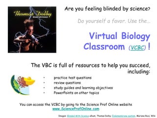
About Science Prof Online
PowerPoint Resources
• Science Prof Online (SPO) is a free science education website that provides fully-developed Virtual Science Classrooms,
science-related PowerPoints, articles and images. The site is designed to be a helpful resource for students, educators, and
anyone interested in learning about science.
• The SPO Virtual Classrooms offer many educational resources, including practice test questions, review questions, lecture
PowerPoints, video tutorials, sample assignments and course syllabi. New materials are continually being developed, so check
back frequently, or follow us on Facebook (Science Prof Online) or Twitter (ScienceProfSPO) for updates.
• Many SPO PowerPoints are available in a variety of formats, such as fully editable PowerPoint files, as well as uneditable
versions in smaller file sizes, such as PowerPoint Shows and Portable Document Format (.pdf), for ease of printing.
• Images used on this resource, and on the SPO website are, wherever possible, credited and linked to their source. Any
words underlined and appearing in blue are links that can be clicked on for more information. PowerPoints must be viewed in
slide show mode to use the hyperlinks directly.
• Several helpful links to fun and interactive learning tools are included throughout the PPT and on the Smart Links slide,
near the end of each presentation. You must be in slide show mode to utilize hyperlinks and animations.
•This digital resource is licensed under Creative Commons Attribution-ShareAlike 3.0:
http://creativecommons.org/licenses/by-sa/3.0/
Alicia Cepaitis, MS
Chief Creative Nerd
Science Prof Online
Online Education Resources, LLC
alicia@scienceprofonline.com
From the Virtual Biology Classroom on ScienceProfOnline.com Image: Compound microscope objectives, T. Port
Tami Port, MS
Creator of Science Prof Online
Chief Executive Nerd
Science Prof Online
Online Education Resources, LLC
info@scienceprofonline.com
Early
Embryonic
Development
From the Virtual Biology Classroom on ScienceProfOnline.com Image: Morula, Wiki.
Two Basic Types of Cells
Eukaryotic Cell
Prokaryotic Cell
Images: Prokaryotic cell diagram &
Eukaryotic cell diagram, M. Ruiz
From the Virtual Biology Classroom on ScienceProfOnline.com
WATCH THIS!
Introduction
to Cells Video
You Started Out As a
Tiny Fertilized Egg!
From the Virtual Biology Classroom on ScienceProfOnline.com
So How Did You
Become This?
Image: Sperm fertilizing egg, Wiki; Goofy kid, T. Port
Mitosis
4 sub-phases:
1st – Prophase
2nd – Metaphase
3rd – Anaphase
4th – Telophase
followed by
Cytokinesis
Secret to remembering phases in order…
From the Virtual Biology Classroom on ScienceProfOnline.com
Image: Mitosis diagram, Marek Kultys
Images: Sperm & egg, Wikipedia;; Morula,
Wiki; My son and daughter, T. Port
Mitosis (Eukaryotic Cell Division) allows
for growth and development in
multicellular organisms.
From the Virtual Cell Biology Classroom on ScienceProfOnline.com
Growth requires
more cells.
Development
requires
specialization of
cells.
In sexually
reproducing
organisms, growth
& development
starts with
fertilization, a
sperm fusing with
an egg (ovum).
From the Virtual Biology Classroom on ScienceProfOnline.com Image: Sperm fertilizing egg, Wiki
Image: Events immediately following fertilization, Wiki
From the Virtual Biology Classroom on ScienceProfOnline.com
A: Egg surrounded by sperm. On right side of egg,
one sperm has just penetrated the egg membranes
and is entering cytoplasm; egg nucleus in the
center. After one sperm has fertilized the egg
(the winner!), chemical changes occur in the egg
membrane that prevent any more sperm from
entering.
B & C: Egg nucleus on left. Sperm nucleus on
right.
D: Chromosomes of egg and sperm nuclei clearly
visible (egg chromosomes in black, sperm
chromosomes shaded).
E: Duplicated chromosomes line up at the cells
equator prior to separating and moving to opposite
poles.
F: Completion of first cleavage, two-celled stage,
each nucleus showing four chromosomes, two from
the egg and two from the sperm.
Only 4 chromosomes are pictured in this
example. Q: How many chromosomes do we
have?
Image: First stages of cell division in mammalian embryo, Wiki.
From the Virtual Biology Classroom on ScienceProfOnline.com
Early stages
of division of
mammalian
embryo.
a. Two-cell stage.
(z.p. Zona striata. p.gl.
Polar bodies.)
b. Four-cell stage.
c. Eight-cell stage.
d & e. Morula stage.
Once the embryo has divided
into 16 cells, it begins to
resemble a mulberry, hence
the name morula (Latin,
morus: mulberry).
Images: Blastulation, Wiki, External view and cross-
section of blastula, Wiki
From the Virtual Biology Classroom on ScienceProfOnline.com
Blastulation
1. Morula
-----
2. Blastula
(“little bladder”)
A. View of the
outside of a
blastula.
B. Cross section of
blastula.
From the Virtual Biology Classroom on ScienceProfOnline.com
Gastrulation
Occurs when a
blastula (made up of
one layer) folds inward
and enlarges to create
a gastrula (“little
stomach”).
A gastrula has 3 germ
layers: the ectoderm,
the mesoderm, and
the endoderm. Some
of the ectoderm cells
from the blastula
collapse inward and
form the endoderm.
The blastopore is the
hole created.
Images: Gastrulation schematic, Wiki
From the Virtual Biology Classroom on ScienceProfOnline.com
Ectoderm
Differentiates into
the entire nervous
system (spine,
peripheral nerves and
brain), as well as
various external
tissues, such as hair,
nails, tooth enamel and
the outer layer of skin
(epidermis).
Also forms the lining
of mouth, anus,
nostrils, sweat glands,
hair and nails.
Image: Ectoderm, Wiki
From the Virtual Biology Classroom on ScienceProfOnline.com
Mesoderm
In vertebrates,
the mesoderm
gives rise to much
of the body’s
mass, including
muscle, connective
tissue, cartilage,
bone and blood.
Image: Mesoderm, Wiki
From the Virtual Biology Classroom on ScienceProfOnline.com
Endoderm
Develops into
interior organs and
tissues, such as
the interior linings
of two tubes in
the body, the
digestive and
respiratory tube
and associated
organs.
Images: Gastrulation schematic, Wiki
Confused?
Here are some links to fun resources
that further explain Embryogenesis:
• Animation of Early
Embryogenesis
• Embryo Development up to
Blastocyst
• IVF Development of Embryo
Up to 5 Days
• Development of a Frog
(You must be in PPT slideshow view to click on links.)
From the Virtual Biology Classroom on ScienceProfOnline.com
Are you feeling blinded by science?
Do yourself a favor. Use the…
Virtual Biology
Classroom (VCBC) !
The VBC is full of resources to help you succeed,
including:
• practice test questions
• review questions
• study guides and learning objectives
• PowerPoints on other topics
You can access the VCBC by going to the Science Prof Online website
www.ScienceProfOnline.com
Images: Blinded With Science album, Thomas Dolby; Endomembrane system, Mariana Ruiz, Wiki

Early-Embryonic-Development-Lecture-PowerPoint-VBC.ppsx

  • 1.
    About Science ProfOnline PowerPoint Resources • Science Prof Online (SPO) is a free science education website that provides fully-developed Virtual Science Classrooms, science-related PowerPoints, articles and images. The site is designed to be a helpful resource for students, educators, and anyone interested in learning about science. • The SPO Virtual Classrooms offer many educational resources, including practice test questions, review questions, lecture PowerPoints, video tutorials, sample assignments and course syllabi. New materials are continually being developed, so check back frequently, or follow us on Facebook (Science Prof Online) or Twitter (ScienceProfSPO) for updates. • Many SPO PowerPoints are available in a variety of formats, such as fully editable PowerPoint files, as well as uneditable versions in smaller file sizes, such as PowerPoint Shows and Portable Document Format (.pdf), for ease of printing. • Images used on this resource, and on the SPO website are, wherever possible, credited and linked to their source. Any words underlined and appearing in blue are links that can be clicked on for more information. PowerPoints must be viewed in slide show mode to use the hyperlinks directly. • Several helpful links to fun and interactive learning tools are included throughout the PPT and on the Smart Links slide, near the end of each presentation. You must be in slide show mode to utilize hyperlinks and animations. •This digital resource is licensed under Creative Commons Attribution-ShareAlike 3.0: http://creativecommons.org/licenses/by-sa/3.0/ Alicia Cepaitis, MS Chief Creative Nerd Science Prof Online Online Education Resources, LLC alicia@scienceprofonline.com From the Virtual Biology Classroom on ScienceProfOnline.com Image: Compound microscope objectives, T. Port Tami Port, MS Creator of Science Prof Online Chief Executive Nerd Science Prof Online Online Education Resources, LLC info@scienceprofonline.com
  • 2.
    Early Embryonic Development From the VirtualBiology Classroom on ScienceProfOnline.com Image: Morula, Wiki.
  • 3.
    Two Basic Typesof Cells Eukaryotic Cell Prokaryotic Cell Images: Prokaryotic cell diagram & Eukaryotic cell diagram, M. Ruiz From the Virtual Biology Classroom on ScienceProfOnline.com WATCH THIS! Introduction to Cells Video
  • 4.
    You Started OutAs a Tiny Fertilized Egg! From the Virtual Biology Classroom on ScienceProfOnline.com So How Did You Become This? Image: Sperm fertilizing egg, Wiki; Goofy kid, T. Port
  • 5.
    Mitosis 4 sub-phases: 1st –Prophase 2nd – Metaphase 3rd – Anaphase 4th – Telophase followed by Cytokinesis Secret to remembering phases in order… From the Virtual Biology Classroom on ScienceProfOnline.com Image: Mitosis diagram, Marek Kultys
  • 6.
    Images: Sperm &egg, Wikipedia;; Morula, Wiki; My son and daughter, T. Port Mitosis (Eukaryotic Cell Division) allows for growth and development in multicellular organisms. From the Virtual Cell Biology Classroom on ScienceProfOnline.com Growth requires more cells. Development requires specialization of cells.
  • 7.
    In sexually reproducing organisms, growth &development starts with fertilization, a sperm fusing with an egg (ovum). From the Virtual Biology Classroom on ScienceProfOnline.com Image: Sperm fertilizing egg, Wiki
  • 8.
    Image: Events immediatelyfollowing fertilization, Wiki From the Virtual Biology Classroom on ScienceProfOnline.com A: Egg surrounded by sperm. On right side of egg, one sperm has just penetrated the egg membranes and is entering cytoplasm; egg nucleus in the center. After one sperm has fertilized the egg (the winner!), chemical changes occur in the egg membrane that prevent any more sperm from entering. B & C: Egg nucleus on left. Sperm nucleus on right. D: Chromosomes of egg and sperm nuclei clearly visible (egg chromosomes in black, sperm chromosomes shaded). E: Duplicated chromosomes line up at the cells equator prior to separating and moving to opposite poles. F: Completion of first cleavage, two-celled stage, each nucleus showing four chromosomes, two from the egg and two from the sperm. Only 4 chromosomes are pictured in this example. Q: How many chromosomes do we have?
  • 9.
    Image: First stagesof cell division in mammalian embryo, Wiki. From the Virtual Biology Classroom on ScienceProfOnline.com Early stages of division of mammalian embryo. a. Two-cell stage. (z.p. Zona striata. p.gl. Polar bodies.) b. Four-cell stage. c. Eight-cell stage. d & e. Morula stage. Once the embryo has divided into 16 cells, it begins to resemble a mulberry, hence the name morula (Latin, morus: mulberry).
  • 10.
    Images: Blastulation, Wiki,External view and cross- section of blastula, Wiki From the Virtual Biology Classroom on ScienceProfOnline.com Blastulation 1. Morula ----- 2. Blastula (“little bladder”) A. View of the outside of a blastula. B. Cross section of blastula.
  • 11.
    From the VirtualBiology Classroom on ScienceProfOnline.com Gastrulation Occurs when a blastula (made up of one layer) folds inward and enlarges to create a gastrula (“little stomach”). A gastrula has 3 germ layers: the ectoderm, the mesoderm, and the endoderm. Some of the ectoderm cells from the blastula collapse inward and form the endoderm. The blastopore is the hole created. Images: Gastrulation schematic, Wiki
  • 12.
    From the VirtualBiology Classroom on ScienceProfOnline.com Ectoderm Differentiates into the entire nervous system (spine, peripheral nerves and brain), as well as various external tissues, such as hair, nails, tooth enamel and the outer layer of skin (epidermis). Also forms the lining of mouth, anus, nostrils, sweat glands, hair and nails. Image: Ectoderm, Wiki
  • 13.
    From the VirtualBiology Classroom on ScienceProfOnline.com Mesoderm In vertebrates, the mesoderm gives rise to much of the body’s mass, including muscle, connective tissue, cartilage, bone and blood. Image: Mesoderm, Wiki
  • 14.
    From the VirtualBiology Classroom on ScienceProfOnline.com Endoderm Develops into interior organs and tissues, such as the interior linings of two tubes in the body, the digestive and respiratory tube and associated organs. Images: Gastrulation schematic, Wiki
  • 15.
    Confused? Here are somelinks to fun resources that further explain Embryogenesis: • Animation of Early Embryogenesis • Embryo Development up to Blastocyst • IVF Development of Embryo Up to 5 Days • Development of a Frog (You must be in PPT slideshow view to click on links.) From the Virtual Biology Classroom on ScienceProfOnline.com
  • 16.
    Are you feelingblinded by science? Do yourself a favor. Use the… Virtual Biology Classroom (VCBC) ! The VBC is full of resources to help you succeed, including: • practice test questions • review questions • study guides and learning objectives • PowerPoints on other topics You can access the VCBC by going to the Science Prof Online website www.ScienceProfOnline.com Images: Blinded With Science album, Thomas Dolby; Endomembrane system, Mariana Ruiz, Wiki