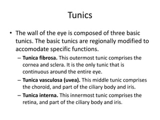
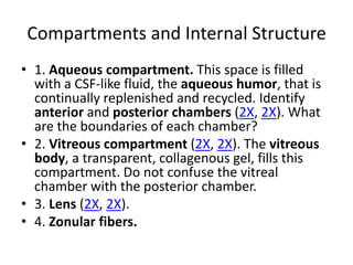
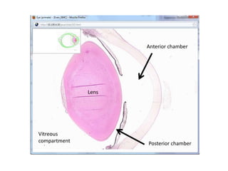
Eye Study Guide

  Samantha Blum
Tunics
• The wall of the eye is composed of three basic
  tunics. The basic tunics are regionally modified to
  accomodate specific functions.
   – Tunica fibrosa. This outermost tunic comprises the
     cornea and sclera. It is the only tunic that is
     continuous around the entire eye.
   – Tunica vasculosa (uvea). This middle tunic comprises
     the choroid, and part of the ciliary body and iris.
   – Tunica interna. This innermost tunic comprises the
     retina, and part of the ciliary body and iris.
Compartments and Internal Structure
• 1. Aqueous compartment. This space is filled
  with a CSF-like fluid, the aqueous humor, that is
  continually replenished and recycled. Identify
  anterior and posterior chambers (2X, 2X). What
  are the boundaries of each chamber?
• 2. Vitreous compartment (2X, 2X). The vitreous
  body, a transparent, collagenous gel, fills this
  compartment. Do not confuse the vitreal
  chamber with the posterior chamber.
• 3. Lens (2X, 2X).
• 4. Zonular fibers.
Anterior chamber


              Lens


Vitreous
compartment          Posterior chamber
Tunica Fibrosa
• This layer is comprised of the sclera
  (posteriorly) and the cornea (anteriorly). Both
  structures are formed predominantly by
  extracellular C.T. components.
Tunica Fibrosa:
                          Sclera
• This region begins at the corneoscleral limbus (2X, the junction
  between the cornea and sclera) and extends posteriorly. Note that
  it is opaque.
• Identify the bulbar conjunctiva (2X, 10X) epithelium that covers the
  anterior portion (analogous to the "white of the eye").
• Identify the limbus at the corneoscleral junction (2X).
• Identify the region containing the lamina cribrosa (2X, 10X) of the
  sclera at the optic disc (the sclera will be obscured here by the optic
  nerve).
• Identify the Canal of Schlemm (4X, 10X, 20X).
• Identify the trabecular meshwork (4X, 10X, 20X), located at the
  iridocorneal angle (4X).
• What is the function of the trabecular meshwork?
• Note the fibrous nature of the scleral stroma (10X).
Tunica Fibrosa:
                         Cornea
• This clear anterior continuation of the tunica fibrosa is responsible
  for the majority of the refraction (bending of light) necessary for
  image focusing. Why? The cornea has five histological layers.
  Anterior epithelium. (20X, 50X)
• Bowman's membrane (anterior limiting membrane). This structure
  is the basement membrane of the anterior epithelium (50X).
• Substantia propria (stroma). Note that this avascular layer accounts
  for 90% of the corneal thickness (20X, 50X).
• Descemet's membrane (posterior limiting membrane). This is the
  basement membrane of the posterior epithelium (100X).
• Posterior epithelium. Note the low cuboidal "endothelial" cells
  (100X).
Tunica Vasculosa (Uvea):
                  Choroid
• The choroid (40X) has four histological layers.
• Suprachoroid. This is an avascular layer of loose
  C.T., melanocytes and nerves.
• Vascular layer. (100X) This layer is characterized
  by prominent veins and small arteries (20X).
• Choriocapillaris (Choriocapillary layer). (100X)
  The capillaries in this layer directly nourish the
  outer half of the retina.
• Basal layer, or Brüch's membrane. (100X) This
  structure is the basement membrane of the
  retinal pigment epithelium.
Choroid


 Vascular layer

                     Retina

Choriocapillaris
Tunica Vasculosa (Uvea):
                  Ciliary Body
• The ciliary ring and ciliary crown are gross structures that are
  best seen in an axial view (remember that they are circular
  structures).
• Locate the ciliary muscle (4X, 40X). What type of muscle is it?
   – Smooth muscle
Tunica Vasculosa (Uvea):
                           Iris
•   This structure (2X) changes shape to regulate the size of the pupil. The iris is
    predominantly uveal; the only non-uveal portion is the epithelium that lines its
    posterior surface.
•   Identify the uveal portion (4X, 20X, 40X) of the iris, and note that it has two layers,
    the anterior border layer and the stroma.
•   Note that the anterior border layer (20X, 40X) is thin and avascular.
•   Identify the stromal layer, and note that it is thick and vascular, and contains a
    variable amount of pigment cells. This pigment is responsible for "eye color".
•   Note that the iris has no epithelial covering (40X) on its anterior surface.
•   Identify the pupil (2X).
•   Identify the sphincter pupillae (4X, 20X, 40X, 50X) muscle in the stroma. These
    smooth muscle fibers are arranged circumferentially around the edge of the iris,
    and are under parasympathetic cholinergic control. By shortening, they reduce the
    diameter of the pupil (schematic).
•   Indentify the dilator pupillae (50X). These myoepithelial fibers are arranged
    radially around the iris, and are under sympathetically innervated. By shortening,
    they increase the diameter of the pupil (schematic).
Tunica Interna:
                               Layers of Retina
•   Retinal neurons and glia are organized into readily-identifiable histological layers. Identify the
    following: Pigmented cell layer (retinal pigment epithelium, RPE). This is the epithelium on which
    the retina rests; it functions to regenerate bleached photopigment, and to phagocytose
    degenerated discs from the photoreceptors.
•   Note that the epithelium is simple cuboidal (50X, 100X).
•   Identify pigment granules (100X).
•   Rod and cone layer.
•   Identify outer segments of rods and cones (schematic; 100X). Membranes within the outer
    segments contain the photosensitive pigment rhodopsin.
•   Outer nuclear layer (20X, 100X). This layer contains the cell bodies and nuclei of the photoreceptor
    cells .
•   Outer plexiform layer (20X, 100X, 100X). The synaptic terminals of photoreceptors make contact
    here with bipolar and horizontal cells.
•   Inner nuclear layer (20X, 100X, 100X). This layer contains the cell bodies of bipolar cells, horizontal
    cells, and amacrine cells.
•   Internal plexiform layer (50X). Several types of retinal neuron interact synaptically in this layer.
    Information about light is transmitted to the ganglion cells, whose dendrites are found here.
•   Ganglion cell layer (20X, 50X, 100X). This layer contains the cell bodies of ganglion cells.
•   Nerve fiber layer (20X, 50X, 100X). The axons of the ganglion cells run on the inner surface of the
    retina, heading toward the optic disk (see below).
•   **THE LAYERS OF THE RETINA DIFFER IN 2 AREAS OF SPECIALIZATION: FOVEA AND OPTIC DISK
Tunica Interna:
                       Fovea
• Within the fovea (4X, 20X), the number of retinal layers is
  reduced, because only the photoreceptors are present. The
  photoreceptor axons make their normal synaptic
  connections (to bipolar cells), but those bipolar cells lie
  outside the foveal region, so the photoreceptor axons must
  run laterally to get to them.
• Look for a thinning of the retina (4X, 20X) at the posterior
  pole, and note the absence of the inner retinal layers at this
  point.
• What effect does this retinal thinning have on visual acuity?
• To what region of the visual field does the fovea
  correspond?
Tunica Interna:
                    Optic Disk
• At this point, the axons of ganglion cells leave the globe
  through a perforated region of the sclera (the lamina
  cribrosa, 10X).
• Look for an indentation in the inner (vitreal) surface of
  the retina, and note the complete absence of any of
  the normal retinal layers here (10X, 20X).
• Note that the optic nerve passes completely through
  the sclera, to exit the globe.
• Note that the sclera is continuous with the sheath of
  the optic nerve (10X, which is, in turn, continuous with
  the dura of the brain).
Tunica Interna:
            Retinal Blood Supply
• Identify capillaries in the retina (50X). These form
  two plexuses, one in the ganglion cell layer and
  one in the outer plexiform layer. These capillaries
  are fed by branches of the central retinal artery,
  which lie on the inner (vitreal) surface of the
  retina. These capillaries nourish the inner half of
  the thickness of the retina.
• Note that the outer half (50X) of the retina is
  avascular. How is it nourished?
Retinal Blood Supply


              Capillaries in GCL
Tunica Interna:
                   Pars ciliaris retinae
• The name means "the ciliary part of the retina". The photosensitive retina
  continues anteriorly, losing its photosensitive properties, and thins down
  to become the double epithelium that covers the ciliary body and the
  posterior surface of the iris. This epithelium also produces aqueous
  humor.
• Identify the outer, pigmented epithelium (10X, 40X).
• Identify the inner, non-pigmented epithelium (10X, 40X).
• Note that these two layers are in intimate contact with one another (as
  are the RPE and the photosensitive retina, more posteriorly).
• Identify ciliary processes (10X, 40X), which are extensions of the ciliary
  epithelium that protrude into the posterior chamber.
• Identify zonule fibers (10X) extending from the ciliary processes to the
  lens epithelium.
• What important substance is produced by the ciliary epithelium?
    – Aqueous humor
Tunica Interna:
                        Pars iridica retinae
•   The name means "the iridical part of the retina". The double epithelium of the
    pars ciliaris continues all the way to the tip of the iris at the pupillary opening.
•   Identify the two epithelial layers (50X, 50X) covering the posterior surface; note
    that both layers contain pigment here.
•   Identify the anterior myo-epithelial layer (50X, 100X), which forms the dilator
    muscle (dilator pupillae) of the iris.
Lens
• The lens (2X) is an avascular, highly specialized epithelial
  structure with three components: lens capsule, lens
  epithelium, and lens fibers.
• The lens capsule (20X, 40X, 40X) surrounds the entire lens;
  this capsule is actually the basement membrane of the lens
  epithelium.
• The lens epithelium (subcapsular epithelium; 40X) is a
  simple cuboidal layer that covers only the anterior portion
  of the lens.
• The body of the lens consists of large numbers of individual
  lens fibers; these are highly specialized cells that develop
  from the anterior subcapsular epithelium, and that extend
  all the way to the posterior surface of the lens (40X; 40X).
Lens

         Capsule


          Epithelium


       Fibers
Zona Ciliaris
                         (Zonule Fibers)
• Identify this suspensory ligament of lens (10X), which takes the form of a
  group of colorless, refractile fibers between ciliary processes and lens.
• What effect does contraction of the ciliary muscle have on the shape of
  the lens?
    – Relaxes (thickens) the lens and improves near vision
Aqueous Humor
•   Aqueous humor is an ultrafiltrate of plasma that circulates through the anterior
    portion of the eye at high pressure. For the maintenance of the correct pressure,
    the rate of synthesis and reuptake must be balanced. A rise in intraocular pressure
    (IOP) can lead to retinal damage and consequent blindness (glaucoma).
•   Identify the following structures or spaces involved in the production, circulation,
    and removal of aqueous humor (2X):
•   Synthesis by the ciliary epithelium (especially the ciliary processes).
•   Release into the posterior chamber.
•   Flow into the anterior chamber via the pupil (and, to some extent, by percolating
    through the body of the iris).
•   Filtration and uptake by the trabecular meshwork. Note that a reduction in the
    iridocorneal angle (e.g. by partial collapse of the iris onto the meshwork) might
    lead to occlusion (blockage) of the trabecular meshwork, and cause an increase in
    intraocular pressure.
•   Drainage into the Canal of Schlemm.
•   Return to the general circulation, via drainage into scleral veins.
Eyelid
• The eyelid is a thin sheet of tissue with a
  muscular core. It covers the front surface of the
  eye, moving vertically over the cornea to keep its
  surface clean and moist. The inner surface lies
  against the cornea, trapping a thin layer of tear
  fluid; this fluid is a mixture of the secretory
  products of the lacrimal (tear) gland, tarsal
  (Meibomian) glands, and glands of Moll. The
  latter two sets of glands are located within the
  eyelid.
Eyelid
•   The external surface of the eyelid has a covering of skin with hair follicles and associated sebaceous
    glands, and sweat glands (1x, 5x).
•   On the inner (conjunctival) surface, note that the lining epithelium varies from simple cuboidal to
    stratified cuboidal, and may contain goblet cells (20x). Bacterial infections of this membrane, called
    conjunctivitis, cause it to be inflamed and red ("pink eye").
•   Identify the cluster of large, sebaceous-like glands that comprise the tarsal plate (5x). These tarsal
    (Meibomian) glands all drain via ducts located at the tip of the eyelid (10x). They secrete an oily substance
    that floats on the surface of the serous tear fluid produced by the lacrimal gland. Inflammation of tarsal
    glands causes swelling of the eyelid ("chalazion").
•   Identify the skeletal muscle fascicles of the orbicularis muscle (5x).
Ear Study Guide

  Samantha Blum
Auricle
• Examine the cartilage framework of the
  auricle, and note that it is comprised of elastic
  cartilage (2X, 20X).
• Note that the auricle is covered by thin skin
  (2X, 10X).
External Auditory Meatus
• Identify elastic cartilage (2X).
• Examine the epidermis of the meatus
  (2X), and note that it is a keratinized stratified
  squamous epithelium (40X).
• Identify sebaceous glands (4X, 20X) and
  ceruminous glands (4X, 40X).
• Identify hair follicles (4X, 10X).
Keratinized, stratified squamous
The Middle Ear
• A. Tympanic cavity.
• B. Ossicles.
  – Malleus.
  – Incus.
  – Stapes.
• C. Auditory Tube.
  – Note that this structure has both bony and
    cartilaginous regions.
Inner Ear
• The two main components of inner ear are the bony labyrinth
  and the membranous labyrinth. The membranous labyrinth is
  a continuous, membrane-enclosed compartment within bony
  labyrinth; it closely follows the shape of the bony labyrinth.
• Membranous labyrinth has 2 divisions:
   – Vestibular division
   – Cochlear (auditory) division
Vestibular Division of the Membranous Labyrinth:
            Utricle & Saccule (Otolith Organs)
• These regions contain receptors responsible for detection of static head
  position and linear acceleration (not all slides contain these regions).
  Identify the sensory epithelium and its specializations: maculae
  (schematic; 4X, 20X, 50X), otolithic membrane (50X) and otoconia (20X,
  50X).
Semicircular Ducts
• Each semicircular canal (4X, 20X, 4X, 10X) has an
  enlargement at its base, in which is located the sensory
  apparatus (the crista ampullaris) that detects
  movement of fluid through the canal. Identify the
  following:
• Crista ampullaris (schematic; 4X, 20X, 40X).
• Sensory hair bundles (40X) extending into the
  gelatinous cupula (20X, 40X).
• What type of motion does the semicircular canal
  detect?
• What cranial nerve (and division) contains the axons of
  sensory neurons in the crista ampullaris?
Cochlear Duct
                    (Scala Media)
• This is the portion of membranous labyrinth that extends
  into the bony labyrinth of the cochlea, like a tunnel within a
  tunnel. It is attached at two sides along its length to the
  cochlea (osseous labyrinth), creating three compartments.
  Identify:
• a. Spiral ligament (10X) and spiral limbus (10X).
• b. Scala vestibuli (10X, 10X) and vestibular membrane
  (10X).
• c. Scala tympani (10X, 10X) and basilar membrane (10X).
• d. Scala media (4X, cochlear duct).
• e. Modiolus (4X)
• f. Bony (osseous) lamina (10X).
Cochlear Duct
Organ of Corti
• This is the site where sensory epithelial cells transduce sound vibrations
  into electrical signals for sound perception. A number of cell types and
  structures are of interest and readily studied on your slides.
• Identify the basilar membrane (50X) that supports the Organ of Corti.
• How do sounds affect the basilar membrane?
• Identify inner and outer hair cells (50X).
• Look for stereocilia (50X).
• Identify the tectorial membrane (50X). What happens to this membrane,
  and to the hair cells, when sound vibrations pass into the cochlea?
• Identify cells of the spiral ganglion (10X). Which cranial nerve (and
  division) is formed by the axons of these cells?
• Which region of the cochlea detects the highest sound frequencies? The
  lowest? How does this arrangement affect the pattern of hearing loss
  caused by exposure to loud noises?
Organ of Corti
Central Nervous System

     Samantha Blum
Central Nervous System
• The major components of the central nervous system (CNS)
  are neurons and neuroglia. Connective tissue is essentially
  absent.
White Matter of Spinal Cord
• Identify myelinated axons in cross-section
  (10X; 40X).
Gray Matter of Spinal Cord
• On the silver-stained slide, identify myelinated axonal fibers (10X) and
  unmyelinated axonal fibers (20X). Why are axons present in gray matter?
    – Axons traverse the gray matter to make connections between white matter
• Note the various nerve fiber sizes.
Neuroglia
• The staining method used here does not
  demonstrate the cytoplasm of neuroglial cells,
  only their nuclei. One can, however, identify
  the first three cell types on the basis of
  nuclear appearance.
Astrocytes
• These cells have large, pale nuclei, and are the
  most numerous of the neuroglia in gray
  matter. Find them in the cerebral cortex of this
  slide (40X; special stain). They occupy most of
  the space between neurons.
Astrocytes (astrocyte stain)
Astrocytes (H&E)
Microglia
• The fine, highly branched processes of these cells are not
  visible in H&E preparations; only their nuclei are visible (atlas
  example). As a result, you will not be able to reliably identify
  neuroglia in your slides.
Oligodendrocytes
• These cells are responsible for forming the myelin
  ensheathment present around some CNS axons.
• Look in areas of white matter first. Note that
  oligodendrocytes are the predominant neuroglial cell
  type of white matter. Why would you expect to find
  them in gray matter?
   – Axons are concentrated in white matter; there are some
     axons present in gray matter, so there will be some (but
     significantly less) oligodendrocytes in gray matter
• Look for their nuclei, which are small, round, and
  darkly-stained (100X). Oligodendrocyte processes do
  not stain with H&E.
Oligodendrocytes
Ependymal Cells
• The cerebral ventricles and aqueduct are lined
  with a simple cuboidal epithelium, the
  ependyma, made up of two types of cells: 1.
  Ependymal cells have cilia, and are attached
  to one-another with belt desmosomes; the
  latter structures prevent leakage of fluids
  between ependymal cells. 2) Tanycytes; these
  specialized ependymal cells send processes
  into the neuropil to make contact with blood
  vessels.
Ependymal Cells:
                 Cerebral Ventricle
• Examine the lining of the ventricle in this section of midbrain
  (1X).
• Note that the simple cuboidal epithelial lining is formed by a
  single layer of ependymal cells (5X).
• Look for cilia at the luminal surface (40X).
Ependymal Cells:
       Central Canal of Spinal Cord
• Examine the lining of the central canal (which
  may have collapsed), identifying ependymal
  cells (1X; 20X; 50X).
Ependymal Cells:
Central Canal of Spinal Cord
Ependymal Cells:
                Choroid Plexus
• Identify regions where blood vessels have
  penetrated into the ventricular space, forming
  tufts of capillaries covered by a layer of cuboidal
  ependymal cells. (1x, 1x, 4x, 10x, 40x).
• Cerebrospinal fluid, which is an ultrafiltrate of
  plasma, is extruded through the permeable walls
  of these blood vessels, then transported into the
  ventricular lumen by the ependymal cells.
• Note that many ependymal cells are ciliated
  (40x). Why?
Choroid Plexus
Meninges
• The brain lies within a three-layered
  connective tissue capsule. The innermost two
  layers, the pia mater and the arachnoid
  mater, are not readily seen in this slide.
Dura Mater
• This thick outermost layer is composed of
  dense fibroelastic connective tissue (50X).

Ear study guide

  • 1.
    Eye Study Guide Samantha Blum
  • 2.
    Tunics • The wallof the eye is composed of three basic tunics. The basic tunics are regionally modified to accomodate specific functions. – Tunica fibrosa. This outermost tunic comprises the cornea and sclera. It is the only tunic that is continuous around the entire eye. – Tunica vasculosa (uvea). This middle tunic comprises the choroid, and part of the ciliary body and iris. – Tunica interna. This innermost tunic comprises the retina, and part of the ciliary body and iris.
  • 3.
    Compartments and InternalStructure • 1. Aqueous compartment. This space is filled with a CSF-like fluid, the aqueous humor, that is continually replenished and recycled. Identify anterior and posterior chambers (2X, 2X). What are the boundaries of each chamber? • 2. Vitreous compartment (2X, 2X). The vitreous body, a transparent, collagenous gel, fills this compartment. Do not confuse the vitreal chamber with the posterior chamber. • 3. Lens (2X, 2X). • 4. Zonular fibers.
  • 4.
    Anterior chamber Lens Vitreous compartment Posterior chamber
  • 5.
    Tunica Fibrosa • Thislayer is comprised of the sclera (posteriorly) and the cornea (anteriorly). Both structures are formed predominantly by extracellular C.T. components.
  • 6.
    Tunica Fibrosa: Sclera • This region begins at the corneoscleral limbus (2X, the junction between the cornea and sclera) and extends posteriorly. Note that it is opaque. • Identify the bulbar conjunctiva (2X, 10X) epithelium that covers the anterior portion (analogous to the "white of the eye"). • Identify the limbus at the corneoscleral junction (2X). • Identify the region containing the lamina cribrosa (2X, 10X) of the sclera at the optic disc (the sclera will be obscured here by the optic nerve). • Identify the Canal of Schlemm (4X, 10X, 20X). • Identify the trabecular meshwork (4X, 10X, 20X), located at the iridocorneal angle (4X). • What is the function of the trabecular meshwork? • Note the fibrous nature of the scleral stroma (10X).
  • 8.
    Tunica Fibrosa: Cornea • This clear anterior continuation of the tunica fibrosa is responsible for the majority of the refraction (bending of light) necessary for image focusing. Why? The cornea has five histological layers. Anterior epithelium. (20X, 50X) • Bowman's membrane (anterior limiting membrane). This structure is the basement membrane of the anterior epithelium (50X). • Substantia propria (stroma). Note that this avascular layer accounts for 90% of the corneal thickness (20X, 50X). • Descemet's membrane (posterior limiting membrane). This is the basement membrane of the posterior epithelium (100X). • Posterior epithelium. Note the low cuboidal "endothelial" cells (100X).
  • 9.
    Tunica Vasculosa (Uvea): Choroid • The choroid (40X) has four histological layers. • Suprachoroid. This is an avascular layer of loose C.T., melanocytes and nerves. • Vascular layer. (100X) This layer is characterized by prominent veins and small arteries (20X). • Choriocapillaris (Choriocapillary layer). (100X) The capillaries in this layer directly nourish the outer half of the retina. • Basal layer, or Brüch's membrane. (100X) This structure is the basement membrane of the retinal pigment epithelium.
  • 10.
    Choroid Vascular layer Retina Choriocapillaris
  • 11.
    Tunica Vasculosa (Uvea): Ciliary Body • The ciliary ring and ciliary crown are gross structures that are best seen in an axial view (remember that they are circular structures). • Locate the ciliary muscle (4X, 40X). What type of muscle is it? – Smooth muscle
  • 12.
    Tunica Vasculosa (Uvea): Iris • This structure (2X) changes shape to regulate the size of the pupil. The iris is predominantly uveal; the only non-uveal portion is the epithelium that lines its posterior surface. • Identify the uveal portion (4X, 20X, 40X) of the iris, and note that it has two layers, the anterior border layer and the stroma. • Note that the anterior border layer (20X, 40X) is thin and avascular. • Identify the stromal layer, and note that it is thick and vascular, and contains a variable amount of pigment cells. This pigment is responsible for "eye color". • Note that the iris has no epithelial covering (40X) on its anterior surface. • Identify the pupil (2X). • Identify the sphincter pupillae (4X, 20X, 40X, 50X) muscle in the stroma. These smooth muscle fibers are arranged circumferentially around the edge of the iris, and are under parasympathetic cholinergic control. By shortening, they reduce the diameter of the pupil (schematic). • Indentify the dilator pupillae (50X). These myoepithelial fibers are arranged radially around the iris, and are under sympathetically innervated. By shortening, they increase the diameter of the pupil (schematic).
  • 14.
    Tunica Interna: Layers of Retina • Retinal neurons and glia are organized into readily-identifiable histological layers. Identify the following: Pigmented cell layer (retinal pigment epithelium, RPE). This is the epithelium on which the retina rests; it functions to regenerate bleached photopigment, and to phagocytose degenerated discs from the photoreceptors. • Note that the epithelium is simple cuboidal (50X, 100X). • Identify pigment granules (100X). • Rod and cone layer. • Identify outer segments of rods and cones (schematic; 100X). Membranes within the outer segments contain the photosensitive pigment rhodopsin. • Outer nuclear layer (20X, 100X). This layer contains the cell bodies and nuclei of the photoreceptor cells . • Outer plexiform layer (20X, 100X, 100X). The synaptic terminals of photoreceptors make contact here with bipolar and horizontal cells. • Inner nuclear layer (20X, 100X, 100X). This layer contains the cell bodies of bipolar cells, horizontal cells, and amacrine cells. • Internal plexiform layer (50X). Several types of retinal neuron interact synaptically in this layer. Information about light is transmitted to the ganglion cells, whose dendrites are found here. • Ganglion cell layer (20X, 50X, 100X). This layer contains the cell bodies of ganglion cells. • Nerve fiber layer (20X, 50X, 100X). The axons of the ganglion cells run on the inner surface of the retina, heading toward the optic disk (see below). • **THE LAYERS OF THE RETINA DIFFER IN 2 AREAS OF SPECIALIZATION: FOVEA AND OPTIC DISK
  • 15.
    Tunica Interna: Fovea • Within the fovea (4X, 20X), the number of retinal layers is reduced, because only the photoreceptors are present. The photoreceptor axons make their normal synaptic connections (to bipolar cells), but those bipolar cells lie outside the foveal region, so the photoreceptor axons must run laterally to get to them. • Look for a thinning of the retina (4X, 20X) at the posterior pole, and note the absence of the inner retinal layers at this point. • What effect does this retinal thinning have on visual acuity? • To what region of the visual field does the fovea correspond?
  • 17.
    Tunica Interna: Optic Disk • At this point, the axons of ganglion cells leave the globe through a perforated region of the sclera (the lamina cribrosa, 10X). • Look for an indentation in the inner (vitreal) surface of the retina, and note the complete absence of any of the normal retinal layers here (10X, 20X). • Note that the optic nerve passes completely through the sclera, to exit the globe. • Note that the sclera is continuous with the sheath of the optic nerve (10X, which is, in turn, continuous with the dura of the brain).
  • 18.
    Tunica Interna: Retinal Blood Supply • Identify capillaries in the retina (50X). These form two plexuses, one in the ganglion cell layer and one in the outer plexiform layer. These capillaries are fed by branches of the central retinal artery, which lie on the inner (vitreal) surface of the retina. These capillaries nourish the inner half of the thickness of the retina. • Note that the outer half (50X) of the retina is avascular. How is it nourished?
  • 19.
    Retinal Blood Supply Capillaries in GCL
  • 20.
    Tunica Interna: Pars ciliaris retinae • The name means "the ciliary part of the retina". The photosensitive retina continues anteriorly, losing its photosensitive properties, and thins down to become the double epithelium that covers the ciliary body and the posterior surface of the iris. This epithelium also produces aqueous humor. • Identify the outer, pigmented epithelium (10X, 40X). • Identify the inner, non-pigmented epithelium (10X, 40X). • Note that these two layers are in intimate contact with one another (as are the RPE and the photosensitive retina, more posteriorly). • Identify ciliary processes (10X, 40X), which are extensions of the ciliary epithelium that protrude into the posterior chamber. • Identify zonule fibers (10X) extending from the ciliary processes to the lens epithelium. • What important substance is produced by the ciliary epithelium? – Aqueous humor
  • 21.
    Tunica Interna: Pars iridica retinae • The name means "the iridical part of the retina". The double epithelium of the pars ciliaris continues all the way to the tip of the iris at the pupillary opening. • Identify the two epithelial layers (50X, 50X) covering the posterior surface; note that both layers contain pigment here. • Identify the anterior myo-epithelial layer (50X, 100X), which forms the dilator muscle (dilator pupillae) of the iris.
  • 22.
    Lens • The lens(2X) is an avascular, highly specialized epithelial structure with three components: lens capsule, lens epithelium, and lens fibers. • The lens capsule (20X, 40X, 40X) surrounds the entire lens; this capsule is actually the basement membrane of the lens epithelium. • The lens epithelium (subcapsular epithelium; 40X) is a simple cuboidal layer that covers only the anterior portion of the lens. • The body of the lens consists of large numbers of individual lens fibers; these are highly specialized cells that develop from the anterior subcapsular epithelium, and that extend all the way to the posterior surface of the lens (40X; 40X).
  • 23.
    Lens Capsule Epithelium Fibers
  • 24.
    Zona Ciliaris (Zonule Fibers) • Identify this suspensory ligament of lens (10X), which takes the form of a group of colorless, refractile fibers between ciliary processes and lens. • What effect does contraction of the ciliary muscle have on the shape of the lens? – Relaxes (thickens) the lens and improves near vision
  • 25.
    Aqueous Humor • Aqueous humor is an ultrafiltrate of plasma that circulates through the anterior portion of the eye at high pressure. For the maintenance of the correct pressure, the rate of synthesis and reuptake must be balanced. A rise in intraocular pressure (IOP) can lead to retinal damage and consequent blindness (glaucoma). • Identify the following structures or spaces involved in the production, circulation, and removal of aqueous humor (2X): • Synthesis by the ciliary epithelium (especially the ciliary processes). • Release into the posterior chamber. • Flow into the anterior chamber via the pupil (and, to some extent, by percolating through the body of the iris). • Filtration and uptake by the trabecular meshwork. Note that a reduction in the iridocorneal angle (e.g. by partial collapse of the iris onto the meshwork) might lead to occlusion (blockage) of the trabecular meshwork, and cause an increase in intraocular pressure. • Drainage into the Canal of Schlemm. • Return to the general circulation, via drainage into scleral veins.
  • 26.
    Eyelid • The eyelidis a thin sheet of tissue with a muscular core. It covers the front surface of the eye, moving vertically over the cornea to keep its surface clean and moist. The inner surface lies against the cornea, trapping a thin layer of tear fluid; this fluid is a mixture of the secretory products of the lacrimal (tear) gland, tarsal (Meibomian) glands, and glands of Moll. The latter two sets of glands are located within the eyelid.
  • 27.
    Eyelid • The external surface of the eyelid has a covering of skin with hair follicles and associated sebaceous glands, and sweat glands (1x, 5x). • On the inner (conjunctival) surface, note that the lining epithelium varies from simple cuboidal to stratified cuboidal, and may contain goblet cells (20x). Bacterial infections of this membrane, called conjunctivitis, cause it to be inflamed and red ("pink eye"). • Identify the cluster of large, sebaceous-like glands that comprise the tarsal plate (5x). These tarsal (Meibomian) glands all drain via ducts located at the tip of the eyelid (10x). They secrete an oily substance that floats on the surface of the serous tear fluid produced by the lacrimal gland. Inflammation of tarsal glands causes swelling of the eyelid ("chalazion"). • Identify the skeletal muscle fascicles of the orbicularis muscle (5x).
  • 28.
    Ear Study Guide Samantha Blum
  • 29.
    Auricle • Examine thecartilage framework of the auricle, and note that it is comprised of elastic cartilage (2X, 20X). • Note that the auricle is covered by thin skin (2X, 10X).
  • 30.
    External Auditory Meatus •Identify elastic cartilage (2X). • Examine the epidermis of the meatus (2X), and note that it is a keratinized stratified squamous epithelium (40X). • Identify sebaceous glands (4X, 20X) and ceruminous glands (4X, 40X). • Identify hair follicles (4X, 10X).
  • 31.
  • 32.
    The Middle Ear •A. Tympanic cavity. • B. Ossicles. – Malleus. – Incus. – Stapes. • C. Auditory Tube. – Note that this structure has both bony and cartilaginous regions.
  • 33.
    Inner Ear • Thetwo main components of inner ear are the bony labyrinth and the membranous labyrinth. The membranous labyrinth is a continuous, membrane-enclosed compartment within bony labyrinth; it closely follows the shape of the bony labyrinth. • Membranous labyrinth has 2 divisions: – Vestibular division – Cochlear (auditory) division
  • 34.
    Vestibular Division ofthe Membranous Labyrinth: Utricle & Saccule (Otolith Organs) • These regions contain receptors responsible for detection of static head position and linear acceleration (not all slides contain these regions). Identify the sensory epithelium and its specializations: maculae (schematic; 4X, 20X, 50X), otolithic membrane (50X) and otoconia (20X, 50X).
  • 35.
    Semicircular Ducts • Eachsemicircular canal (4X, 20X, 4X, 10X) has an enlargement at its base, in which is located the sensory apparatus (the crista ampullaris) that detects movement of fluid through the canal. Identify the following: • Crista ampullaris (schematic; 4X, 20X, 40X). • Sensory hair bundles (40X) extending into the gelatinous cupula (20X, 40X). • What type of motion does the semicircular canal detect? • What cranial nerve (and division) contains the axons of sensory neurons in the crista ampullaris?
  • 36.
    Cochlear Duct (Scala Media) • This is the portion of membranous labyrinth that extends into the bony labyrinth of the cochlea, like a tunnel within a tunnel. It is attached at two sides along its length to the cochlea (osseous labyrinth), creating three compartments. Identify: • a. Spiral ligament (10X) and spiral limbus (10X). • b. Scala vestibuli (10X, 10X) and vestibular membrane (10X). • c. Scala tympani (10X, 10X) and basilar membrane (10X). • d. Scala media (4X, cochlear duct). • e. Modiolus (4X) • f. Bony (osseous) lamina (10X).
  • 37.
  • 38.
    Organ of Corti •This is the site where sensory epithelial cells transduce sound vibrations into electrical signals for sound perception. A number of cell types and structures are of interest and readily studied on your slides. • Identify the basilar membrane (50X) that supports the Organ of Corti. • How do sounds affect the basilar membrane? • Identify inner and outer hair cells (50X). • Look for stereocilia (50X). • Identify the tectorial membrane (50X). What happens to this membrane, and to the hair cells, when sound vibrations pass into the cochlea? • Identify cells of the spiral ganglion (10X). Which cranial nerve (and division) is formed by the axons of these cells? • Which region of the cochlea detects the highest sound frequencies? The lowest? How does this arrangement affect the pattern of hearing loss caused by exposure to loud noises?
  • 39.
  • 40.
  • 41.
    Central Nervous System •The major components of the central nervous system (CNS) are neurons and neuroglia. Connective tissue is essentially absent.
  • 42.
    White Matter ofSpinal Cord • Identify myelinated axons in cross-section (10X; 40X).
  • 43.
    Gray Matter ofSpinal Cord • On the silver-stained slide, identify myelinated axonal fibers (10X) and unmyelinated axonal fibers (20X). Why are axons present in gray matter? – Axons traverse the gray matter to make connections between white matter • Note the various nerve fiber sizes.
  • 44.
    Neuroglia • The stainingmethod used here does not demonstrate the cytoplasm of neuroglial cells, only their nuclei. One can, however, identify the first three cell types on the basis of nuclear appearance.
  • 45.
    Astrocytes • These cellshave large, pale nuclei, and are the most numerous of the neuroglia in gray matter. Find them in the cerebral cortex of this slide (40X; special stain). They occupy most of the space between neurons.
  • 46.
  • 47.
  • 48.
    Microglia • The fine,highly branched processes of these cells are not visible in H&E preparations; only their nuclei are visible (atlas example). As a result, you will not be able to reliably identify neuroglia in your slides.
  • 49.
    Oligodendrocytes • These cellsare responsible for forming the myelin ensheathment present around some CNS axons. • Look in areas of white matter first. Note that oligodendrocytes are the predominant neuroglial cell type of white matter. Why would you expect to find them in gray matter? – Axons are concentrated in white matter; there are some axons present in gray matter, so there will be some (but significantly less) oligodendrocytes in gray matter • Look for their nuclei, which are small, round, and darkly-stained (100X). Oligodendrocyte processes do not stain with H&E.
  • 50.
  • 51.
    Ependymal Cells • Thecerebral ventricles and aqueduct are lined with a simple cuboidal epithelium, the ependyma, made up of two types of cells: 1. Ependymal cells have cilia, and are attached to one-another with belt desmosomes; the latter structures prevent leakage of fluids between ependymal cells. 2) Tanycytes; these specialized ependymal cells send processes into the neuropil to make contact with blood vessels.
  • 52.
    Ependymal Cells: Cerebral Ventricle • Examine the lining of the ventricle in this section of midbrain (1X). • Note that the simple cuboidal epithelial lining is formed by a single layer of ependymal cells (5X). • Look for cilia at the luminal surface (40X).
  • 53.
    Ependymal Cells: Central Canal of Spinal Cord • Examine the lining of the central canal (which may have collapsed), identifying ependymal cells (1X; 20X; 50X).
  • 54.
  • 55.
    Ependymal Cells: Choroid Plexus • Identify regions where blood vessels have penetrated into the ventricular space, forming tufts of capillaries covered by a layer of cuboidal ependymal cells. (1x, 1x, 4x, 10x, 40x). • Cerebrospinal fluid, which is an ultrafiltrate of plasma, is extruded through the permeable walls of these blood vessels, then transported into the ventricular lumen by the ependymal cells. • Note that many ependymal cells are ciliated (40x). Why?
  • 56.
  • 57.
    Meninges • The brainlies within a three-layered connective tissue capsule. The innermost two layers, the pia mater and the arachnoid mater, are not readily seen in this slide.
  • 58.
    Dura Mater • Thisthick outermost layer is composed of dense fibroelastic connective tissue (50X).