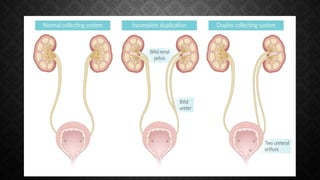
DUPLEX RENAL SYSTEM
DR.JAWAD SAIFULLAH
PGR UROLOGY AND RENAL
TRANSPLANTATION
ALLIED HOSPITAL , FAISALABAD
LEARNING
OBJECTIVES:
2. Identify its
anatomical
features.
1. Define
duplex renal
system.
3. Recognize
potential
complications.
4. Learn how to
diagnose and
manage related
disorders.
WHAT IS A DUPLEX
RENAL SYSTEM:
A duplex renal system occurs when there is a
duplication of the ureter, resulting in two
ureters draining a single kidney.
The exact prevalence of this anomaly is
difficult to ascertain because most patients
are asymptomatic, and the abnormality is
frequently detected incidentally. Prevalence
1-2%.
EMBRYOLOGY
The ureteric bud grows towards the metanephric
blastema (nephrogenic cord), which later develops
into the nephrons of the kidney.
The collecting system of the kidney develops from
the ureteric bud, a small outgrowth from the
mesonephric duct (Wolffian duct) .
The interaction between the ureteric bud and the
metanephric blastema is crucial for the formation
of the collecting system.
INCOMPLETE DUPLEX URETER (BIFID URETER):
This anomaly develops when a single
ureteral bud branches before it
reaches the metanephric blastema.
01
02
• The duplicated ureters unite at a
variable distance from the kidney, and
only one ureteral orifice is present on
the affected side.
COMPLETE DUPLEX URETER:
In the case of complete duplication of the
ureter, a developmental abnormality occurs
where two ureteric buds arise from the
mesonephric duct instead of the normal single
bud.
This leads to the formation of two ureters
originating from a single kidney rather than
the typical single ureter.
01
02
COMPLICATIONS:
INCOMPLETE
DUPLEX
URETER:
Ureteroureteral
reflux, also known
as yo-yo, saddle,
or seesaw reflux,
is a common
phenomenon
This is commonly associated with
vesicoureteral reflux as a result of a
lack of sufficient submucosal tunnel
that leads to pyelonephritis,
scarring and a so-called “drooping
lily” appearance on IVPs
COMPLETE
DUPLICATION:
Ureters that arise from a position either
lower or higher than the normal position
are associated with ectopic insertions,
renal dysplasia, and related complications
Common complications of complete
duplication are vesicoureteral reflux,
ectopic ureterocele, ectopic ureteral
insertion, ureteropelvic junction
obstruction of the lower pole.
MOST COMMON OF
ECTOPIC URETERIC
ORIFICE:
Male : Orifice in prostatic urethra
and in proximal to external
urinary sphincter.
Female: Orifice of upper moiety
ureter is in urethra distal to urinary
sphincter.
INVESTIGATIONS
Ultrasound is often the first-line
imaging modality used to diagnose
a duplex renal system.
• Anatomical Assessment
• Complications Detection
• Monitoring
• Guidance for Procedures
Sonography:
INTRAVENOUS
UROGRAPHY:
• Evaluation of
the function of
each kidney
• Identifying
any structural
abnormalities
• Assessing the
drainage of
urine
• DTPA(Diethylenetriaminepentaacetic acid)
• CT Urography
MANAGEMENT:
With complications Without complications
No intervention
required
What are the
symptoms/complications?
COMPLICATIONS
AND TREATMENTS:
01
Urolithiasis:
URS , RIRS ,
PCNL,
ESWL ,pyelolitho
tomy,
ureterolithotomy
02
Reflux: Re-
implantation
of ureter.
03
Obstruction :
Pyeloplasty
04
Ectopic
ureter: Re-
implantation
05
Nonfunctionin
g kidney:
Nephrectomy,
partial
nephrectomy
THANK YOU

Duplex renal systemComplete/Partial Duplex Renal System, .pptx

  • 2.
    DUPLEX RENAL SYSTEM DR.JAWADSAIFULLAH PGR UROLOGY AND RENAL TRANSPLANTATION ALLIED HOSPITAL , FAISALABAD
  • 3.
    LEARNING OBJECTIVES: 2. Identify its anatomical features. 1.Define duplex renal system. 3. Recognize potential complications. 4. Learn how to diagnose and manage related disorders.
  • 4.
    WHAT IS ADUPLEX RENAL SYSTEM: A duplex renal system occurs when there is a duplication of the ureter, resulting in two ureters draining a single kidney. The exact prevalence of this anomaly is difficult to ascertain because most patients are asymptomatic, and the abnormality is frequently detected incidentally. Prevalence 1-2%.
  • 5.
    EMBRYOLOGY The ureteric budgrows towards the metanephric blastema (nephrogenic cord), which later develops into the nephrons of the kidney. The collecting system of the kidney develops from the ureteric bud, a small outgrowth from the mesonephric duct (Wolffian duct) . The interaction between the ureteric bud and the metanephric blastema is crucial for the formation of the collecting system.
  • 8.
    INCOMPLETE DUPLEX URETER(BIFID URETER): This anomaly develops when a single ureteral bud branches before it reaches the metanephric blastema. 01 02 • The duplicated ureters unite at a variable distance from the kidney, and only one ureteral orifice is present on the affected side.
  • 9.
    COMPLETE DUPLEX URETER: Inthe case of complete duplication of the ureter, a developmental abnormality occurs where two ureteric buds arise from the mesonephric duct instead of the normal single bud. This leads to the formation of two ureters originating from a single kidney rather than the typical single ureter. 01 02
  • 10.
    COMPLICATIONS: INCOMPLETE DUPLEX URETER: Ureteroureteral reflux, also known asyo-yo, saddle, or seesaw reflux, is a common phenomenon This is commonly associated with vesicoureteral reflux as a result of a lack of sufficient submucosal tunnel that leads to pyelonephritis, scarring and a so-called “drooping lily” appearance on IVPs
  • 12.
    COMPLETE DUPLICATION: Ureters that arisefrom a position either lower or higher than the normal position are associated with ectopic insertions, renal dysplasia, and related complications Common complications of complete duplication are vesicoureteral reflux, ectopic ureterocele, ectopic ureteral insertion, ureteropelvic junction obstruction of the lower pole.
  • 13.
    MOST COMMON OF ECTOPICURETERIC ORIFICE: Male : Orifice in prostatic urethra and in proximal to external urinary sphincter. Female: Orifice of upper moiety ureter is in urethra distal to urinary sphincter.
  • 14.
    INVESTIGATIONS Ultrasound is oftenthe first-line imaging modality used to diagnose a duplex renal system. • Anatomical Assessment • Complications Detection • Monitoring • Guidance for Procedures Sonography:
  • 15.
    INTRAVENOUS UROGRAPHY: • Evaluation of thefunction of each kidney • Identifying any structural abnormalities • Assessing the drainage of urine
  • 16.
  • 17.
    MANAGEMENT: With complications Withoutcomplications No intervention required What are the symptoms/complications?
  • 18.
    COMPLICATIONS AND TREATMENTS: 01 Urolithiasis: URS ,RIRS , PCNL, ESWL ,pyelolitho tomy, ureterolithotomy 02 Reflux: Re- implantation of ureter. 03 Obstruction : Pyeloplasty 04 Ectopic ureter: Re- implantation 05 Nonfunctionin g kidney: Nephrectomy, partial nephrectomy
  • 19.