BY:  NASREEN SULTANA M-PHARM (PHARMACOLOGY) 10T21S0111 .  UNDER THE GUIDANCE OF: Dr. T. VEDAVATHI M.PHARM, PhD
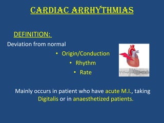
CARDIAC ARRHYTHMIAS DEFINITION:  Deviation from normal  Origin/Conduction Rhythm Rate Mainly occurs in patient who have  acute M.I. , taking  Digitalis  or in  anaesthetized patients.
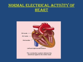
NORMAL ELECTRICAL ACTIVITY OF HEART
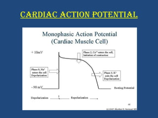
CARDIAC ACTION POTENTIAL
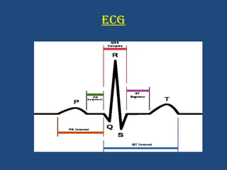
ECG
CLASSIFICATION OF ARRHYTMIAS Normal heart rate – 60-100 beats/min. Based on the heart rate : Tachyarrhythmia - rapid heart beat – >100/min Bradyarrhythmia – slow heart beat-<60/min
TACHYARRHYTHMIAS Caused by 2 mechanisms: Increased automaticity Re-entry Depending on ectopic focus tachyarrhythmias are classified as  Atrial tachyarrhythmias Ventricular tachyarrhythmias
ATRIAL TACHYARRYTHMIAS Atrial tachycardia: abnormal P wave Atrial flutters: the heart rate (atrial) is approximately 300/min Atrial fibrillation: irregularly spaced QRS complex with absent P waves.
VENTRICULAR ARRHYTHMIAS Ventricular tachycardia: abnormal QRS complex and heart rate-  >120beats/min . Torsade de pointes:   QT prolongation . Ventricular fibrillation:  VF produces rapid, uncoordinated, movement of the ventricles which produces no pulse. The ECG shows chaotic, bizarre, irregular ventricular complexes.
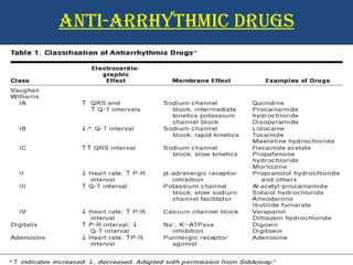
ANTI-ARRHYTHMIC DRUGS
NEW DRUGS Conventional anti arrhythmic drugs - ↑risk of proarrhythmias. Dronedrone   - analogue of amiodrone (MULTAQ)-> LESS TOXIC. Vernalakant  - atrial selective (↓proarrhythmic risk) Ranolazine  – anti-anginal atrial selective
HERBAL DRUGS Haw thorn – strengthens heart muscle improves blood circulation. Magnesium – relaxes and calms the heart. Angelica  Astragalus – heart tonic Barberry – heart tonic
SCREENING OF ANTI-ARRHYTHMICS DRUG SCREENING: Pre clinical testing of drugs in experimental animals or in vitro for their biological and toxic effects and potential clinical applications The screening of anti-arrhythmics is done by two different methods.  In-vitro models  In-vivo models
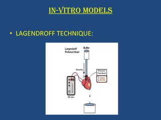
IN-VITRO MODELS LAGENDROFF TECHNIQUE:
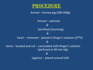
PROCEDURE Animal – Guinea pig (300-500g) Animal – selected  Sacrificed (stunning)  heart – removed – placed in Ringer’s solution (37⁰C)  Aorta – located and cut – cannulated with Ringer’s solution  (perfused at 40 mm Hg)  Ligature – placed around LAD
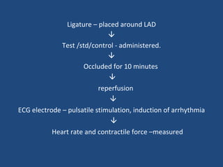
Ligature – placed around LAD  Test /std/control - administered.  Occluded for 10 minutes  reperfusion  ECG electrode – pulsatile stimulation, induction of arrhythmia  Heart rate and contractile force –measured
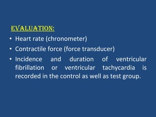
EVALUATION: Heart rate (chronometer) Contractile force (force transducer) Incidence and duration of ventricular fibrillation or ventricular tachycardia is recorded in the control as well as test group.
IN VIVO MODELS In vivo models used to screen anti arrhythmic drugs can be divided into five groups: Chemically induced arrhythmia Electrically induced arrhythmia Exercise induced ventricular fibrillation Mechanically induced arrhythmia Genetically induced arrhythmia
CHEMICALLY INDUCED ARRHYTHMIA A large number of agents are capable of inducing arrhythmias. Administration of anesthetics like chloroform, ether, halothane (sensitizing agents) followed by a precipitating stimulus such as adrenaline, ouabain alkaloids cause arrhythmia.
PURPOSE AND RATIONALE: Aconitine – acts persistently on sodium channels and activates them – ventricular arrhythmias. REQUIREMENTS:  Animals - Male Ivanovas rats (300-400g) Anesthetic – Urethane  Standard drug – Procainamide i.v (5mg/kg) Lidocaine i.v (5mg/kg)
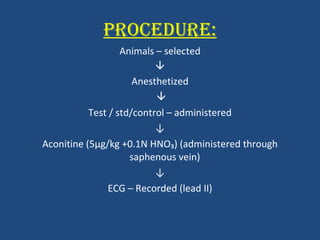
PROCEDURE: Animals – selected  Anesthetized  Test / std/control – administered ↓ Aconitine (5 μ g/kg +0.1N HNO₃) (administered through saphenous vein)  ↓ ECG – Recorded (lead II)
EVALUATION:  Anti-arrhythmic effect of test is measured by the amount of Aconitine/100g animal. Includes: Ventricular extra systoles Ventricular tachycardia Ventricular fibrillation Death
ELECTRICALLY INDUCED ARHYTHMIAS
PURPOSE AND RATIONALE:  Electrical stimulations – flutters and fibrillation. REQUIREMENTS : Animals – adult dogs (8-12kg) Anesthetic – sodium pentobarbital (35mg/kg)
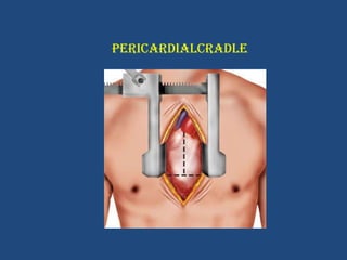
PROCEDURE Animal – selected ↓ Anesthetized ↓ Artificial respiration – Harvard respiratory pump B.P – monitored Body temperature – maintained by thermal blanket ↓ Chest cavity is opened ↓ Heart suspended in pericardial cradle ↓
PERICARDIALCRADLE
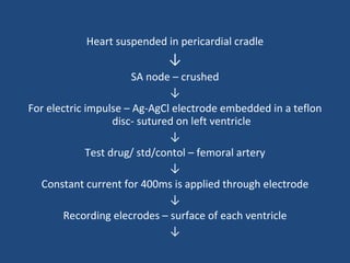
Heart suspended in pericardial cradle ↓ SA node – crushed ↓ For electric impulse – Ag-AgCl electrode embedded in a teflon disc- sutured on left ventricle ↓ Test drug/ std/contol – femoral artery ↓ Constant current for 400ms is applied through electrode ↓ Recording elecrodes – surface of each ventricle ↓
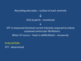
Recording elecrodes – surface of each ventricle ↓ ECG (Lead II) – monitored ↓ VFT is measured (minimal current intensity required to induce sustained ventricular fibrillation) When VF occurs – heart is defibrillated – recovered. EVALUATION: VFT - determined
EXERCISE INDUCED VENTRICULAR FIBRILLATION PURPOSE AND RATIONALE: Tests combining coronary constriction with physical exercise may resemble most closely the situation in coronary patients
REQUIREMENTS: Animals – Mongrel dogs (15 -19 kg) Anesthetic – Sodium pentobarbitone (10mg/kg) i.v. SURGICAL PREPARATION: Animals – selected ↓ Anesthetized ↓ Chest cavity – opened ↓ Heart  suspended in pericardial cradle ↓ around left circumflex artery – Doppler flow inducer- to measure blood  pressure - hydraulic occluder  - to occlude coronary artery are placed
↓ Pair of insulated silver coated wires – sutured on left and right ventricles- measure HR  ↓ Occlusion of LAD ↓ Myocardial infarction ↓ After 24 hrs- test drug/std/control - administered During this experiment: Transdermal fentanyl patch (↓post operative discomfort) Bupivacaine HCl Antibiotic therapy(amoxicillin)
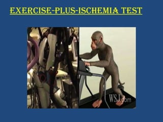
EXERCISE-PLUS-ISCHEMIA TEST
3-4 weeks after the production of MI ↓ Animals – run on a motor –driven treadmill ↓ Work load - ↑every 3 min for total of 18 mins ↓ During last minute-treadmill is stopped-  left circum flex artery- occluded for 2 mins ↓ After 10-20 sec of VF – defibrillation is achieved without any delay by placing large metal plates across animal’s chest. EVALUATION: HR is measured
MECHANICALLY  INDUCED  ARRHYTHMIA PURPOSE AND RATIONALE: Arrhythmias –induced directly by ischemia and reperfusion Coronary artery ligation in anesthetized dog results in: ↑  in HR ↑ in heart contractility ↑  in BP Ventricular arrhythmias
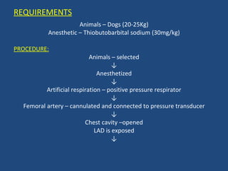
REQUIREMENTS Animals – Dogs (20-25Kg) Anesthetic – Thiobutobarbital sodium (30mg/kg) PROCEDURE: Animals – selected ↓ Anesthetized ↓ Artificial respiration – positive pressure respirator ↓ Femoral artery – cannulated and connected to pressure transducer ↓ Chest cavity –opened LAD is exposed ↓
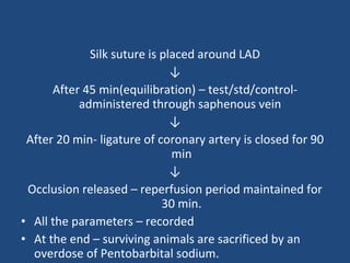
Silk suture is placed around LAD ↓ After 45 min(equilibration) – test/std/control- administered through saphenous vein  ↓ After 20 min- ligature of coronary artery is closed for 90 min ↓ Occlusion released – reperfusion period maintained for 30 min. All the parameters – recorded At the end – surviving animals are sacrificed by an overdose of Pentobarbital sodium.
EVALUATION Mortality Hemodynamics Arrhythmia Ventricular fibrillation % animals with VF.
CONCLUSION Species  differences do exist with respect to factors  that determine arrhythmogenesis and  no animal model will accurately mimic the human suffering of arrhythmia.  Nevertheless, the knowledge gathered from animal studies undoubtedly has been  instrumental in devising diagnostic  and  therapeutic strategies  both in supraventricular and ventricular arrhythmias. In the future, new knowledge will be obtained from experiments performed at many levels.
REFERENCES Vogel.G.H: Drug Discovery and Evaluation (Pharmacological Assays): Second Edition: McGraw- Hill, New York, 2002, Chapter-Cardiovascular activity, A.5, Anti-arrhythmic activity, Page 209-227.   Gupta.S.K: Drug Screening Methods (Pre Clinical Evaluation of New Drug): Second Edition: Jaypee, New Delhi, 2009, Chapter-18, Antiarrhythmics, pg-285-296. Websites: Circ.ahajournals.org Oxfordjournals.org Onlinejacc.org Japphysiology.org