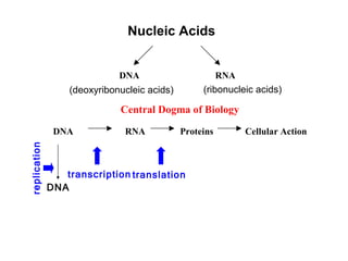
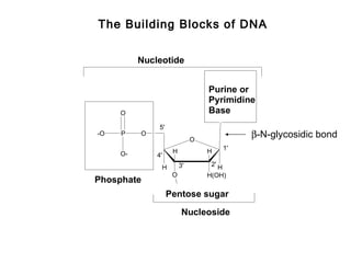
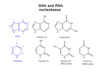
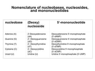
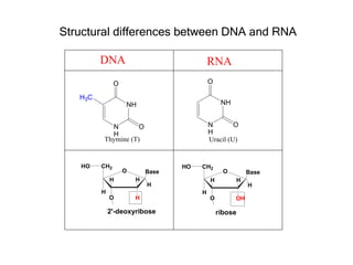
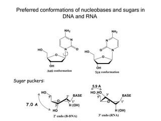
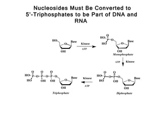
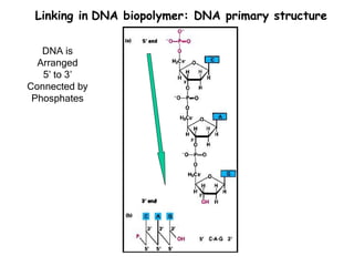
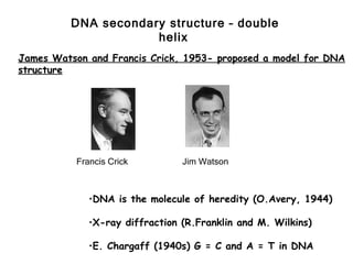
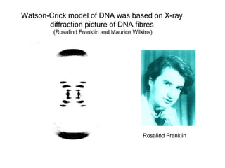
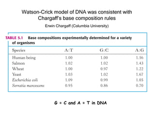
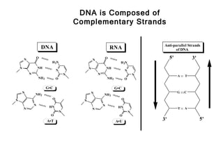
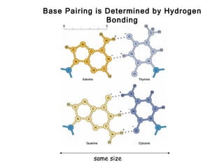
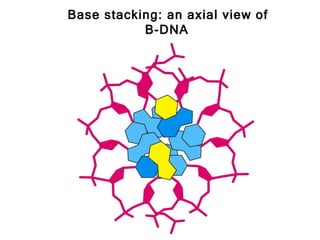
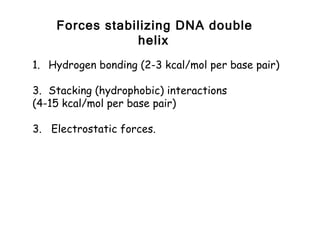
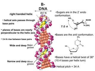
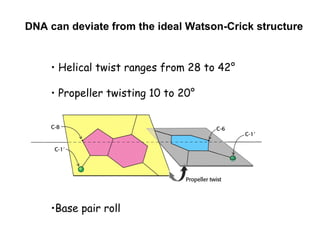
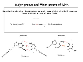
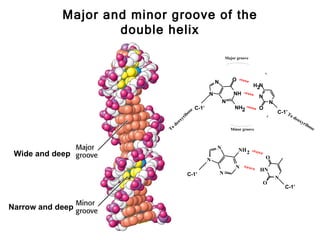
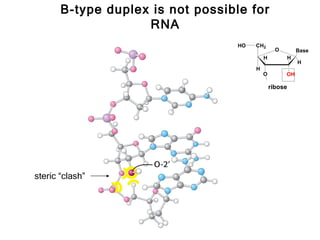
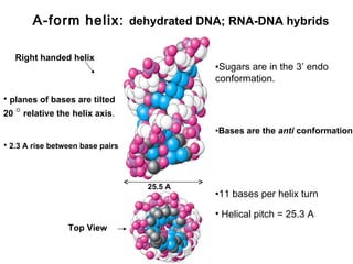
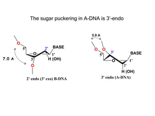
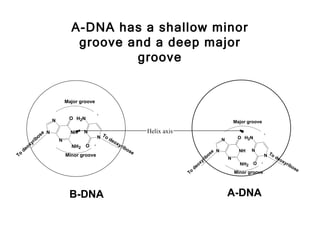
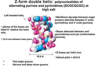
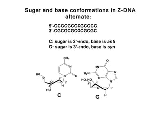
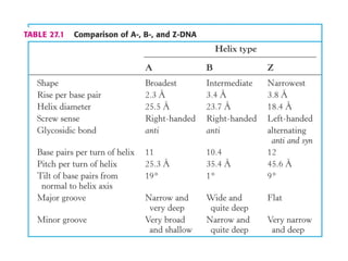
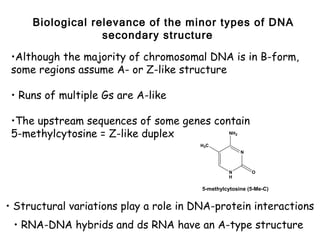
Biochemistry of Medicinals I – Nucleic
Acids
Instructor: Natalia Tretyakova, Ph.D.
760E CCRB (Cancer Center)
Tel. 6-3432
e-mail trety001@umn.edu

Lecture: MWF 2:30-3:20 7-135 WDH

Web page: see “Web enhanced courses”
Chapter 1. DNA Structure.
Required reading: Stryer 5th Edition p. 117-125, 144-146, 152, 745-750,
754-762, 875-877)
 (or Stryer’s Biochemistry 4th edition p. 75-77,80-88, 119-122, 126-128,
787-799, 975-980)
DNA Structure: Chapter outline

1. Biological roles of DNA. Flow of genetic
   information.

3. Primary and secondary structure of DNA.

5. Types of DNA double helix. Sequence-
   specific DNA recognition by proteins.

7. Biophysical properties of DNA.

9. DNA topology. Topoisomerases.

11.Restriction Endonucleases. Molecular Cloning
Nucleic Acids


                             DNA                        RNA
                  (deoxyribonucleic acids)        (ribonucleic acids)

                             Central Dogma of Biology

               DNA            RNA            Proteins         Cellular Action
replication


                 transcription translation
              DNA
Why ?

Questions?

                 •How is genetic information transmitted to progeny cells?
                 •How is DNA synthesis initiated?
                 •What causes DNA defects and what are their biological an
                 physiological consequences?
                 •What causes the differences between cells containing the same
                 genetic information?

Relevance:

                  •Cancer: ex. Xeroderma pigmentosum
                  •Genetic diseases: ex., cystic fibrosis, sickle cell anemia, inborn
        errors of metabolism
                  •Genetic typing: ex., drug metabolism
                  •Rational drug design: ex., antitumor and antimicrobial drugs
                  •Biotechnology: ex., growth hormones
The Building Blocks of DNA


          Nucleotide


                                  Purine or
                                  Pyrimidine
     O                            Base
               5'
-O   P    O
                              O
                                               β-N-glycosidic bond
                                          1'
     O-              H            H
              4'
               H         3'        2' H
                     O            H(OH)
Phosphate
                    Pentose sugar
                          Nucleoside
DNA and RNA
                                        nucleobases
                                               NH2                         O

        7                 6
         N        5           1   N                            N
                              N                      N                           NH
8

                  4           2   N                            N
        9N                N                    N               H
                                                                           N            NH2
         H                        H
                          3
             Purine                   Adenine (A)                  Guanine (G)
                                         NH2                                                  O
                                                                       O
              4
                          3                              H3C
    5                 N                        N                                                   NH
                                                                               NH

    6                     2
              N1                         N           O                                        N            O
                                         H                             N            O
              H                                                        H                      H

        Pyrimidine                     Cytosine (C)                 Thymine (T)               Uracil (U)
                                                                    (DNA only)                (RNA only)
Purine Nucleotides
Pyrimidine Nucleotides
Nomenclature of nucleobases, nucleosides,
                and mononucleotides


nucleobase     (Deoxy)                   5’-mononucleotide
               nucleoside

Adenine (A)    2’-Deoxyadenosine    Deoxyadenosine 5’-monophosphate
               (dA)                 (5’-dAMP)
Guanine (G)    2’- Deoxyguanosine   Deoxyguanosine 5’-monophosphate
               (dG)                 (5’-dGMP)
Thymine (T)    2’- Deoxythymidine   Deoxythymidine 5’-monophosphate
               (dT)                 (5’-dTMP)
Cytosine (C)   2’- Deoxycytidine    Deoxycytidine 5’-monophosphate
               (dC)                 (5’-dCMP)
Uracil (U)     Uridine (U)          Uridine 5’-monophosphate (5’-UMP)
Structural differences between DNA and RNA

         DNA                               RNA
               O                           O

   H3C
                   NH                           NH


           N        O                      N        O
           H                               H
         Thymine (T)                       Uracil (U)


   HO    CH2                      HO   CH2
                   O       Base                  O          Base
             H         H                   H            H
                           H                                H
         H                             H
             O         H                   O         OH

             2'-deoxyribose                    ribose
Preferred conformations of nucleobases and sugars in
                   DNA and RNA

                               NH2                                     NH2

                                      N                           N


             HO                 N          O              O            N
                                                 HO
                          O
                                                              O


                    OH
                                                         OH
                   Anti conformation                    Syn conformation


Sugar puckers:
                                                      5.9 A
              HO                                      HO HO
                                          BASE                    3'         BASE
                    5'           2'                      5'
                           O            1'                        O          1'
     7.0 A                3'          H (OH)
                         HO                                          H (OH)
                         2' endo (B-DNA)                  3' endo (RNA)
Nucleosides Must Be Converted to
5’-Triphosphates to be Part of DNA and
                 RNA

                                               O
                                            HO
           HO                                   P O              Base
                          Base
                   O             Kinase      HO             O
                                 ATP
                 OH                                    OH
                                                  Monophosphate

                                                      ATP       Kinase


   O   O   O                                  O   O
HO P O P O P O                             HO P O P O
                          Base    Kinase                                Base
 HO    OH OH       O                        HO    OH             O
                                   ATP
                 OH                                             OH
           Triphosphate                               Diphosphate
Linking in DNA biopolymer: DNA primary structure

   DNA is
  Arranged
   5’ to 3’
Connected by
 Phosphates
DNA secondary structure – double
                      helix
James Watson and Francis Crick, 1953- proposed a model for DNA
structure


           Francis Crick         Jim Watson


              •DNA is the molecule of heredity (O.Avery, 1944)

              •X-ray diffraction (R.Franklin and M. Wilkins)

              •E. Chargaff (1940s) G = C and A = T in DNA
Watson-Crick model of DNA was based on X-ray
       diffraction picture of DNA fibres
        (Rosalind Franklin and Maurice Wilkins)


                                           Rosalind Franklin
Watson-Crick model of DNA was consistent with
      Chargaff’s base composition rules
         Erwin Chargaff (Columbia University)


              G = C and A = T in DNA
DNA is Composed of
                                             Complementary Strands


                                                                                             Anti-parallel Strands
                     DNA                                            RNA                            of DNA

        N
                O
                                                       N
                                                               O                                 5'            3'
                             H 2N                                           H 2N
    N               NH                             N               NH
                                 N                                              N
            N                            N                 N                            N             A :: T
                    NH 2         O                                 NH 2         O

                           G•C                                            G•C
        N                                              N                                              G :::C
                NH 2                                           NH 2
N                            O                                              O
                                               N
                N                                              N
                            HN                                             HN                         T :: A
        N                                              N
                                     N                                              N
                             O                                              O
                           A•T                                            A•U               3'                      5'
Base Pairing is Determined by Hydrogen
                Bonding


            same size
Base stacking: an axial view of
           B-DNA
Forces stabilizing DNA double
                helix
1. Hydrogen bonding (2-3 kcal/mol per base pair)

3. Stacking (hydrophobic) interactions
(4-15 kcal/mol per base pair)

3. Electrostatic forces.
B-
                                   DNA
                                   23.7 A    •Sugars are in the 2’ endo
        right handed helix
                                            conformation.
                                                          HO
 • helical axis passes through                                 5'            2'     BASE

 base pairs                                                          O              1'
                                                                    3'            H (OH)
                                                 7.0 A
                                                                    HO
• planes of bases are nearly
perpendicular to the helix axis.
                                            •Bases are the anti conformation.
                                                                     NH2
• 3.4 A rise between base pairs
                                                                         N


        Wide and deep                                HO              N       O
                                                                O


                                                           OH

                                            •Bases have a helical twist of 36º
      Narrow and deep                       (10.4 bases per helix turn)

                                            • Helical pitch = 34 A
DNA can deviate from the ideal Watson-Crick structure


    • Helical twist ranges from 28 to 42°

    • Propeller twisting 10 to 20°


    •Base pair roll
Major groove and Minor groove of DNA

Hypothetical situation: the two grooves would have similar size if dR residues
                     were attached at 180° to each other


                    To deoxyribose-C1’                    Base       Base                   C1’ -To deoxyribose


                                   Major groove                                                          Major groove


                                                                                                     N       NH 2
                                       O
                               N
                                                  H 2N                                                                   O
                                                                                                 N
                           N            NH                                                                  N
                                                      N                                                                 HN
                                   N                                                  C-1’           N
                                                            N                                                                N
                    C-1’                 NH 2         O                                                                 O
                   e                                             C-1’                                                            C-1’
                 os                                                 To
              rib                                                      d   eox
         xy                                                                   yri
     deo                                                                            bo
                                                                                       se                Minor groove
To
                                       Minor groove
Major and minor groove of the
                    double helix

                                                                       Major groove


                                                               N          O
                                                                                      H2N
                                                           N               NH
                                                                                        N
                                                                   N                            N
                                                  e C-1’                    NH2         O
                                               os                                                   C-1’T
                                        y   rib                                                             od
                                                                                                                 eox
                                      ox                                                                            yri
                                 de                                                                                     bo
                            To                                           Minor groove                                     se


                                                               N              NH 2
 Wide and deep                                                                              O
                                                           N
                                                                            N
                                                                                        HN
                                             C-1’                  N
                                                                                                    N
                                                                                            O
                                                                                                        C-1’


Narrow and deep
B-type duplex is not possible for
                    RNA
                                   HO   CH2
                                                  O          Base
                                            H            H
                                                             H
                                        H
                                            O         OH

                                                ribose


steric “clash”
A-form helix: dehydrated DNA; RNA-DNA hybrids

   Right handed helix
                                           •Sugars are in the 3’ endo
                                           conformation.

• planes of bases are tilted
20 ° relative the helix axis.
                                           •Bases are the anti conformation.
• 2.3 A rise between base pairs


                                  25.5 A
                                           •11 bases per helix turn

                                           • Helical pitch = 25.3 A
                 Top View
The sugar puckering in A-DNA is 3’-endo


                                           5.9 A

        O
            5'             2'     BASE
                                            O     O
                    O                                  3'          BASE
                                  1'            5'
7.0 A              3'           H (OH)                             1'
                                                        O
                   O                                          2'
                                                              H (OH)
                 2' endo (3' exo) B-DNA            3' endo (A-DNA)
A-DNA has a shallow minor
                                       groove and a deep major
                                               groove


                                    Major groove


                            N        O H2N
                                                                                                                      Major groove


                  os
                       eN             NH•   N
                                                   N To d
                                                                      Helix axis                                         •
                                                                                                                       O H2N
                ib              N                        eo                                                   N
              yr                                           xy
         e ox                         NH2   O                rib
                                                                os
     d                                                            e                                   e   N             NH    N
To                                  Minor groove                                                   os                                N To d
                                                                                                 ib               N                         eo
                                                                                              yr                                               xy
                                                                                           ox                           NH2   O                  rib
                                                                                        de                                                             os
                                                                                                                                                         e
                                                                                   To                                 Minor groove


                                     B-DNA                                                                        A-DNA
Z-form double helix: polynucleotides of
        alternating purines and pyrimidines (GCGCGCGC) at
                              high salt
   Left handed helix
                                           • Backbone zig-zags because sugar
                                           puckers alternate between 2’ endo
                                           pyrimidines and 3’ endo (purines)
• planes of the bases are
tilted 9° relative the helix
axis.                                      • Bases alternate between anti
                                           (pyrimidines) and syn conformation
                                           (purines).
• 3.8 A rise between base pairs


                                           •12 bases per helix turn
                                  18.4 A   • Helical pitch = 45.6 A
  •        Flat major groove
  •        Narrow and deep minor groove
Sugar and base conformations in Z-DNA
             alternate:
              5’-GCGCGCGCGCGCG
              3’-CGCGCGCGCGCGC

              C: sugar is 2’-endo, base is anti
              G: sugar is 3’-endo, base is syn

                        NH2
                                                       O

                                 N               HN

                                          H2N                   N
   HO
                        N            O
        5'         2'                              N
                                         HO HO
              O             1'                   3'        N
                                            5'
             3'         H
                                                  O        1'
             HO

                  C                              G     H
Biological relevance of the minor types of DNA
                  secondary structure

 •Although the majority of chromosomal DNA is in B-form,
 some regions assume A- or Z-like structure

 • Runs of multiple Gs are A-like

 •The upstream sequences of some genes contain
 5-methylcytosine = Z-like duplex         NH          2


                                       H3C
                                                          N


                                                  N           O
                                                  H

                                        5-methylcytosine (5-Me-C)


• Structural variations play a role in DNA-protein interactions
 • RNA-DNA hybrids and ds RNA have an A-type structure

Dna and rna

  • 1.
    Biochemistry of MedicinalsI – Nucleic Acids Instructor: Natalia Tretyakova, Ph.D. 760E CCRB (Cancer Center) Tel. 6-3432 e-mail trety001@umn.edu Lecture: MWF 2:30-3:20 7-135 WDH Web page: see “Web enhanced courses”
  • 3.
    Chapter 1. DNAStructure. Required reading: Stryer 5th Edition p. 117-125, 144-146, 152, 745-750, 754-762, 875-877) (or Stryer’s Biochemistry 4th edition p. 75-77,80-88, 119-122, 126-128, 787-799, 975-980)
  • 4.
    DNA Structure: Chapteroutline 1. Biological roles of DNA. Flow of genetic information. 3. Primary and secondary structure of DNA. 5. Types of DNA double helix. Sequence- specific DNA recognition by proteins. 7. Biophysical properties of DNA. 9. DNA topology. Topoisomerases. 11.Restriction Endonucleases. Molecular Cloning
  • 5.
    Nucleic Acids DNA RNA (deoxyribonucleic acids) (ribonucleic acids) Central Dogma of Biology DNA RNA Proteins Cellular Action replication transcription translation DNA
  • 6.
    Why ? Questions? •How is genetic information transmitted to progeny cells? •How is DNA synthesis initiated? •What causes DNA defects and what are their biological an physiological consequences? •What causes the differences between cells containing the same genetic information? Relevance: •Cancer: ex. Xeroderma pigmentosum •Genetic diseases: ex., cystic fibrosis, sickle cell anemia, inborn errors of metabolism •Genetic typing: ex., drug metabolism •Rational drug design: ex., antitumor and antimicrobial drugs •Biotechnology: ex., growth hormones
  • 7.
    The Building Blocksof DNA Nucleotide Purine or Pyrimidine O Base 5' -O P O O β-N-glycosidic bond 1' O- H H 4' H 3' 2' H O H(OH) Phosphate Pentose sugar Nucleoside
  • 8.
    DNA and RNA nucleobases NH2 O 7 6 N 5 1 N N N N NH 8 4 2 N N 9N N N H N NH2 H H 3 Purine Adenine (A) Guanine (G) NH2 O O 4 3 H3C 5 N N NH NH 6 2 N1 N O N O H N O H H H Pyrimidine Cytosine (C) Thymine (T) Uracil (U) (DNA only) (RNA only)
  • 9.
  • 10.
  • 11.
    Nomenclature of nucleobases,nucleosides, and mononucleotides nucleobase (Deoxy) 5’-mononucleotide nucleoside Adenine (A) 2’-Deoxyadenosine Deoxyadenosine 5’-monophosphate (dA) (5’-dAMP) Guanine (G) 2’- Deoxyguanosine Deoxyguanosine 5’-monophosphate (dG) (5’-dGMP) Thymine (T) 2’- Deoxythymidine Deoxythymidine 5’-monophosphate (dT) (5’-dTMP) Cytosine (C) 2’- Deoxycytidine Deoxycytidine 5’-monophosphate (dC) (5’-dCMP) Uracil (U) Uridine (U) Uridine 5’-monophosphate (5’-UMP)
  • 12.
    Structural differences betweenDNA and RNA DNA RNA O O H3C NH NH N O N O H H Thymine (T) Uracil (U) HO CH2 HO CH2 O Base O Base H H H H H H H H O H O OH 2'-deoxyribose ribose
  • 13.
    Preferred conformations ofnucleobases and sugars in DNA and RNA NH2 NH2 N N HO N O O N HO O O OH OH Anti conformation Syn conformation Sugar puckers: 5.9 A HO HO HO BASE 3' BASE 5' 2' 5' O 1' O 1' 7.0 A 3' H (OH) HO H (OH) 2' endo (B-DNA) 3' endo (RNA)
  • 14.
    Nucleosides Must BeConverted to 5’-Triphosphates to be Part of DNA and RNA O HO HO P O Base Base O Kinase HO O ATP OH OH Monophosphate ATP Kinase O O O O O HO P O P O P O HO P O P O Base Kinase Base HO OH OH O HO OH O ATP OH OH Triphosphate Diphosphate
  • 15.
    Linking in DNAbiopolymer: DNA primary structure DNA is Arranged 5’ to 3’ Connected by Phosphates
  • 16.
    DNA secondary structure– double helix James Watson and Francis Crick, 1953- proposed a model for DNA structure Francis Crick Jim Watson •DNA is the molecule of heredity (O.Avery, 1944) •X-ray diffraction (R.Franklin and M. Wilkins) •E. Chargaff (1940s) G = C and A = T in DNA
  • 17.
    Watson-Crick model ofDNA was based on X-ray diffraction picture of DNA fibres (Rosalind Franklin and Maurice Wilkins) Rosalind Franklin
  • 18.
    Watson-Crick model ofDNA was consistent with Chargaff’s base composition rules Erwin Chargaff (Columbia University) G = C and A = T in DNA
  • 19.
    DNA is Composedof Complementary Strands Anti-parallel Strands DNA RNA of DNA N O N O 5' 3' H 2N H 2N N NH N NH N N N N N N A :: T NH 2 O NH 2 O G•C G•C N N G :::C NH 2 NH 2 N O O N N N HN HN T :: A N N N N O O A•T A•U 3' 5'
  • 20.
    Base Pairing isDetermined by Hydrogen Bonding same size
  • 21.
    Base stacking: anaxial view of B-DNA
  • 22.
    Forces stabilizing DNAdouble helix 1. Hydrogen bonding (2-3 kcal/mol per base pair) 3. Stacking (hydrophobic) interactions (4-15 kcal/mol per base pair) 3. Electrostatic forces.
  • 23.
    B- DNA 23.7 A •Sugars are in the 2’ endo right handed helix conformation. HO • helical axis passes through 5' 2' BASE base pairs O 1' 3' H (OH) 7.0 A HO • planes of bases are nearly perpendicular to the helix axis. •Bases are the anti conformation. NH2 • 3.4 A rise between base pairs N Wide and deep HO N O O OH •Bases have a helical twist of 36º Narrow and deep (10.4 bases per helix turn) • Helical pitch = 34 A
  • 24.
    DNA can deviatefrom the ideal Watson-Crick structure • Helical twist ranges from 28 to 42° • Propeller twisting 10 to 20° •Base pair roll
  • 25.
    Major groove andMinor groove of DNA Hypothetical situation: the two grooves would have similar size if dR residues were attached at 180° to each other To deoxyribose-C1’ Base Base C1’ -To deoxyribose Major groove Major groove N NH 2 O N H 2N O N N NH N N HN N C-1’ N N N C-1’ NH 2 O O e C-1’ C-1’ os To rib d eox xy yri deo bo se Minor groove To Minor groove
  • 26.
    Major and minorgroove of the double helix Major groove N O H2N N NH N N N e C-1’ NH2 O os C-1’T y rib od eox ox yri de bo To Minor groove se N NH 2 Wide and deep O N N HN C-1’ N N O C-1’ Narrow and deep
  • 27.
    B-type duplex isnot possible for RNA HO CH2 O Base H H H H O OH ribose steric “clash”
  • 28.
    A-form helix: dehydratedDNA; RNA-DNA hybrids Right handed helix •Sugars are in the 3’ endo conformation. • planes of bases are tilted 20 ° relative the helix axis. •Bases are the anti conformation. • 2.3 A rise between base pairs 25.5 A •11 bases per helix turn • Helical pitch = 25.3 A Top View
  • 29.
    The sugar puckeringin A-DNA is 3’-endo 5.9 A O 5' 2' BASE O O O 3' BASE 1' 5' 7.0 A 3' H (OH) 1' O O 2' H (OH) 2' endo (3' exo) B-DNA 3' endo (A-DNA)
  • 30.
    A-DNA has ashallow minor groove and a deep major groove Major groove N O H2N Major groove os eN NH• N N To d Helix axis • O H2N ib N eo N yr xy e ox NH2 O rib os d e e N NH N To Minor groove os N To d ib N eo yr xy ox NH2 O rib de os e To Minor groove B-DNA A-DNA
  • 31.
    Z-form double helix:polynucleotides of alternating purines and pyrimidines (GCGCGCGC) at high salt Left handed helix • Backbone zig-zags because sugar puckers alternate between 2’ endo pyrimidines and 3’ endo (purines) • planes of the bases are tilted 9° relative the helix axis. • Bases alternate between anti (pyrimidines) and syn conformation (purines). • 3.8 A rise between base pairs •12 bases per helix turn 18.4 A • Helical pitch = 45.6 A • Flat major groove • Narrow and deep minor groove
  • 32.
    Sugar and baseconformations in Z-DNA alternate: 5’-GCGCGCGCGCGCG 3’-CGCGCGCGCGCGC C: sugar is 2’-endo, base is anti G: sugar is 3’-endo, base is syn NH2 O N HN H2N N HO N O 5' 2' N HO HO O 1' 3' N 5' 3' H O 1' HO C G H
  • 34.
    Biological relevance ofthe minor types of DNA secondary structure •Although the majority of chromosomal DNA is in B-form, some regions assume A- or Z-like structure • Runs of multiple Gs are A-like •The upstream sequences of some genes contain 5-methylcytosine = Z-like duplex NH 2 H3C N N O H 5-methylcytosine (5-Me-C) • Structural variations play a role in DNA-protein interactions • RNA-DNA hybrids and ds RNA have an A-type structure

Editor's Notes

  • #12 In addition to these common bases, unusual nucleosides exist. Involved in gene regulation, Can be modified – 6-methyladenine, 5-methylcytosine As we will discuss in biochem 2 nucleosides have many other physiological functions dTMP = TMP
  • #19 Base composition of a given organism is independent of the cell type
  • #21 Only two types of base pairs can be accommodated without altering the positions of the sugars.
  • #22 Each base is rotated 36  relative to its neighbor base (like a spiral staircase), so that 10 base pairs correspond to 1 helix turn.
  • #23 Cooperative forces: each contributes a little, but adds up because DNA chains can be millions od nculeotides long.
  • #27 .