1. Elastic fibers are key components of connective tissue that provide resilience and elasticity to skin. Disorders impacting elastic fibers can produce changes in skin appearance and structure.
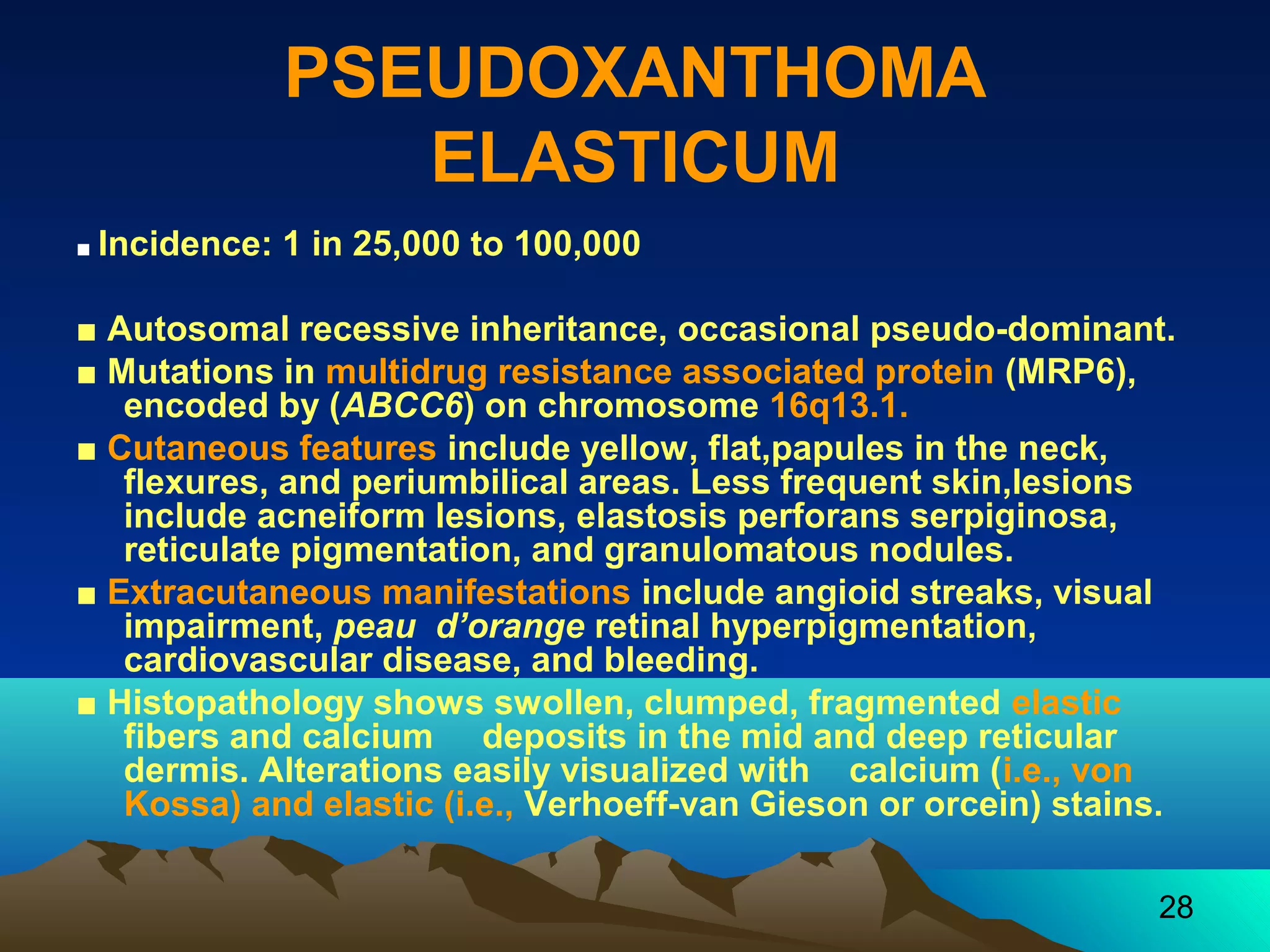
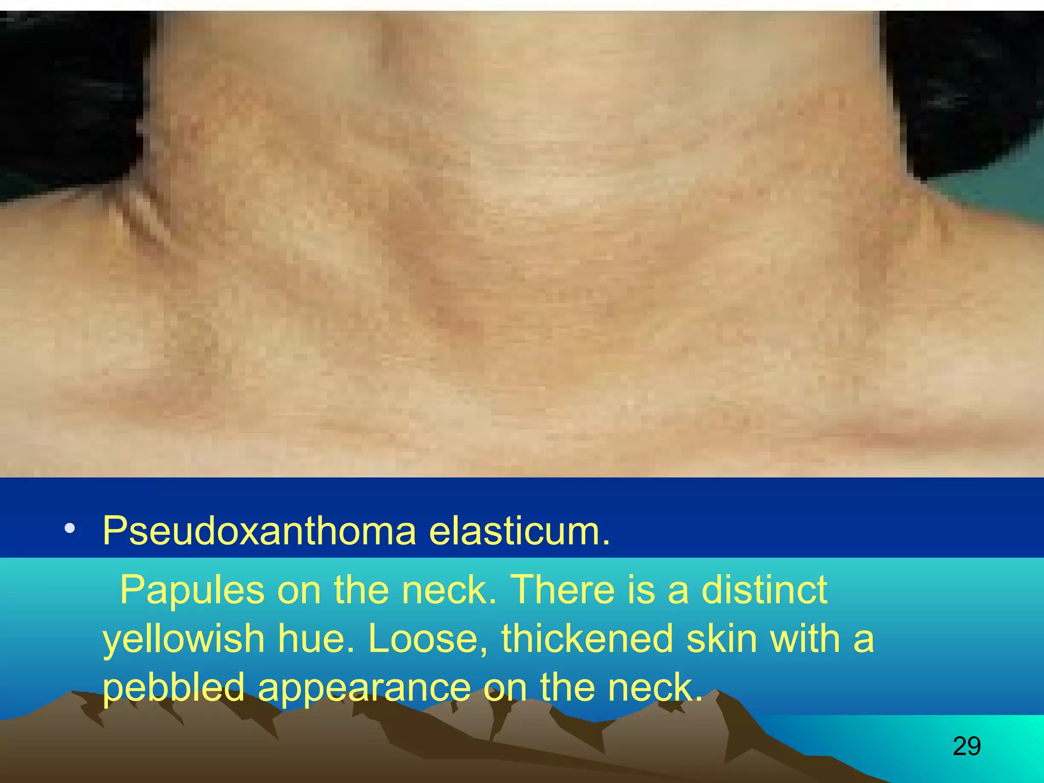
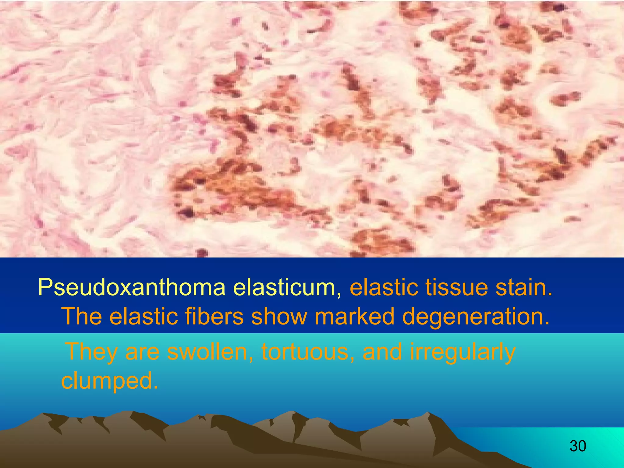
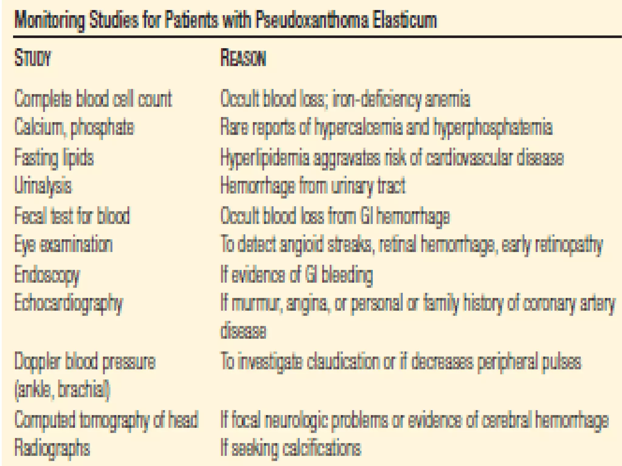
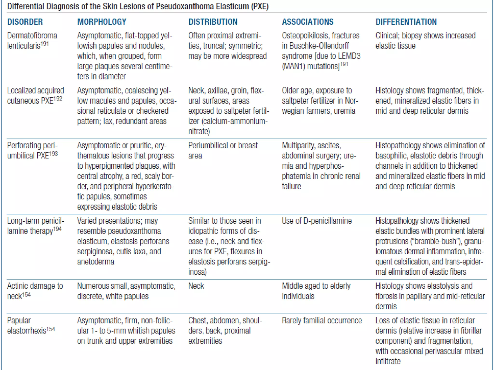
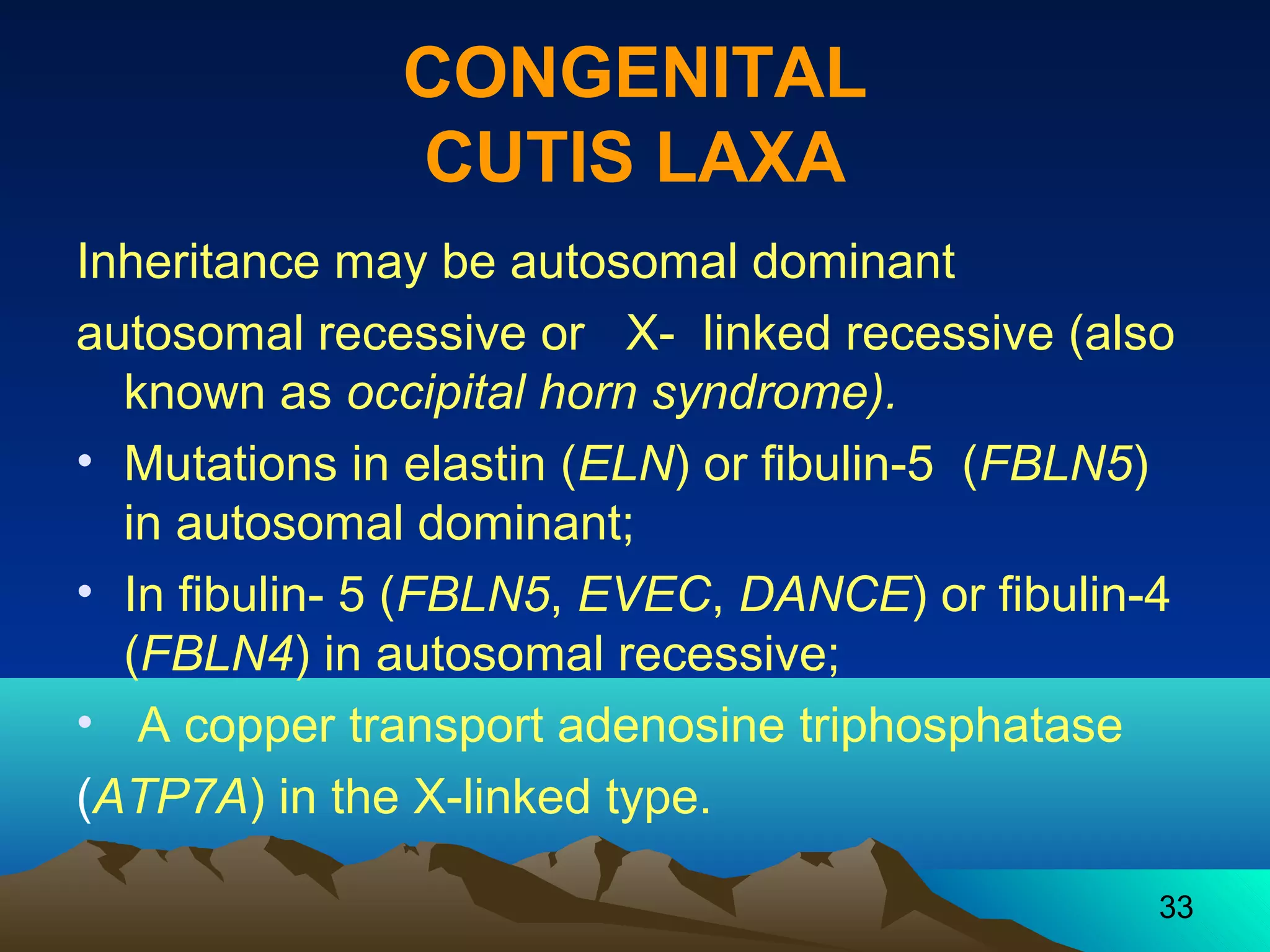
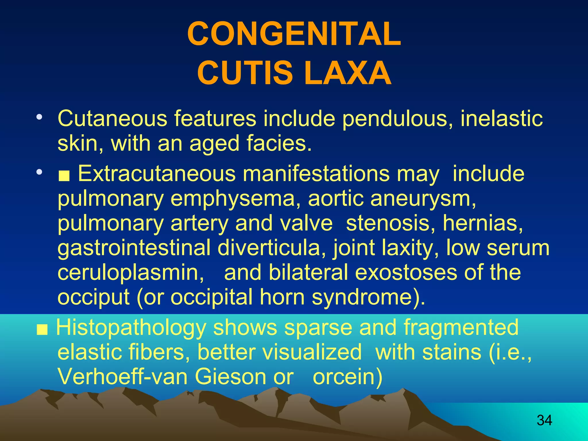
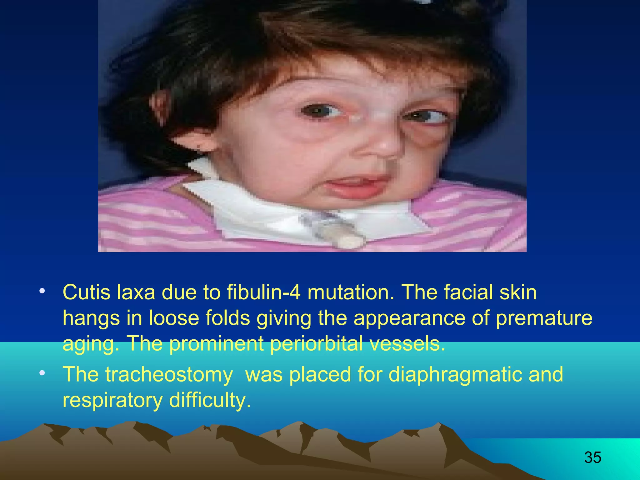
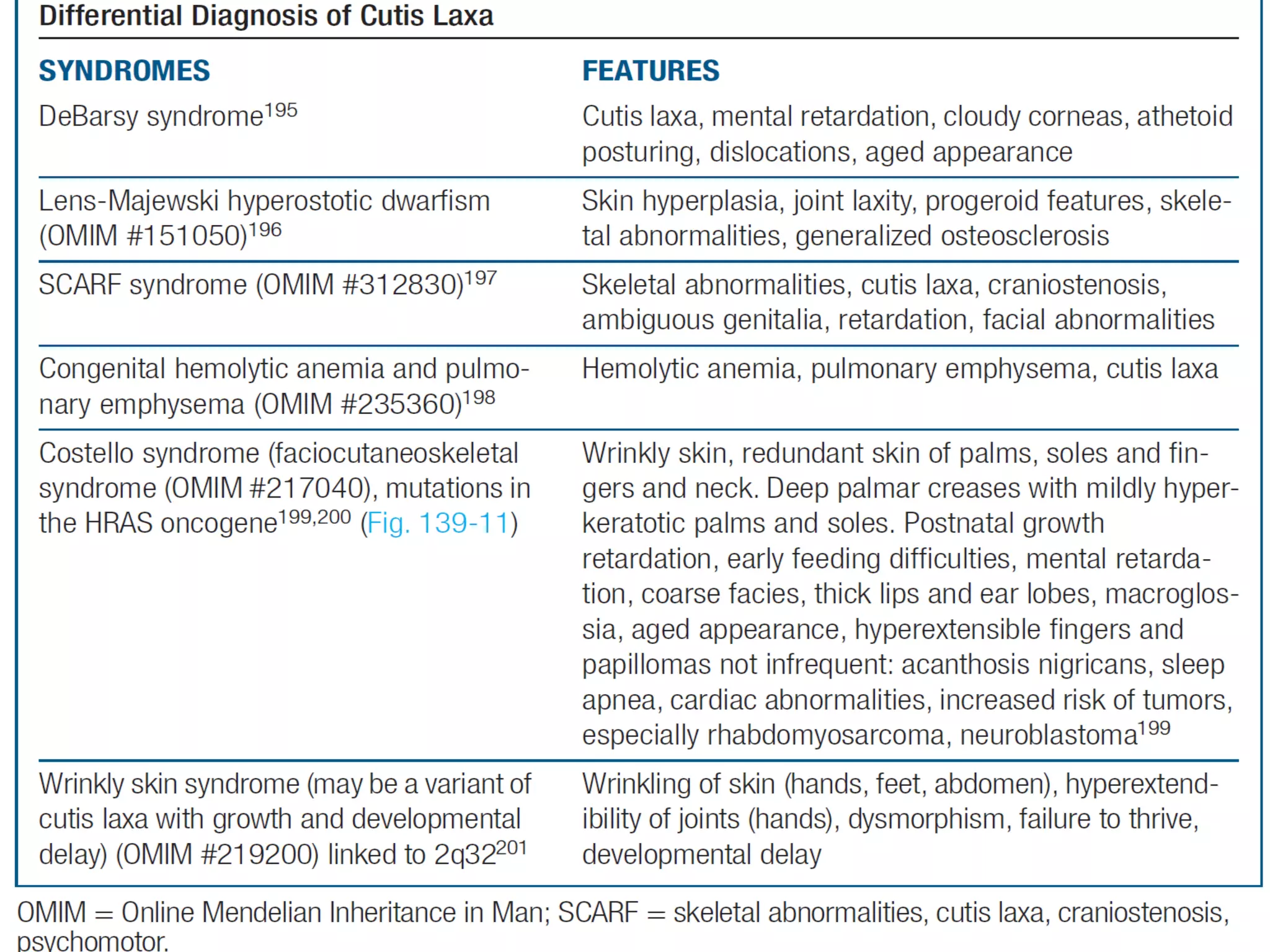
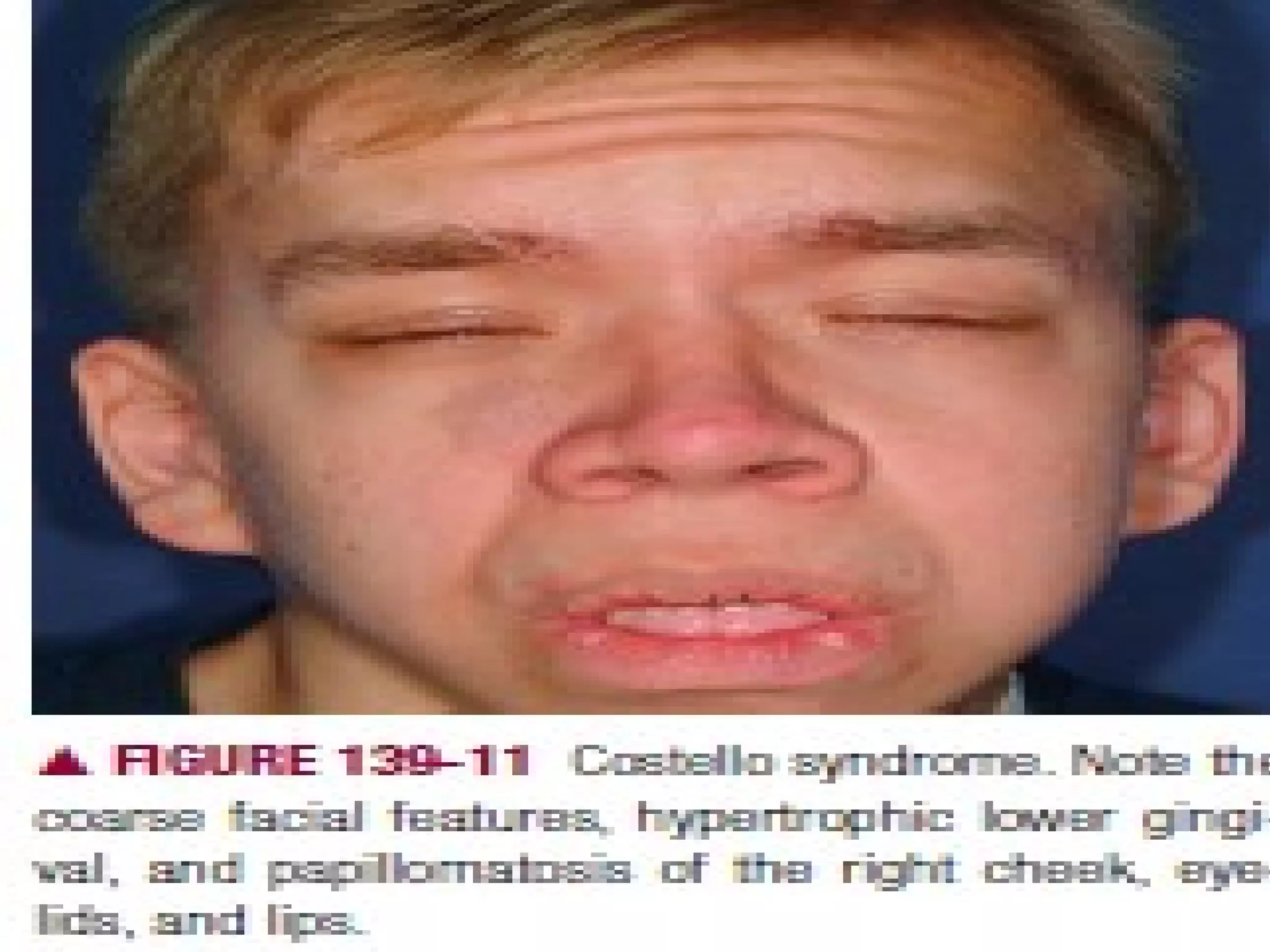
2. The document describes several such disorders, including pseudoxanthoma elasticum where elastic fibers are abnormally clumped and fragmented, and congenital cutis laxa where elastic fibers are sparse and fragmented leading to loose, inelastic skin.
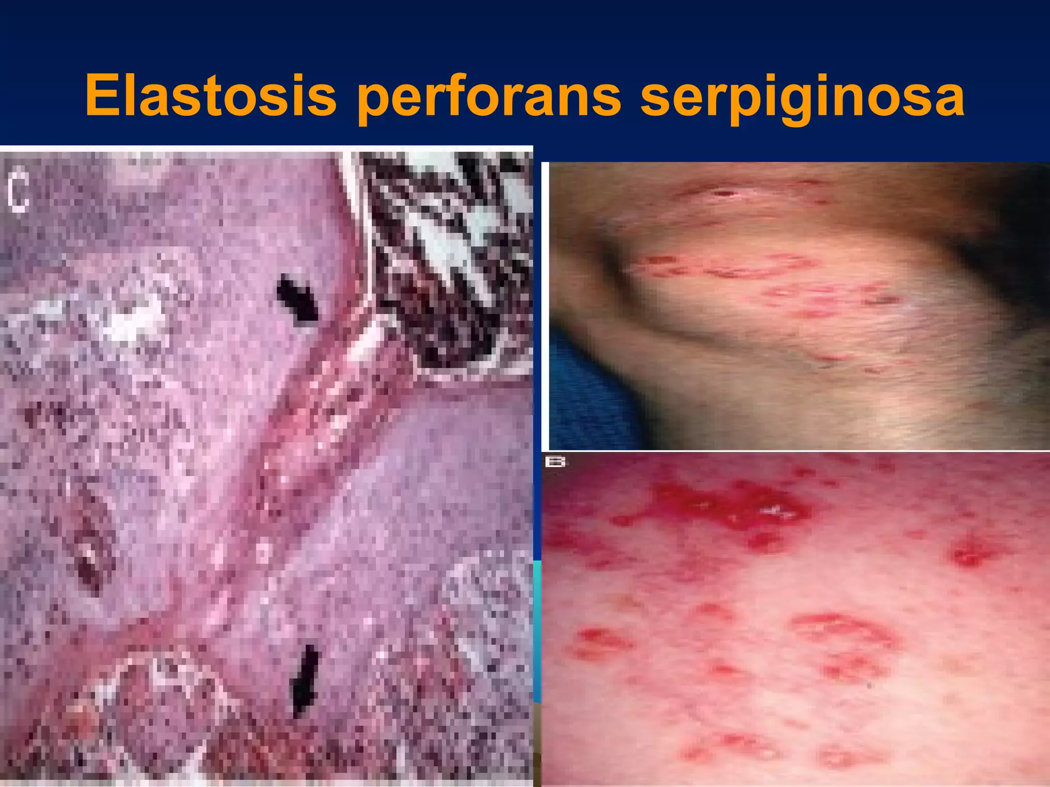
3. Elastosis perforans serpiginosa is characterized by the extrusion of abnormal elastic fibers through the epidermis, often triggered by dermal irritation or D-penicillamine use.


![Extracellular matrix [autosaved]](https://cdn.slidesharecdn.com/ss_thumbnails/extracellularmatrixautosaved-210313065413-thumbnail.jpg?width=640&height=640&fit=bounds)


