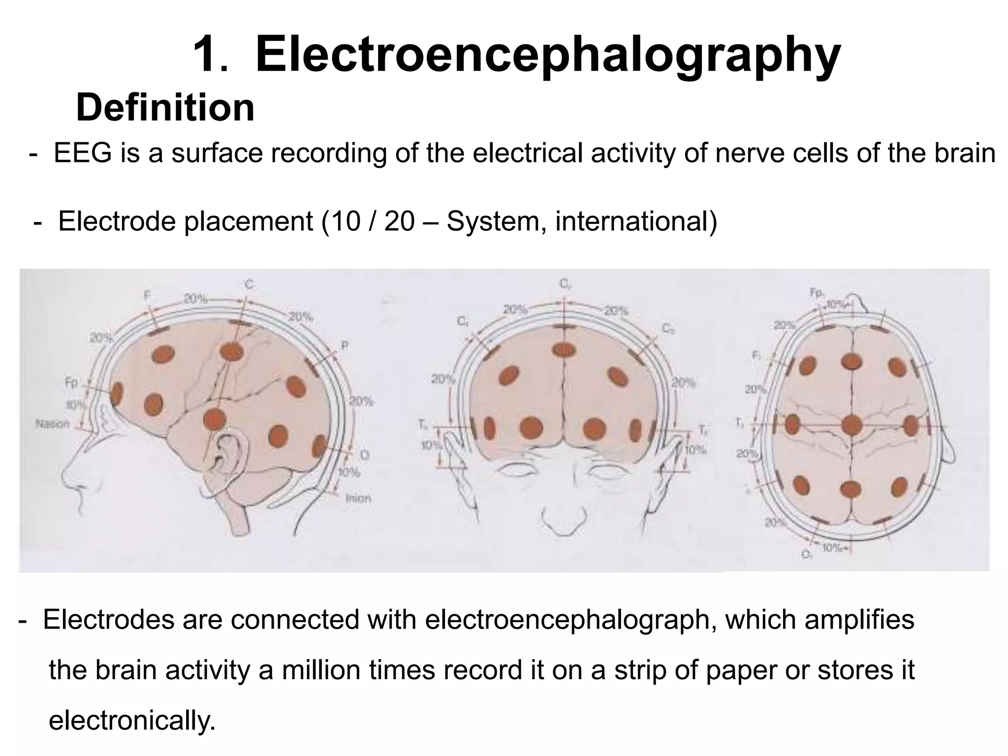
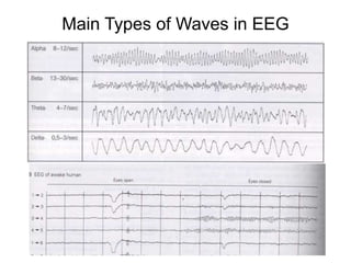
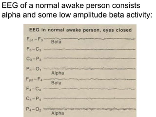
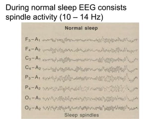
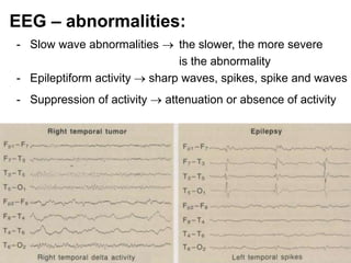
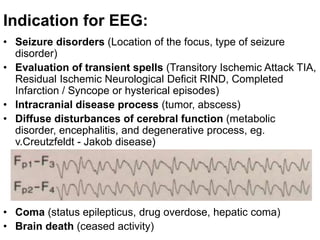
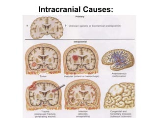
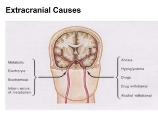
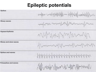
1. Electroencephalography
Definition
- EEG is a surface recording of the electrical activity of nerve cells of the brain
- Electrode placement (10 / 20 – System, international)
- Electrodes are connected with electroencephalograph, which amplifies
the brain activity a million times record it on a strip of paper or stores it
electronically.
Main Types of Waves in EEG
EEG of a normal awake person consists
alpha and some low amplitude beta activity:
During normal sleep EEG consists
spindle activity (10 – 14 Hz)
EEG – abnormalities:
- Slow wave abnormalities  the slower, the more severe
is the abnormality
- Epileptiform activity  sharp waves, spikes, spike and waves
- Suppression of activity  attenuation or absence of activity
Indication for EEG:
• Seizure disorders (Location of the focus, type of seizure
disorder)
• Evaluation of transient spells (Transitory Ischemic Attack TIA,
Residual Ischemic Neurological Deficit RIND, Completed
Infarction / Syncope or hysterical episodes)
• Intracranial disease process (tumor, abscess)
• Diffuse disturbances of cerebral function (metabolic
disorder, encephalitis, and degenerative process, eg.
v.Creutzfeldt - Jakob disease)
• Coma (status epilepticus, drug overdose, hepatic coma)
• Brain death (ceased activity)
Intracranial Causes:
Extracranial Causes
Simple Partial Seizures
Complex Partial Seizures
Epileptic potentials
Epileptic Focus
NMDA = N-methyl – D – aspartate
AMPA = α – amino – 3 – hydroxy – 5 – methylisoxazole – 4 – propionate

Diagrams-Eeg-Epilepsy.ppt

  • 1.
    1. Electroencephalography Definition - EEGis a surface recording of the electrical activity of nerve cells of the brain - Electrode placement (10 / 20 – System, international) - Electrodes are connected with electroencephalograph, which amplifies the brain activity a million times record it on a strip of paper or stores it electronically.
  • 2.
    Main Types ofWaves in EEG
  • 3.
    EEG of anormal awake person consists alpha and some low amplitude beta activity:
  • 4.
    During normal sleepEEG consists spindle activity (10 – 14 Hz)
  • 5.
    EEG – abnormalities: -Slow wave abnormalities  the slower, the more severe is the abnormality - Epileptiform activity  sharp waves, spikes, spike and waves - Suppression of activity  attenuation or absence of activity
  • 6.
    Indication for EEG: •Seizure disorders (Location of the focus, type of seizure disorder) • Evaluation of transient spells (Transitory Ischemic Attack TIA, Residual Ischemic Neurological Deficit RIND, Completed Infarction / Syncope or hysterical episodes) • Intracranial disease process (tumor, abscess) • Diffuse disturbances of cerebral function (metabolic disorder, encephalitis, and degenerative process, eg. v.Creutzfeldt - Jakob disease) • Coma (status epilepticus, drug overdose, hepatic coma) • Brain death (ceased activity)
  • 7.
  • 8.
  • 9.
  • 10.
  • 12.
  • 14.
  • 15.
    NMDA = N-methyl– D – aspartate AMPA = α – amino – 3 – hydroxy – 5 – methylisoxazole – 4 – propionate