1. 101 級牙放共筆 W5-1
Normal Radiographic Anatomy
● 牙齒(Teeth)
a. 牙釉質:96%的礦物質化,對 X-ray 有最多的稀釋(衰減,attenuation),故
呈現最明顯且均勻的 Radiopaque。
b. 牙本質:75%(70%)的礦物質化,曝光程度跟骨頭相近,但牙本質較平滑
(smooth)且均質(homogeneous)。
c. 牙骨質:50%的礦物質化,但因為太薄而且和牙本質的對比不高,故在片
子上很難分辨出來
鄰接面蛀牙可以 tracing 連續性
● 齒頸部照蝕(Cervical burn-out):
a. 定義:常出現在Mesial和Distal的CEJ處,多呈圈狀(collar)或楔型(wedge),
模糊的不透線區域
b. “an ill-defined wedge-shaped radiolucency on the mesial or distal root
surfaces near the CEJ, result in decreased x-ray absorption in the areas in
queation”
並非 caries,是廣泛性的出現。臨床上要辨別是 caries 或是 cervical
burn-out 可用探針去勾
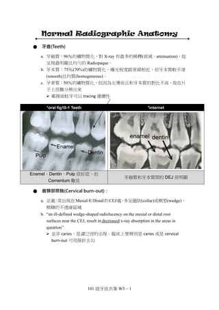
*oral fig10-1 Teeth *internet
Enamel、Dentin、Pulp 很好認,但
Cementum 難見
牙釉質和牙本質間的 DEJ 很明顯
2. 101 級牙放共筆 W5-2
*oral fig10-2 Cervical burn-out
*internet Cervical
burn-out
介於 enamel 和 alveolar crest 間的過曝區域 前牙也可見到這種現象
c. 原理:” Because of the difference in densities of adjacent tissues.” 相較於其
上下區域,齒頸部所含的組織量較少,故 X-ray 通過這個緊縮的區域時,
被稀釋的程度較低而呈現 radiolucent。
d. 齒頸部照蝕是人為因素所造成的正常現象,在所有牙齒都有可能發生。臨
床上要避免和 Caries 混淆。
*essential fig21.7A *essential fig21.6A
Cervical burn-out 示意圖
【小余】:Cervical burn-out 有時會連 crest 也黑掉,導致無法正確判讀骨頭的高度。
如有需要,可以調低劑量再照看看。
【國考】99-2
下列關於齒頸部照蝕(cervical burn-out)之敘述何者錯誤?
(A) 為周界明顯的 X 光透性陰影(radiolucent shadow)
(B) 發生的原因是牙釉質與齒槽骨的放射線不透性(radiopaque)較齒頸部
高
(C) 並非齒頸部蛀牙
(D) 前牙及後牙都會發生 Ans: (A)
11. 101 級牙放共筆 W5-11
b. 鼻中膈的放射影像往往比預期的來的更寬大,是因為重疊了其它解剖構造
(EX:梨骨、鼻軟骨)。此外也常見鼻中膈偏離正中線;梨骨彎曲等現象。
c. 依照射角度方向的不同,鼻孔骨性下緣的白線也可能出現在 canine 上方,
甚至是與上頷竇重疊。這種狀況往往會很難由單一張 X 光片去判斷兩個構造
間的相對關係。
*oral fig10-21 floor of nasal aperture
*oral fig10-22 floor of nasal
aperture
側門齒和犬齒的上方常常可以看到鼻
孔底部造成的不透光影像(這張是由前
往後拍)
與上頷竇重疊,易使人誤判相對關
係(這張是由後往前拍)
TCKJ 提醒您:一般來說 nasal fossa
會比 sinus 高
正中縫線(Median suture = Intermaxillary suture)
a. 定義:由前牙的口內根尖片觀察,可見一條 radiolucent 細黑線自#11 和#21
間的 crest 中點向上走,經前鼻棘(Anterior nasal spine)、上頷腭突(Maxillary
palatine process)到硬腭後板。
b. 其位於 crest 的開口可能呈圓形或 V 字型的擴大。
c. 能否照出 Middle suture 取決於解剖構造的型態和 X-ray 照射的角度。
前鼻棘(Anterior nasal spine)
a. 定義:前牙口內根尖片最常照出的解剖構造,V 字型、Radiopaque 的骨性
構造。
The radiopaque pattern of anterior nasal spine is usually V shape.
b. 位於正中線上,距離正中門齒 Crest 約 1.5~2.0 cm,即鼻中膈與骨性鼻孔下
緣的交界處。
12. 101 級牙放共筆 W5-12
*oral fig10-16 intermaxillary suture *slide 20、21
正中縫線與前鼻棘 骨性解剖圖
門齒孔(Incisive foramen=nasopalatine foramen =anterior palatine foramen)
a. 定義:位於腭板上、門齒後方的鼻腭管開口,其中有支配供應上頷正中門齒
的鼻顎神經血管。
*oral fig10-23 A incisive foramen *oral fig10-23 B incisive foramen
出現在正中門齒牙根的中 1/3 到根尖
1/3 的 radiolucent
注意這張門齒孔的邊界有些微擴散,但
仍在安全範圍內
b. 門齒孔的大小、位置、形狀都因人而異,但大多出現在正中門齒牙根的中 1/3
到根尖 1/3 處。
c. 臨床上具重要意義,因它可能形成 cyst 的潛在病發部位,若 radiolucent 的
區域寬度超過 1cm,或者擴張到牙根之下時,就要特別注意。
The presence of an incisive canal cyst is presumed if the width of the
incisive foramen exceeds 1mm or if enlargement can be demonstrated on
successive radiographs.
13. 101 級牙放共筆 W5-13
鼻腭管(Nasopalatine canal)
a. 定義:源自鼻腔底部的兩個孔洞,上部開口於鼻中膈兩側、接近鼻腔前下緣
處,之後 canals 往前下方走,最後匯集並開口於門齒孔。
*oral fig10-24 A 鼻腭管管壁 *oral fig10-25 鼻腭管開口-門齒孔
自鼻腔底部延伸至門齒孔的兩條白線 位於鼻中膈兩側的上部開口
b. 通常為圓形或鵝卵石型(oval),取決於 X-ray 攝影的角度
門齒窩(Lateral fossa = incisive fossa)
a. 定義:常見於側門齒根尖附近的 radiolucent,為該位置骨頭上的壓跡
(depression)所造成。
The incisive fossa is a gentle depression in the maxilla near the apex of the
lateral incisor.
b. 通常與病理狀況扯不上關係,在診斷時記得將它視為骨頭型態造成的常態影
像。
17. 101 級牙放共筆 W5-17
*oral fig10-35 bony nodule
均質的骨性結節出現在 sinus 中是屬正常現象,要和含根管的根尖碎片做鑑別診斷
● Maxillary Premolar 上頷小臼齒區
顴突(Zygomatic process)
a. 定義:上頷骨約位於第一和第二大臼齒的根尖部位,向外延伸與顴骨連結。
放射線圖上呈現 U shaped radiopaque line。
*oral fig10-36A Zygomatic process *oral fig10-36B Zygomatic process
small with thick borders 小厚 large with thin borders 大薄
18. 101 級牙放共筆 W5-18
*D94
TCKJ 提醒您:根尖片、分角線法易拍進去
鼻唇溝(Nasolabial fold)
a. 定義:鼻翼嘴角連線這區域的頰部組織較厚而稀釋 X-ray,在放射影像上形
成一較白的區塊並呈現明顯分界線,這條分界的斜線就是鼻唇溝。
Nasolabial fold: The opaque veil is the thick cheek tissue superimposed on
the teeth and the alveolar process.
b. 臨床上要避免將其誤診 fracture line
c. fold 會因年齡(重覆提唇、顴骨頭、口輪匝肌全都嵌入皮膚)而更明顯,彈性
纖維的退化最終導致這條線越 變越深。
d. 此構造在上顎無牙且沒有其它解剖特徵區域的 Case 上,對於左右的判斷上
是有助益的
*oral fig10-38 nasolabial fold *internet folds
30. 101 級牙放共筆 W5-30
下頷骨下方邊界Inferior Border of the Mandible [white 170]
*inferior border of the mandible
[fig.10-59]就是下頷骨下方的邊界(沒什
麼好解釋的…)
冠狀突 Coronoid Process [white 170]
*coronoid process
(1) 通常見於上頷臼齒區域,呈現三角形的 X 光不透區約在第三大臼齒處(下右圖編號 10)
(2) 因為下頜位置或者是 X 光束照射的角度,有些 case 出更往前到第二大臼齒,也有可能又
更往前些
(3) 看起來有點像手指頭,若有影響到片子判讀,一定要重拍 X 光
(4) 當嘴巴張開下頜往前或往下時,coronoid process 可能會出現在上頜 molar 的位置,影響
X-ray 的判讀,這時就必須將嘴巴闔小一點再拍一次
(5) 沒有經驗的醫師可能會將 coronoid process 誤以為是牙根
[fig.10-60]跟上頷 tuberosity 重疊照出來的 coronoid process
※[補充臨床小技巧]當看見一張 x-ray 時如何分上下頜:上頜trabecular
bone 多、下頜cortical bone 多
31. 101 級牙放共筆 W5-31
Panoramic radiographic anatomy
● Maxilla and skull
Maxillary sinus 上頷竇
1.A radiolucent area of space demarcated by thin radiopaque lines 外圍由 radiopaque 細線所包圍
2.Important to identify all of its borders 邊緣位置的界定非常重要
3.在拔牙跟植牙手術中,sinus 的邊界就很重要。
Pterygomaxillary fissure 翼顎窩
1.Outlined anterioly by the posterior border of the maxillary sinus and posteriorly by the lateral pterygoid plate
前界:上頷竇的後界;後界:翼突外側版
2.Teardrop-shaped radiolucency 淚滴狀型態
3.此處有很多重要結構,神經血管都在這個地方,左右兩邊都要會看,且左右兩邊不會很對稱。
33. 101 級牙放共筆 W5-33
Zygomatic arch 顴弓
1.A triangular-shaped radiopacity 三角形
2.Extends from near the posterior region of the maxillary sinus toward the upper corner of
the radiograph
師說:會跟 sinus 重疊,從上頷竇的後方區域往 x 光片的左上方延伸
Malar process 顴突
1.A c-shaped or j-shaped thick radiopaque line 一條 C 型或 J 型的粗線
2.Located just above the apices of the maxillary first or second molar 上頷第一或第二大臼
齒 apices 的上方
3.Should be distinguished from the posterior border of the maxillary sinus 應與上頷竇的後
界做辨別
4.最前緣的地方有一個 C shape(右臉),或者 J shape(左臉)。有點像 sinus,不過他並不
是完整的一圈。
34. 101 級牙放共筆 W5-34
Glenoid fossa(articular fossa)關節窩
通常不是很清楚
Articular eminence 關節結節
External auditory meatus 外聽道
1.Located posterior to the glenoid fossa 關節窩後方
2.A round to ovoid radiolucency image 圓形或卵圓形的影像
35. 101 級牙放共筆 W5-35
Mastoid process 乳突
Is filled with air cells,giving it a bubbly or waffled appearance
師說:乳突是胸鎖乳突肌附著的地方。乳突裡面有很多充滿空氣的空腔,所以會有泡
泡的外觀。除非病患臉很小才看的見,一般 pano 是看不到的。
Middle cranial fossa 中顱窩
1.Located superior to the zygomatic arch in the upper corner of the panoramic radiograph 位
於顴弓上方
師說:在 condyle 上方擠在一起
36. 101 級牙放共筆 W5-36
Orbit 眼眶
1.A circular-shaped radiolucent space that surrounded by a thin radiopaque line
被一條細的radiopaque 線圍成的circular-shaped radiolucent 空間。
2.Located superior to maxillary sinus 在上頷竇的上面。
3.在一個正常 Pano 影像會出現 1/2~1/3 的眼眶。
4.眼眶這邊很複雜,C shape、sinus、middle cranial fossa 都重疊在這邊
Infraorbital ridge 眼眶下緣
1.A radiopaque line along the inferior border of the orbit 一條沿著眼眶下邊界的
radiopaque X 光線。
2.Usually located near the superior aspect of the maxillary sinus 通常在上頷竇的上面附近
37. 101 級牙放共筆 W5-37
Infraorbital foramen 眶下孔
The opening of the infraorbital canal 眶下管(infraorbital canal)的開口。
Infraorbital canal 眶下管
Faint pair of parallel radiopaque lines
師說:看起來像裂開但其實是 canal
39. 101 級牙放共筆 W5-39
Nasal septum 鼻中隔
1.The thin wall of bone in the midline of the face 一片薄的骨頭,位在臉部的中線上。
2.Not always straight or symmetric 不是總是直的且有對稱性,有時會有彎曲的現象。
Inferior nasal turbinate 下鼻甲
1.Thin shelves of bone projecting off the lateral wall of the nasal fossa
為一塊薄得突起的骨頭投射在鼻窩的側壁上。在實際的解剖構造上,下鼻甲是往鼻窩
後方延伸的,但因為pano 拍攝是旋轉方式拍攝,故投影出來的影像為往側面延伸。
2.Tend to be superimposed on the maxillary sinus 有與上頷竇重疊的傾向。
師說:看起來是常常的 shadow
40. 101 級牙放共筆 W5-40
Incisive foramen 門齒窩
1.An opening in the middle of the palate just posterior to the central incisor
一個位在正中門齒正後方與硬顎正中線上的開口。
2.A pear-shaped rediolucency between the apices of the central incisors
一個梨子狀的 radiolucency 位在於兩個正中門齒的根尖處。
[小余補充] pano 在中間部分因為根脊椎重疊,所以比較不清楚,所以相對來說,要看
見門齒孔的機會比根尖片來得小,所以前牙的部分還是用根尖片來看會比較好
Hard palate/floor of nasal fossa 硬顎
1.A thick radiopaque band 下面那一條較粗的 radiopaque band。
2.Double line image:superimposition of the contralateral ghost image(superior to the true
hard palate image) 影像中好像有兩條band 重疊,下面那條是hard palate 所形成的影
像,在上面那條是contralateral ghost image 。
師說:若兩邊水平高度不一樣就會有 double line
41. 101 級牙放共筆 W5-41
Torus palatinus 顎隆突
Variable amounts of increased radiopacity and thickness of the radiopaque band
為一個上顎往外突的骨頭構造
Maxillary tuberosity 上頷粗隆
1.Just posterior to the most distal molar
2.A rounded bony eminence
位於最後一顆 molar 的後方,為一個圓形的骨頭突出物
42. 101 級牙放共筆 W5-42
● Mandible and neck
Condyle 髁狀突
Rests in the glenoid fossa of the temporal bone
髁突 rest in 顳骨(temporal bone)的 glenoid fossa 中形成TMJ。
Coronoid process 冠狀突
A thin triangular prominence of the upper part of the mandible
冠狀突是位在下頷骨較上端的一個三角形薄突起(thin triangular prominence)。
43. 101 級牙放共筆 W5-43
Sigmoid notch 下頷切跡
Between the condyle and coronoid process
乙狀切跡位在髁突(condyle)與冠狀突(coronoid process)之間。
師說會考~~~
Medial sigmoid depression(sigmoid notch 凹陷)
1.10% in panoramic radiographs
約 10%的 pano 圖片可以看得見 Medial sigmoid depression。
2.An ovoid or wedge shaped radiolucency
在 pano 上可以看到一個卵圓形或楔形(ovoid or wedge shaped)的 radiolucency。
3.Due to a concavity in the medial surface of the ramus
會有這樣的現象是因為下頷骨上升支的內側(近心測)面有凹陷。
師說:notch 再往底部會有 radiolucency 的凹陷
44. 101 級牙放共筆 W5-44
Ramus of mandible 下頷骨"枝"部
The vertical component of the mandible 下頷骨垂直走向的部分。
師說:ramus ghost image
Styloid process 莖突
A slender projection of bone arising out of the temporal bone
源於顳骨外的細長突起狀骨頭。
師說:口病學的 eagle syndrome 就是莖突鈣化所致
45. 101 級牙放共筆 W5-45
Cervical vertebrae 頸椎
師說:不用特別記 C3,知道是頸椎就好
Inferior border of mandible 下頷下緣
Have to visualize the extent of this structure fully and to recognize any changes which might
occur due to pathology such as border expansion(a benign process)or border destruction(a
possibly-malignant process)
在看pano 時,必須要去注意下頷骨邊緣的連續性。而且要能夠去辨認任何因為病理因
素而改變的外型,如:邊緣膨脹 → 可能為一個良性的病理過程(a benign process);
邊緣被破壞 → 就可能為一個惡性的病理現象(a possibly-malignant process)。
C-
46. 101 級牙放共筆 W5-46
External oblique ridge 外斜嵴
A radiopaque line extending from the anterior border of the ramus to the coronal aspect of the
roots
一條radiopaque line,從下頷骨支部前緣(anterior border of the ramus) 一直延伸至冠狀
面根部(coronal aspect of the roots)。
Mandibular foramen 下頷孔
大約在 ramus 的中間,左右兩邊都有。
48. 101 級牙放共筆 W5-48
Submandibular fossa 下頷下窩
1.A rounded depression in the lingual aspect of the bodt of the mandible
一個位在下頷骨體舌側的圓形凹陷。
2.The location of submandibular gland
此處為下頷下腺(submandibular gland)的位置所在。
師說:因為這個大範圍的 radiolucency 有對稱性所以不要誤以為是 lesion
Internal oblique ridge 內斜嵴
1.Also called Mylohyoid line 下頷舌骨線
2.A varibly well defined radiopaque band extending from the mid-root area of the third
molar to the apical area of the premoalrs
一條 varibly well defined radiopaque band,從第三大臼齒牙根中段延伸至小臼齒的根
尖部。
49. 101 級牙放共筆 W5-49
Mental ridge 頦嵴
1.A thick radiopaque bilateral structure near the midline
一個厚 radiopaque 雙側性結構,非常靠近中線。
2.Are elevated ridges of bone located along the anterior aspect of the mandible
為一個隆起的骨脊,位於下頷骨的前緣。
Genial tubercles 頦棘
1.Often difficult to visualize on panoramic films 在 pano 影像中一般很難看到此構造。
2.Small bony spine found on lingual aspect of the mandible adjacent to the midline 一個小
的骨性脊 Small bony spine ,可以在下頷骨的舌側、鄰近中線處找到此構造。
50. 101 級牙放共筆 W5-50
Lingual foramen
A tiny opening surrounded by genial tubercles 一個被 genial tubercles 包圍的小開口。
Hyoid bone 舌骨
師說:可能會重疊在下顎骨角,當重疊時可能會誤判多一顆牙齒,所以臨床上要小心
51. 101 級牙放共筆 W5-51
Epiglottis 會厭
一個均質(homogenous) 的 radiopaque arch,由 x 光片的下緣延伸上來然後彎向前方。
● Airway and soft tissue(TCKJ 驚嘆:三個 space!)
Tongue 舌頭
圖中箭頭所指即是 Tongue,在 X 光下呈現較模糊的 radiopaque,為軟組織。
52. 101 級牙放共筆 W5-52
Palatoglossal air space 顎舌間氣道
上顎和舌頭之間的 space。圖為一全口無牙患者。
Soft palate 軟顎
A well defined homogenous ovoid radiopacity originating at the posterior hard palate and
extending posteriorly and inferiorly
一個邊緣清晰的卵圓形均質radiopacity 區域,起源於硬腭並向後向下延伸。palate 往
後延伸就是Soft palate,和舌頭很像。
師說:會跟硬顎相連在一起
53. 101 級牙放共筆 W5-53
Posterior pharyngeal wall 咽部後壁
An homogeneous soft tissue density overlying the cervical spine
緊密覆蓋在頸椎(cervical spine)上的一個均質軟組織。
Nasopharyngeal air space 鼻咽氣道
Naso cavity 和 pharyngeal wall 之間的區域。
54. 101 級牙放共筆 W5-54
Glossopharyngeal air space 舌咽氣道
舌頭延伸線的地方。Palatoglossal air space、Nasopharyngeal air space、
Glossopharyngeal air space ,這3 個space 位置相近要加以區分。
Ear soft tissue 耳朵軟組織
Less radiopaque and usually less denser than mastoid process
通常比乳突(mastoid process) 較不radiopaque 也較不緻密。
耳朵前緣是 condyle 的後緣,condyle 後面是耳屏,和骨頭比起來較 radiolucent,為
軟組織。
56. 101 級牙放共筆 W5-56
Nasolabial fold 鼻唇褶
左邊箭頭為 nasolabial fold
白線: nasolabial fold,約在canine 的位置。
黑線:對側 ramus 的 ghost image,見下述。
通常是無牙嵴的人看得比較清楚。
● Artifacts
Anterior positioning guide(赳 4 拍片要咬的東西)
Guides the anterior-posterior alignment of the patient in the panoramic machine
拍 pano 時校準患者的前、後側的工具。
57. 101 級牙放共筆 W5-57
Lateral positioning guide
To center the mid-sagittal plane of the patient in the panoramic unit
拍pano 時有個大圈圈在耳朵旁邊,下面接一個桿子,塑膠製。使拍pano 時使正中矢
狀平面(mid-sagittal plane) 位於中央。圈起來的那個不是眼睛喔~是拍片固定用的,
嘻嘻。
Ramus ghost 下頷骨枝的鬼影幢幢
方形的樣子。
58. 101 級牙放共筆 W5-58
Hard palate ghost 硬顎的鬼影幢幢
Progressively higher above the real image of the hard palate as the patient's chin is tipped
increasingly farther down
當患者的下巴向下壓時,在原本硬顎的位置之上有 ghost image(即 double line image)
Spine ghost 脊椎的鬼影幢幢
Is least accentuated when the spine is perpendicular(呈直角) to the central rays of the x-ray
beam
有時會影響判讀,像前牙的lesion 最好拍periapical film。 pano主要在看整體結構,細
微部分如cyst、tumor、caries 等還是要拍periapical film,老師的經驗是有時在pano 看
到caries在periapical下卻是正常的牙齒。
解決方法:在脊椎垂直於 x-ray beam 的中央射線(perpendicular to the central rays)
時較不明顯。
59. 101 級牙放共筆 W5-59
Hyoid ghost 舌骨的鬼影幢幢
Often projects across the midline of the mandible常會越過下頷中線。
兩邊連在一起跑到apical去,有時看起來像bone的病變但其實不是。
● TCKJ 關心您的統整~