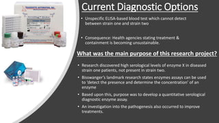
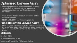
Development of a diagnostic enzyme assay to detect and diagnose
Yellow Eye Disease alongside an investigation into the pathogenesis
Adam Boulger
BMS 1
27040016
Introduction to Leuser Virus
Currently ravaging rural communities in South-East Asia
First investigated in the 1970’s due to animal epidemics.
Only caused mild symptoms in humans.
OLD Strain one
non-fatal, mild symptoms & no
treatment required
NEW Strain two
rapidly fatal & treatment necessary
(fairly effective)
RECENTLY mutated to form two strains;
Current Diagnostic Options
• Unspecific ELISA-based blood test which cannot detect
between strain one and strain two
• Consequence: Health agencies stating treatment &
containment is becoming unsustainable.
• Research discovered high serological levels of enzyme X in diseased
strain one patients, not present in strain two.
• Bisswanger’s landmark research states enzymes assays can be used
to ‘detect the presence and determine the concentration’ of an
enzyme
• Based upon this, purpose was to develop a quantitative serological
diagnostic enzyme assay.
• An investigation into the pathogenesis also occurred to improve
treatments.
What was the main purpose of this research project?
Research Project Experimental Methodology
• Urinalysis, haemolysis and H&E staining followed standard practice.
• Patient Samples provided by Public Health England
• Optimised Enzyme Assay (EA)
• ELISA Assay
• Urinalysis
• Haemotoxylin & Eosin Staining
• Haemolysis Assay
Optimised Enzyme Assay
• Following optimization based experiments analysing
the variables of; temperature, pH, water-bath
incubation duration and wavelength to measure
absorbance.
• It was decided that the optimum conditions for the
enzyme assay were.
• 311.5K, pH 8, 1200s and 415nm respectively.
Principles of this Enzyme Assay
• 10mM enzyme X substrate assay buffer solution was heated to
311.5K for 300s. After, 100μl of patient serum sample was
added and incubated for 1200s and the absorbance read at
415nm.
Materials:
SA Buffer – 43529
SA Enzyme X substrate P4744
Patient Sample provided by Public Health England
ELISA Principles
25 microlitres of samples added to plate in
duplicates alongside 100ul detection antibody and
followed standard incubations. 100 microlitres poly-
HRP streptavidin was added and then the plate was
washed. 100 microlitres TMB substrate solution was
added and then stop solution (H2SO4) was added
and the plate read at 450nm
Materials:
TF – Plates – 88-50650-22
AL – Detection Antibody – DIA HE1-01
TF – Poly-HRP streptavidin – N200
SA – Wash Buffer – N301
Samples (patient, control and blank)
provided by Public Health England
Determination of an enzyme assay cut off point
Graph produced using
manipulated data.
Statistical normal
distribution was plotted
against enzyme
concentration (mg/L).
Graphical manipulation of
a scatter graph was
performed to produce
bell-curves which were
extrapolated with a
polynomial trendline until
cross over point (cut off
point – 1.69mg/L)
Investigation into Proof of Principle
Analysis of 40 unknown
patient samples occurred
using optimised
methodology.
Patient samples were
plotting on a scatter graph
and analysed.
New method displayed
93% success rate, with 1
false negative and 2 false
positives.
True diagnostic success
rate of 97.5%
Investigation into Proof of Principle
Analysis of 40 unknown
patient samples occurred
using optimised
methodology.
Patient samples were
plotting on a scatter graph
and analysed.
New method displayed
93% success rate, with 1
false negative and 2 false
positives.
True diagnostic success
rate of 97.5%
Comparison between enzyme assay and current diagnostic
ELISA
Currently; ELISA
diagnosed both
as positive,
requiring
treatment.
Future: EA
diagnosed C1 as
positive for strain
two and C2 as
negative for
strain two.
Haemolysis Assay to further investigate mechanisms of
Leuser virus
Major haemolysis
of RBC found in
diseased strain
two patients.
Result of 1.49
absorbance and
the cut off
haemolysis level
was 0.997.
Urinalysis and H&E staining
Urinalysis was performed due to
indicative nature. Results present
abnormal bilirubin, urobilinogen and
blood
Following the urinalysis, the indicators
link to hepatic malfunction.
H&E stain displays morphological loss
of hepatocytes and hepatic vessels
alongside an increased density of
nuclei.
Haemolysis Assay
Identified major
haemolysis which
directly links to
hepatic malfunction.
Diseased strain two
patient shows severe
breakdown of
erythrocytes, links to
high levels of
bilirubin and
urobilinogen.
Conclusion of Results and Indicative Discussion
• Optimised enzyme performed better compared to the current diagnostic
ELISA. Has a true success rate of 97.5% alongside 90% specify and 95%
sensitivity
• Urinalysis indicates hepatic, renal and haemolysis
• H&E indicates morphological loss & increased density of nuclei
• Haemolysis assay indicates major haemolysis of erythrocytes
Report Conclusion and Futuristic Scope
This report established foundations to improve both treatment and
diagnosis of strain two Leuser Virus.
• Future treatment research should focus on the viral genomic with
mechanism investigation, transmission mechanisms and enzyme X
proteonomics.
• Future diagnostic research should focus on further optimisation, PCR
investigation and the potential for futuristic approaches such as Crispr
and controlled non-disease viral integration

d

  • 1.
    Development of adiagnostic enzyme assay to detect and diagnose Yellow Eye Disease alongside an investigation into the pathogenesis Adam Boulger BMS 1 27040016
  • 2.
    Introduction to LeuserVirus Currently ravaging rural communities in South-East Asia First investigated in the 1970’s due to animal epidemics. Only caused mild symptoms in humans. OLD Strain one non-fatal, mild symptoms & no treatment required NEW Strain two rapidly fatal & treatment necessary (fairly effective) RECENTLY mutated to form two strains;
  • 3.
    Current Diagnostic Options •Unspecific ELISA-based blood test which cannot detect between strain one and strain two • Consequence: Health agencies stating treatment & containment is becoming unsustainable. • Research discovered high serological levels of enzyme X in diseased strain one patients, not present in strain two. • Bisswanger’s landmark research states enzymes assays can be used to ‘detect the presence and determine the concentration’ of an enzyme • Based upon this, purpose was to develop a quantitative serological diagnostic enzyme assay. • An investigation into the pathogenesis also occurred to improve treatments. What was the main purpose of this research project?
  • 4.
    Research Project ExperimentalMethodology • Urinalysis, haemolysis and H&E staining followed standard practice. • Patient Samples provided by Public Health England • Optimised Enzyme Assay (EA) • ELISA Assay • Urinalysis • Haemotoxylin & Eosin Staining • Haemolysis Assay
  • 5.
    Optimised Enzyme Assay •Following optimization based experiments analysing the variables of; temperature, pH, water-bath incubation duration and wavelength to measure absorbance. • It was decided that the optimum conditions for the enzyme assay were. • 311.5K, pH 8, 1200s and 415nm respectively. Principles of this Enzyme Assay • 10mM enzyme X substrate assay buffer solution was heated to 311.5K for 300s. After, 100μl of patient serum sample was added and incubated for 1200s and the absorbance read at 415nm. Materials: SA Buffer – 43529 SA Enzyme X substrate P4744 Patient Sample provided by Public Health England
  • 6.
    ELISA Principles 25 microlitresof samples added to plate in duplicates alongside 100ul detection antibody and followed standard incubations. 100 microlitres poly- HRP streptavidin was added and then the plate was washed. 100 microlitres TMB substrate solution was added and then stop solution (H2SO4) was added and the plate read at 450nm Materials: TF – Plates – 88-50650-22 AL – Detection Antibody – DIA HE1-01 TF – Poly-HRP streptavidin – N200 SA – Wash Buffer – N301 Samples (patient, control and blank) provided by Public Health England
  • 7.
    Determination of anenzyme assay cut off point Graph produced using manipulated data. Statistical normal distribution was plotted against enzyme concentration (mg/L). Graphical manipulation of a scatter graph was performed to produce bell-curves which were extrapolated with a polynomial trendline until cross over point (cut off point – 1.69mg/L)
  • 8.
    Investigation into Proofof Principle Analysis of 40 unknown patient samples occurred using optimised methodology. Patient samples were plotting on a scatter graph and analysed. New method displayed 93% success rate, with 1 false negative and 2 false positives. True diagnostic success rate of 97.5%
  • 9.
    Investigation into Proofof Principle Analysis of 40 unknown patient samples occurred using optimised methodology. Patient samples were plotting on a scatter graph and analysed. New method displayed 93% success rate, with 1 false negative and 2 false positives. True diagnostic success rate of 97.5%
  • 10.
    Comparison between enzymeassay and current diagnostic ELISA Currently; ELISA diagnosed both as positive, requiring treatment. Future: EA diagnosed C1 as positive for strain two and C2 as negative for strain two.
  • 11.
    Haemolysis Assay tofurther investigate mechanisms of Leuser virus Major haemolysis of RBC found in diseased strain two patients. Result of 1.49 absorbance and the cut off haemolysis level was 0.997.
  • 12.
    Urinalysis and H&Estaining Urinalysis was performed due to indicative nature. Results present abnormal bilirubin, urobilinogen and blood Following the urinalysis, the indicators link to hepatic malfunction. H&E stain displays morphological loss of hepatocytes and hepatic vessels alongside an increased density of nuclei.
  • 13.
    Haemolysis Assay Identified major haemolysiswhich directly links to hepatic malfunction. Diseased strain two patient shows severe breakdown of erythrocytes, links to high levels of bilirubin and urobilinogen.
  • 14.
    Conclusion of Resultsand Indicative Discussion • Optimised enzyme performed better compared to the current diagnostic ELISA. Has a true success rate of 97.5% alongside 90% specify and 95% sensitivity • Urinalysis indicates hepatic, renal and haemolysis • H&E indicates morphological loss & increased density of nuclei • Haemolysis assay indicates major haemolysis of erythrocytes
  • 15.
    Report Conclusion andFuturistic Scope This report established foundations to improve both treatment and diagnosis of strain two Leuser Virus. • Future treatment research should focus on the viral genomic with mechanism investigation, transmission mechanisms and enzyme X proteonomics. • Future diagnostic research should focus on further optimisation, PCR investigation and the potential for futuristic approaches such as Crispr and controlled non-disease viral integration