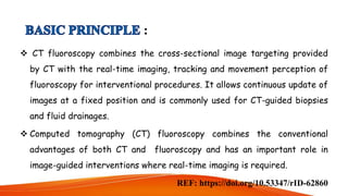
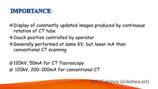
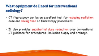
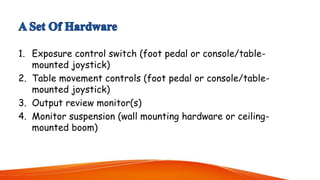
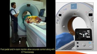
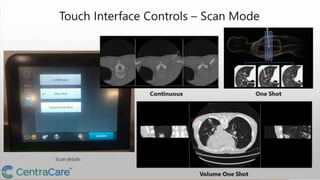
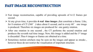
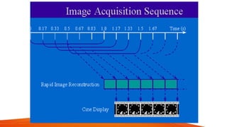
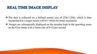
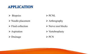
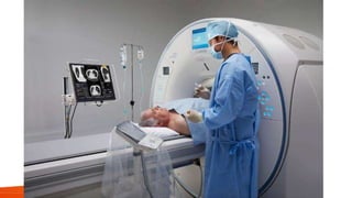
3. :
CT fluoroscopy combines the cross-sectional image targeting provided
by CT with the real-time imaging, tracking and movement perception of
fluoroscopy for interventional procedures. It allows continuous update of
images at a fixed position and is commonly used for CT-guided biopsies
and fluid drainages.
Computed tomography (CT) fluoroscopy combines the conventional
advantages of both CT and fluoroscopy and has an important role in
image-guided interventions where real-time imaging is required.
REF: https://doi.org/10.53347/rID-62860
4. The first scanner with CT fluoroscopic capability was developed in 1993
by Dr. Katada
FDA approved the use of CTF in 1995.
Since its introduction , real time CT has been used for optimization of
scan timing in contrast studies, as well as for guidance in biopsy or
drainage procedures.
https://thancguide.org/2021/03/the-journey/patients-journey/a-surprising-new-
imaging-technique-fluoroscopic-ct-scanning
5.
6. :
Display of constantly updated images produced by continuous
rotation of CT tube
Couch position controlled by operator
Generally performed at same KV, but lower mA than
conventional CT scanning
@120kV, 50mA for CT fluoroscopy
@ 120kV, 200-300mA for conventional CT
Ref:CT History (slideshare.net)
7. • CT fluoroscopy can be an excellent tool for reducing radiation
dose and saving time on fluoroscopy procedures
• It also provides substantial dose reduction over conventional
CT guidance for procedures like lesion biopsy and drainage.
8. • CT fluoro is enabled through a combination of
A software suite and
A set of hardware
REF:https://info.blockimaging.com
9. 1. Exposure control switch (foot pedal or console/table-
mounted joystick)
2. Table movement controls (foot pedal or console/table-
mounted joystick)
3. Output review monitor(s)
4. Monitor suspension (wall mounting hardware or ceiling-
mounted boom)
10.
11.
12.
13.
14. How it works?
• Activation of touch interface panel
• Setting the couch speed and gantry position
• Setting the target
• Move in / Move out
• Activate laser
• Scan
• Fine Tuning with joystick
• Image or series review
15. Fluoroscopy software suite of real time CT is dependent on following
factors :
Fast continuous scanning
Fast image reconstruction
Real time image display
REF:http://www.impactscan.org/slides/ctfluoroscopy/sld005.htm
16. The continuous scanning is possible because of Slip Ring Technology, with
no interscan delay for unwinding of connecting cables.
It thus reduces the scan time also and subsequently helps decrease the patient
dose
SYSTEM REQUIRMENTS :
Slip rings for continuous scanning
Fast tube rotation (1s or less)>>some newer systems have 0.5s rotation
High heat capacity tube to allow extended scanning times if necessary
17. Fast image reconstruction, capable of providing upwards of 6-12 frames per
second .
At any given time, it provides 6 real time images ,that constitute a frame. Like,
for 1 rotation of CT (360°) takes about 1 second, and in every 60° one image
is produced. Therefore six images are produced for one gantry rotation.
After one rotation in one second , the CT performs the second rotation and
produces the seventh real time image. Now, this image is added and the 1st image
is discarded .Thus 6 images or frames are obtained at a time.
Sometimes motion artefacts may be seen on the images and appear as streaks ,
however these do not restrict the visualisation of important structure.
18.
19. The data is collected on a defined matrix size of (256×256), which is then
interpolated to a larger matrix (1024×1024) for better resolution.
Images are subsequently displayed on the monitor kept in the operating room
on the Cine mode with a frame rate of 6-12 per second.
21. Low MillIampher Technique for reduction in radiation dose.
Reduction of procedure time.
Quick Check Method
Real Time Imaging
Dedicated High Speed Array Processor
Potential for increased procedure effeciency and accuracy leading to higher
diagnostics yeild .
22. Overlapping structures can be removed, providing accurate spatial information
Real-time display of images
Consequent reduction in complications through finer needle control
Reduced procedure time
Increased operator confidence
23. Large radiation doses to both patient and operator.
Limited FOV / scanning range.
Poor image quality due to low mA , because of the appearance of noise.
Increased investment in more powerful hardware and specialized software
needed.
24. The Doctor may want to obtain highly accurate imaging
They may wish to provide the patient with shorter intervention time which
may decrease the radiation exposure
Real time view of the body may give the doctor more information to make
better decisions
https://thancguide.org/2021/03/the-journey/patients-journey/a-surprising-new-imaging-
technique-fluoroscopic-ct-scanning
25. Video monitor will need to be displayed in the scanning room
An operator panel is required in the scanning room – with controls available for table
movement, gantry lift, laser light control and fluoroscopic factors. Exposures will
usually be activated using a footswitch
Involves an x-ray tube current of 30-50 ma
Need for additional beam filtration to decrease patient dose exposure.
Multislice machines have finer z-axis resolution.
CT fluoroscopy requires special techniques for image reconstruction, due to the need for
rapid imaging.
26. Manufacturer /
Model
CT Flouro
System
KV / ma Settings Rotation Time (s)
Patient Skin
Dose Rate
(mGy/s)
IGE HiSpeed CT/i Smart View 120 / 50 0.8 / 1.0 3.2
Picker UltraZ CCT / BolusPro 120 / 30 1 4.1
Siemens Somaton CARE Vision 120 / 50 0.75 / 1 4 .0
Toshiba Asteion* Aspire CI 120 / 50 0.75 / 1 3.3
REF: https://doi.org/10.53347/rID-62862
31. • CT fluoroscopy is a recently developed acquisition mode that allows faster
imaging reconstruction, near continuous image update and convenient in room
table control and image viewing during procedure .
• Acceptable radiation dose rate to patient and operator, is user friendly and
guides interventional procedures with rapidity.
• Operators have to adopt techniques that will minimize the risk of putting their
fingers in the beam