Basic physics of multidetector computed tomography ( CT Scan) - how ct scan works, different generations of ct, how image is generated and displayed and image artifacts related to CT Scan.
Computed tomography (CT scan) is a medical imaging procedure that uses computer-processed X-rays to produce tomographic images or 'slices' of specific areas of the body. These cross-sectional images are used for diagnostic and therapeutic purposes in various medical disciplines.
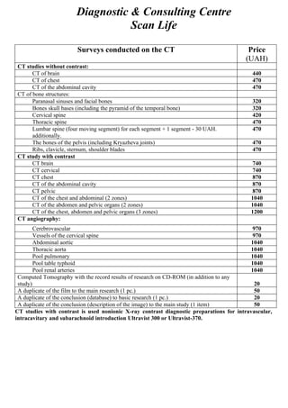
1. Diagnostic & Consulting Centre
Scan Life
Surveys conducted on the CT Price
(UAH)
CT studies without contrast:
CT of brain 440
CT of chest 470
CT of the abdominal cavity 470
CT of bone structures:
Paranasal sinuses and facial bones 320
Bones skull bases (including the pyramid of the temporal bone) 320
Cervical spine 420
Thoracic spine 470
Lumbar spine (four moving segment) for each segment + 1 segment - 30 UAH. 470
additionally.
The bones of the pelvis (including Kryazheva joints) 470
Ribs, clavicle, sternum, shoulder blades 470
CT study with contrast
CT brain 740
CT cervical 740
CT chest 870
CT of the abdominal cavity 870
CT pelvic 870
CT of the chest and abdominal (2 zones) 1040
CT of the abdomen and pelvic organs (2 zones) 1040
CT of the chest, abdomen and pelvic organs (3 zones) 1200
CT angiography:
Cerebrovascular 970
Vessels of the cervical spine 970
Abdominal aortic 1040
Thoracic aorta 1040
Pool pulmonary 1040
Pool table typhoid 1040
Pool renal arteries 1040
Computed Tomography with the record results of research on CD-ROM (in addition to any
study) 20
A duplicate of the film to the main research (1 pc.) 50
A duplicate of the conclusion (database) to basic research (1 pc.) 20
A duplicate of the conclusion (description of the image) to the main study (1 item) 50
CT studies with contrast is used nonionic X-ray contrast diagnostic preparations for intravascular,
intracavitary and subarachnoid introduction Ultravist 300 or Ultravist-370.