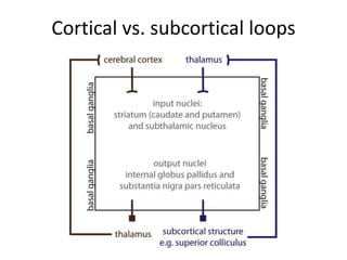
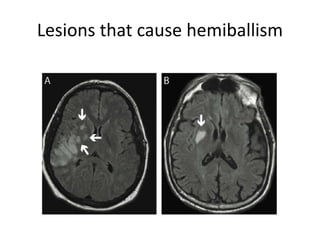
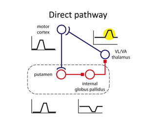
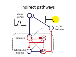
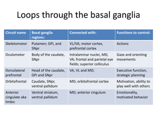
Cortical vs. subcortical loops
Lateral inhibition in striatum
Switching from one action to another
Direct and indirect pathways
Hyperdirect pathway
Lesions that cause hemiballism
Direct pathway
Indirect pathways
Loops through the basal ganglia
Circuit name Basal ganglia
regions:
Connected with: Functions to control:
Skeletomotor Putamen; GPi, and
SNpr
VL/VA; motor cortex,
prefrontal cortex
Actions
Oculomotor Body of the caudate,
SNpr
Intralaminar nuclei, MD,
VA; frontal and parietal eye
fields; superior colliculus
Gaze and orienting
movements
Dorsolateral
prefrontal
Head of the caudate,
GPi and SNpr
VA, VL and MD; Executive function,
strategic planning
Orbitofrontal Caudate, SNpr,
ventral pallidum
MD; orbitofrontal cortex Motivation, ability to
play well with others
Anterior
cingulate aka
limbic
Ventral striatum,
ventral pallidum
MD; anterior cingulum Emotionality,
motivated behavior

Cortical vs. subcortical loops.ppt

  • 1.
  • 2.
  • 3.
    Switching from oneaction to another
  • 4.
  • 5.
  • 6.
    Lesions that causehemiballism
  • 7.
  • 8.
  • 9.
    Loops through thebasal ganglia Circuit name Basal ganglia regions: Connected with: Functions to control: Skeletomotor Putamen; GPi, and SNpr VL/VA; motor cortex, prefrontal cortex Actions Oculomotor Body of the caudate, SNpr Intralaminar nuclei, MD, VA; frontal and parietal eye fields; superior colliculus Gaze and orienting movements Dorsolateral prefrontal Head of the caudate, GPi and SNpr VA, VL and MD; Executive function, strategic planning Orbitofrontal Caudate, SNpr, ventral pallidum MD; orbitofrontal cortex Motivation, ability to play well with others Anterior cingulate aka limbic Ventral striatum, ventral pallidum MD; anterior cingulum Emotionality, motivated behavior

Editor's Notes

  • #2 Figure 25-1. Cortical loops (brown on left) involve direct projections to the basal ganglia coupled with indirect projections, via thalamus, from basal ganglia back to cortex. In contrast, subcortical structures reach the basal ganglia only through a synapse in thalamus but receive output from the basal ganglia directly (blue on right).
  • #3 Figure 25-2. A cartoon of lateral inhibition in the striatum is illustrated. Indecision could result if striatal cells supporting two candidate actions receive similar levels of excitation. To decrease the likelihood of indecision, lateral inhibitory circuits facilitate the winning margin between the leading candidate and the runner-up. Incoming excitatory input excites target striatal cells, leading to a smile of enjoyment (black pathway). The same cortical cells indirectly inhibit, via local inhibitory interneurons (red cells), off-target striatal cells favoring a volitional smile (blue pathway). The result is that the close competition between the two inputs becomes a landslide victory for the target cells
  • #4 Figure 25-3A-B: A functional overview of basal ganglia pathways is illustrated. In each graph, the amount of movement (y axis) for a number of discrete movement possibilities (x axis) is plotted. In the starting condition (A), two movements are in progress. Movements that occur simultaneously are typically both well practiced and use different muscles, for example, walking and chewing gum. When a high-priority movement arises, the first pathway engaged is the hyperdirect pathway (B). The effect of the hyperdirect pathway is to quickly stop movements in process. Dopamine (DA) facilitates (upward blue arrow) ongoing movements. More dopamine means more movement, and less dopamine means less movement. In part, dopamine’s facilitation of movement stems from a facilitation of the direct pathway and a net inhibition (downward blue arrow) of the indirect pathways.
  • #5 Figure 25-3C-D: In each graph, the amount of movement (y axis) for a number of discrete movement possibilities (x axis) is plotted. Immediately following the hyperdirect pathway, the direct pathway is engaged (C), leading to the focal disinhibition of a salient action. This disinhibition may not be perfectly focused on the chosen action. Indirect pathways provide an annulus, or donut, of inhibition around the chosen action (D). Thus, the indirect pathways sharpen the disinhibition produced by the direct pathway and also keep other potential movements from occurring. Dopamine (DA) facilitates (upward blue arrow) ongoing movements. More dopamine means more movement, and less dopamine means less movement. In part, dopamine’s facilitation of movement stems from a facilitation of the direct pathway and a net inhibition (downward blue arrow) of the indirect pathways.
  • #6 Figure 25-4. The hyperdirect pathway within the skeletomotor circuit is illustrated. Next to each neuron is a cartoon showing the firing activity in that neuron. Neurons in somatomotor cortex are normally inactive but discharge before initiating a movement. The myelinated axons of motor cortex neurons excite neurons in the subthalamic nucleus at short latency. Excitation of subthalamic neurons in turn causes an increase in the discharge of tonically active neurons in the internal globus pallidus. GABAergic cells in the internal globus pallidus inhibit the tonically active neurons in ventral anterior (VA) and ventral lateral (VL) thalamus. Since thalamic cells project to somatomotor cortex, the inhibitory effect of the hyperdirect pathway on thalamic firing (yellow highlight) is passed on to motor cortex. Thus, we can use thalamic firing as a proxy for the effect of basal ganglia circuits. In sum, the hyperdirect pathway has an immediate but short-lasting effect of widespread suppression of motor cortex. GABAergic neurons are shown in red, and their inhibitory terminals are shown as red squares. Excitatory neurons are shown in blue.
  • #7 Figure 25-5. Magnetic resonance images (MRIs) from two individuals with hemiballism, neither of whom has a lesion in the subthalamus. A: A 34-year-old man with a stroke in the right middle cerebral artery presented with left-sided hemiballism. Affected areas include cerebral cortex and several small areas within the striatum and pallidum (arrows). B: A 69-year-old man with Parkinson’s disease presented with left-sided hemiballism. A lesion in the right striatum (arrow) was found. The left-sided hemiballism coincided with an amelioration of this patient’s symptoms of Parkinson’s disease on the left side. Note that radiological convention is that the left side of the brain is illustrated on the right and right side of the brain on the left. Modified from Posturna, R.B., and Lang, A.E. Hemiballism: Revisiting a classic disorder. Lancet Neurol 2: 661–8, 2003, with permission of the publisher, Elsevier.
  • #8 Figure 25-6. The direct pathway within the skeletomotor circuit is illustrated with the same conventions as previously. The direct pathway starts with activity in cortical neurons possessing unmyelinated axons. A burst of activity in these motor cortex neurons excites neurons in the putamen at longer latency than is involved in the hyperdirect pathway. Neurons in the putamen are GABAergic medium spiny neurons. Therefore, a burst of activity in neurons of the putamen inhibits the discharge of tonically active neurons in the internal globus pallidus. The inhibition of GABAergic pallidal output neurons leads to a disinhibition of thalamic cells (yellow highlight). As a result, the direct pathway serves to facilitate somatomotor cortex and ultimately movement.
  • #9 Figure 25-7. A family of indirect pathways within the skeletomotor circuit is illustrated with the same conventions as in previous slides. Thick lines mark the conventional indirect pathway. Yet, many additional routes through the basal ganglia also exist, some of which are illustrated here in faint lines. The conventional indirect pathway involves a projection from the motor cortex to the putamen. Neurons in the putamen inhibit tonically active cells in the external globus pallidus (GPe), leading to the disinhibition of subthalamic neurons. An increase in subthalamic neuronal firing excites cells in the internal globus pallidus (GPi). An increase in the discharge of neurons in the internal globus pallidus leads to an inhibition of thalamic cells (yellow highlight). As a result, the conventional indirect pathway serves to suppress somatomotor cortex and ultimately movement. The net effect of the family of indirect pathways also appears to be net inhibition of thalamic cells and consequently a suppression of movement.