SlideShare a Scribd company logo
1 of 23
Download to read offline
Treatment
a
with
ASD
may
not
require
treatment
it
he
or
e
nas
tew
or
no
symptoms,
or
if
the
defect
is
small.
towever,
medical
management
usually
is
done
with
gOxin
and
diuretics.
If
bacterial
endocarditis
is
there,
t
s
managed
with
antibiotics
and
other
methods
or
preventing
intection.
ardiac
catheterization
may
be
recommended
in
severe
cases.
An
umbrella-shaped
patch
is
closed
and
inserted
into
the
damaged
area
througha
small
tube,
called
a
catheter.
The
umbrella
patch
is
then
opened
to
cover
the
hole.
t
requires
a
small
incision,
avoiding
the
need
for
open-heart
surgery.
Surgery
It
may
be
option
if
child
cannot
have
a
cardiac
catheterization.
Surgeons
stitch
or
patch
the
hole
in
the
wall.
Child
will
be
connected
to
a
heart-lung
machine,
which
will
do
the
work
of
the
heart
during
surgery.
Patent
Ductus
Arteriosus
Patent
ductus
arteriosus
(PDA)
is
a
congenital
disorder
in
the
heart
wherein
a
neonate's
ductus
arteriosus
fails
to
close
after
birth.
The
ductus
arteriosus
(DA)
is
a
normal
fetal
blood
vessel
that
closes
soon
after
birth.
In
a
PDA,
the
vessel
does
not
close
and
connection
remains
patent'
(open)
resulting
in
irregular
transmission
of
blood
between
two
of
the
most
important
arteries
close
to
the
heart,
the
aorta
and
the
pulmonary
artery.
Within
minutes
or
up
to
a
few
days
(5-7
days)
after
birth,
the
DA
is
supposed
to
close.
This
change
is
normal
in
newborns.
If
the
closure
of
the
DA
does
not
occur
even
after
two
to
three
weeks
of
age
it
is
called
as
PDA
(Fig.19.5).
Incidence:
PDA
is
c
o
m
m
o
n
in
neonates
with
persistent
respiratory
problems
such
as
hypoxia,
and
has
a
high
o
c
c
u
r
r
e
n
c
e
in
premature
children.
In
hypoxic
newborns,
too
little
oxygen
reaches
the
lungs
to
produce
sufficient
levels
of
bradykinin
and
subsequent
closing
of
the
DA
Premature
children
are
more
likely
to
be
hypoxic
and
thus
have
PDA
because
of
their
underdeveloped
heart
and
lungs.
Pathophysiology
and
Altered
Hemodynamics
A
PDA
allows
a
portion
of
the
oxygenated
blood
from
the
left
heart
to
flow
back
to
the
lungs
by
Tlowing
from
the
aorta
(which
has
higher
pressure)
to
the
puimonary
artery.
If
this
shunt
is
Contd..
Fig. 19.5: Patont duuctus arlerionuns (PDA)
ontd.
substantial, the neonate becomes short of broath. The
additional
blood is
re-circulated through the lungs and
roturned to the
left atrium and left ventricle. This causes increased workload
on the left side of the heart and increased pulmonary vascular
congestion and possibly resistance, and
potentialy increased
right ventricular pressure and
hypertrophy. The additional fluid
returning to the lungs increases lung pressure to the pointthat
the neonate has greater difficulty inflating the lungs. This uses
more calories than normal and often interferes with feeding in
infancy. This condition, as a
constellation of findings, is called
congestive heart failure.
In some cases, such as in
transposition of the greatvessels (the
pulmonary arlery and the aorta), a PDA may need to remain
open. In this cardiovascular condition, the PDA is the only way
that oxygenated blood can mix with
deoxygenated blood. In
| these cases, prostaglandins are used to keep the DA open
Clinical Features
The
symptoms of PDA depend on the size of the DA
and how much blood flow it carries. Babies with a large
PDA might experience symptoms such as a
bounding
(strong and forceful) pulses result from runoff of bloou
from the aorta to the pulmonary artery, fast breathing
poor feeding habits, shortness of breath, sweating whie
feeding, tiring very easily, poor growth.
Patients may be
are at risk
be asymptom or
show sign
The
N l o n a r y V a s c u l a
h r o n i ce x c o
Signs of
for bacterial endocarditis
and
ructive disease in later life due
ssive pulmonary blood flow.
0eutic Management
Therapeut
ciatheter-based,
ethacin
thebased procedures, and
surgery. Administration
(prostaglane
inhibitor) has
proved
e
t r e a t m e n t
options tor PDA are
medication,
sing PDA in
premature infants
ful in clo
newborns. It is
usually given orally or
l indomethaci
stuessti
i n s o m e
svenously in dose ot0.2 mg/kg of body weight and
int the ageof ten days. The dose may be
repeated up
ree times, at an interval of 12 to 24 hours.
based procedure: A catheter (a thin, flexible
through their veins using special X-rays
a c r o s s
thePDAto
reaches
the heart.A special plug is then inserted
remain in position wi
ithin the ductus. This is called
1PDAs to prevent the risk of infective endocarditis,
C a t h e t e r
is fed
until
it
toblockblood tlow to the heart. The plug
scatheter device closure.' Sometimes it is done on
smallPE
e procedure
will be able to go home the same day.
ction of the lining of the heart, valves, or arteries.
lasts approximately two hours and child
Surgery
Gurgery of PDA is done if the size of the opening is large
enough that the lungs could become overloaded with
hlood, a condition that can lead to an enlarged heart. A
PDAalsomightbe closed to reducethe riskofdeveloping
a heart infection known as endocarditis, which affects
the tissue lining the heart and blood vessels.
Atrioventricular Septal Defect
An atrioventricular septal defect (AVSD) is a birth defect
of the heart in which important parts at the center of
the heart are not fully formed. There are holes between
chambers of the heart, and the valves that control the
flow of blood between these chambers may not be
Tormed correctly. This means that blood flows where it
OTmally should not be able to, and extra blood flows to
e
lungs. This defect is also known as
atrioventricular
Lanal (AV canal) defect or endocardial cushion deteet.
In A septal defect, there is a hole in the wall
the
prum between the right and left ventricles.
nave just one largervalve openingin the middle,
instead
between the right and left atria. There is also a hole in
Inaddition, the
tormed correctly.
wo
a t r i o v e n t r i c u l a r
valves are not
tone on eachside oftheheart.
A baby with AV septal
detect may
Ogether these problems may ereate a hole in the
enter ot the baby's heart. As a result, blood does not
tlow the wav it should between the chambers. So the
neart has to work harder to pump blood to the lungs
and the rest of the body.
This condition is also known as atrioventricular
canal detect or endocardial cushion delect.
Clinical Features
Usually these patients have moderate to severe Ci
Ihere is a characteristic murmur. Mild cyanoSIS 1s
observed, which increases in crying. These children
are at high risk for developing pulmonary vascular
obstructive disease.
Treatments
All AVSDs, both partial and complete types, usually
require surgery. Surgery depends on how sick the child
IS and the specific structure of the AVSD. If possible,
surgery should be done before there is pernmanent
damage to the lungs. As short-term method medication
may be used to treat congestive heart failure, but it is
only a short-term measure until the infant is strong
enough for surgery.
During surgery, any hole in the septa is closed
using patches.
Sometimes the mitral valve does not
close completely, allows blood to flow backwards
and make the heart work harder to get enough blood
to the rest of the body. This leaky mitral valves need
to be repaired or replaced. For a complete AVSD, the
common
valve is separated into two distinct valves-
one on the right side and one on the left. With proper
treatment, most babies with AVSD grow up to lead
healthy, productive lives.
OBSTRUCTIVE HEART DISEASES
In an
obstructive disorder, the blood flow is restricted
or completely
blocked. This blockage or narrowing can
occur in any of the tour heart valves or above or below
the valve. The blockage (atresia) or narrowing (stenosis)
can occur in vessels returning blood to the heart (veins)
or in vessels pumping
blood out of the heart (arteries).
Unfortunately,
even the most impressive
structures,
such as the heart, can have problems. Valves do not
always
work the way they should. If a valve is not
formed properly
from birth (congenital valve disease)
or if it is damaged at some point after birth from age or
disease (acquired
valve disease), then vital organs,
such
as the brain and kidneys, may not get the oxygen-rich
blood they need to function.
B0 Pediatric Nursing
Heart valve disease (sometimes called valvular
heart disease) can strain the heart, too. But when the
valves are defective or do not work the way they should,
it can put the heart and other organs at risk. Valve
disease can affect one or more of the four valves in the
heart (mitral, aortic, tricuspid, and pulmonary). Most
often it causes one or both of the following problems:
Regurgitation: The valves tissue flaps, or leaflets, that
control the flow and direction of the blood, do not fully
close, which causes blood to leak back into the heart.
Stenosis:The leaflets cannotopen fully to allow enough
blood to flow through.
The heart has to work harder to compensate for the
faulty valve, which can weaken the heart and increase
the risk of heart failure (a condition where the heart
does not fill up with enough blood or
pump enough
blood to
supply the body with the oxygen and nutrients
that it needs) or sudden cardiac arrest (when the heart
stops beating). A heart valve problem can also increase
the risk of blood clots, which can cause stroke.
Valve defects
Defective valves may be caused by:
Stenosis (narrowing): The valve is not able to open
completely. As a result, the heart has to work harder to pump
blood through it.
Regurgitation: The valve does not close correctly and
allows blood to leak backward.
Atresia: The valve is missing a hole for the blood to pass
through. This is considered a more complex defect.
Pulmonary Stenosis
Pulmonary stenosis accounts for approximately 5 to
8% of all CHD. Pulmonary stenosis is a
narrowing
at the entrance to the pulmonary artery (pulmonary
valve), and the pulmonary blood flow is decreased.
The pulmonary valve is within the heare's right
ventricle. Because of the narrowing of the valve, the
right ventricle needs to work harder to get blood past
the blockage and get hypertrophied.
Pulmonary atresia is the extreme form of PS in
which the
pulmonary valve orifice fails to develop.
The valve is
completely closed thereby obstructing the
outflow of blood from the heart to the lungs. The right
ventricle may be
hypoplastic.
Pathophysiology and Altered Hemodynamics
The main pathophysiological consequence of PS is RV strain
and an increase in RV pressure. The cellular effect on the
RV depends on the timing of obstruction and on the size of
Contd..
obstruction. If the obstruction is present in a fo.
the myocardial response is hyperplasia and a eona
vascularity. There is an increase in myocyte siza
without an increase in capillary network
develops within mature myocardium.
Generally children with mild to moderate obstrs
increase in
ypertophyy
when the obstruction
are
hemodynamically well tolerated and are not a
iated
on a
with
cyanosis or cardiac symptoms. In the severe to critica form of
PS, right ventricular pressure increases a
and causes
thickening
of right ventricular wall. Right atrial pressure
increases i
hypertrophy is severe, resulting to right to left shuntiheR
foramen of ovale. Cyanosis becomes apparent when
re is a
right-to-left shunt at the atrial level via an atria septal defectand
exaggerated by limited blood flow to the
pulmonary vascu
bed. If there is no atrial or ventricular level shunt, sudt ncular
death
may ensue due to compromised cardiac output
Clinical Manifestations
The most obvious symptom 1s blue, or
cyanotic skin
in a newborn wh can be noted shortly after bith
or several weeks later as the ductus arteriosus closec
Clinical symptoms such as
dyspnea, heart murmur
exercise intolerance, cyanosis, and
syncope may occur. In
severe PS, the RV will eventually fail as the myocardium
becomes unable to support the enhanced work load
imposed by the stenosis, and patients develop jugular
venous distension, peripheral edema, pleural effusion,
ascites, and hepatomegaly. Severe and critical PS may
be differentiated clinically by the presence of cyanosis
(more pronounced with critical PS) and symptoms of
profound heart failure (present with critical PS).
Diagnosis
Cardiac examination: A systolic ejection mumur is
heard best at upper sterna border.
Electrocardiogram: The ECG may be normal or show
right ventricular hypertrophy with mild to moderate
PS. In severe PS, the ECG shows right ventricular
hypertrophy and enlargement of right atrium.
Chest radiograph: Chest X-ray shows right ventricular
hypertrophy and post-stenotic pulmonary artery
dialatation.
Echocardiogram: The structure of the pulmonary valve
the location and severity of the narrowing (stenosis),an
the size of the RV and its out flow tract can be visualized
d
Other imaging tests: Magnetic resonance imaginga
CT scans are sometimes used to confirm the diagnosi
pulmonary valve stenosis.
Cardiac catheterization: During the procedure the
.pressure and oxygen measurements are taken
the
ambers theiheart, as well as the
pulmonary
isualize the ructures inside the heart. This
u r
a r t e r ya n d
nd aorta. Contrast
dye is also
injected to more
h a r l y
generally only done whern child will need balloon
pulmonary valve stenosis because
be done at the same time as cardiac
s t
valvuloplasBy to treat
that
catheterization.
M a n a g e m e n t
cases of pulmona stenosis are mild and do not
S o m e
uire.
treatment
exceptfor routine checkups. However,
is more serious child will most likely be admitted
once symptoms are noted. Initially, child may be placed
ifcasei
intensive care unit
(ICU) or
special care
nursery
the
Oxygen, and possib even on a ventilator, to assist
his or her breathing. V medicati may be given to
ofpulmonary
atresia, as it stops the ductus arteriosus from
closing, wing mixing of the pulmonary and systemic
asit can cause apnea. Another example of preliminary
on
lungs function more efficiently. An IV
helpthe heart and.
m e d i c a t i
ation called prostaglandin E1 is used for treatment
circulations But prostaglandin E1 can be
dangerous
atment is heartcatheterization to evaluate the defect or
efects of the heart; this procedure is much more invasive.
Surgery
Surgical repair may be needed for child. The procedure
aims at relieving the obstruction of blood flow through
the pulmonary valve. This procedure requires heart-
lung bypass. Postoperative hospital stay averages 5 to
7days.
Child may need either balloon valvuloplasty or
open-heart surgery. The decision to perform a balloon
vavuloplasty or open-heart surgery depends on the
extent to which the pulmonary valve is obstructed.
Pulmonary stenosis is classified as mild, moderate
0r severe, depending on a measurement of the blood
Presure difference between the right ventricle and
pulmonary artery.
nav aseries of surgeries to improve the blood flow
Ultimately, however, the patient will needto
ently. The first surgery will likely be pertormed
hortly after birth. A
aorta and the
pulmonary
How to the lungs. As the child grows, so does the heart
shunt can be created between the
artery to help increaseblood
andthe shunt
body's requirements.
The type of surgery recommended depends on the
Size of the rightventricleand the pulmonaryartery
may need revised in order to meet the
If they are norm in size
1s able to
pump blood, open heart surgery can
performed to make blood flow through the heart "d
normal pattern.
and the right ventricle
be
IT the right ventricle is small and unable to act as a
PLmp, doctors may perform another type of operatIO
Caled the Fontan procedure. In this three-stag8
procedure, the right atrium is disconnected from tne
Puimonary circulation. The systemic venous return gOes
directly to the lungs, by-passing the heart.
Another treatment option involves the electrosurgical
puncture of the atretic valve usinga wire introduced into
the patient percutaneously. The puncture is then dilatea
using a balloon catheter to allow blood flow from the
right ventricle into the pulmonary artery.
The outcome varies for every child. If the condition
is left uncorrected it may be fatal, but the prognosis
has greatly improved over the years for babies with
pulmonary atresia. Some factors that affect, how wel
the child does include how well the heart is beating
the condition of the blood vessels that supply the heart,
and how leaky the other heart valves are. Most cases of
pulmonary atresia can be helped with surgery. If the
patient's right ventricle is exceptionally small, many
Surgeries will be needed in order to help stimulate
normal circulation of blood to the heart.
Aortic Stenosis
Aortic stenosis acounts for approximately 5% of all
CHD. It is more common in males than females. The
aorta is the large artery that supplies oxygen-rich blood
to the body. The aortic valve is within the heart's left
ventricle and acts as the entrance to the aorta. Narrowing
of the aortic valve, which controls blood flow between
left ventricle and aorta, is known as aortic stenosis.
Depending on the severity of the stenosis, the symptoms
at birth can vary from none to decreased blood flow and
decreased oxygenation to the body. As the PDA closes,
the symptoms usually become more acute.
Aortic sternosis is three types:
1. The commonest valvular type is the stricture of
aortic valve
2. In subvalvular type, the narrowing remains below
the aortic valve
3. In supravalvular, the stenosis remains above the
aortic valve.
Pathophysiology
and
Altered
Hemodynamics
e
human
aortic
valve
normally
consists
of
three
leatlets
neaflefs)
and
has
an
orifice
of
3.0-4.0
square
centimeters.
aortic
stenosis,
the
opening
of
the
aortic
valve
becomes
narrowed
or
constricted
(stenotic)
S8
Consequence
of
this
stenosis,
the
left
ventricle
must
generate
a
higher
pressure
with
each
contraction
to
ettectively
move
blood
forward
into
the
aorta.
Increased
pressure
in
tne
LV
has
direct
correlation
with
the
degree
of
obstruction
in
aortic
valve.
Ihitially,
the
LV
generates
this
increased
pressure
by
thickening
its
muscular
walls
(myocardial
hypertropny).
he
type
of
hypertrophy
usually
seen
in
aortic
stenosIS
IS
Known
as
concentric
hypertrophy,
in
which
the
walls
of
the
Lv
are
(approximately)
equally
thickened.
In
the
later
stages,
the
eft
ventricle
dilates,
the
wall
thins,
and
the
systolic
function
deteriorates
(resulting
in
impaired
ability
to
pump
blood
forwara)
Heart
failure
can
develop
due
to
this
heavy
workload.
In
severe
cases,
pulmonary
edema
may
occur
as
a
result
of
increased
ert
ventricular
pressure
that
may
cause
a
backflow
of
blood
into
the
lungs.
Sometimes,
a
percutaneous
balloon
valvuloplasty
(opening
of
a
valve)
can
effectively
relieve
the
valve
obstruction.
This
procedure
involves
a
special
catheter
(tube)
containing
a
balloon
being
passed
through
the
aortic
valve.
The
surgeon
inflates
the
balloon
to
stretch
the
valve
open.
The
surgical
repair
aims
to
relieve
the
obstruction
of
blood
flow
through
the
aortic
valve.
This
procedure
requires
heart-lung
bypass.
Postoperative
hospital
stay
averages
7
to
10
days.
Clinical
Manifestation
Less
severe
forms
of
the
disease
may
be
'silent
-
causing
no
visible
symptoms-until
later
in
life.
As
the
child
grows
older,
increased
physical
growth
requires
additional
cardiac
output.
Children
may
present
the
following
symptoms
in
severe
obstruction.
Fatigue
and
exercise
intolerance
Having
pain,
pressure
or
tightness
in
their
chest
Exertional
dyspnea
Fainting
or
feeling
weak
or
dizzy
when
active
Having
palpitations
Babies
born
with
severe
aortic
stenosis
may
have
to
work
hard
to
breathe,
have
poor
appetite
or
trouble
feeding,
and
failure
to
thrive.
They
may
also
show
signs
of
shock
because
their
heart
cannot
pump
enough
blood
to
the
rest
of
their
body.
Signs
of
cardiogenic
shock
include
less
frequent
urination,
cool
limbs,
increased
heart
rate,
fussiness,
poor
feeding,
rapid
breathing,
lethargy,
mottled
skin.
They
have
decreased
perfusion
and
pulmonary
congestion.
Diagnosis
Cardiac
examination:
A
harsh
systolic
murmur,
click,
or
other
abnormal
sound
is
almost
always
heard
at
the
upper
right
sternal
border,
radiating
to
the
upper
left
sternal
border and neck. A vibration or moue
can
when placing a hand over the hcart. T re
ht
may
car
be
faint
the pulse in the
nee
pulse or changes in the
quality of the pule
neck.
be
Electrocardiogram: The ECG may
normal
demonstrate left ventriculan
hypertrophy.
Tmal heart svze is
seen.
Chest radiograph: Usually normal heart.
In severe case of sternosis, dilatation of
may be seen.
n of
ascending aorta
well as presence
Echocardiogram: Type of stenosis as well
of other cardiac defects can be seen. The
function of left
ventricle and thickness ot its wall is also
assessed.
Management
Regular checkups by a health
provider m
may be all
no
symptoms or
only mild
should ask
about
that is needed if there is
symptoms. The health care provider
child's health history, do a
physical exam, and
perform
an echocardiogram.
People with severe aortic stenosis may be told not
not to
play competitive sports, even if
they have no
sympto
If symptoms do occur, strenuous
activity must ofteni
limited.
Medicines are used to treat
symptoms of heart
failure or abnormal heart
rhythms (most commonlv
atrial fibrillation). These include diuretics, nitrates,
and beta-blockers. High blood pressure should also be
treated. If aortic stenosis is severe, this treatment must
be done carefully so blood pressure does not drop to
dangerously low levels.
Treatment may be done in the cardiac catheterization
lab using a balloon procedure. A balloon is inserted
across the valve. When the balloon is inflated, the valve
is stretched open. Then the balloon is removed. This is
called balloon valvuloplasty.
Some children need surgery to
replace their aortic
valve with an artificial valve. In some cases the child's
n De
own
pulmonary valve can be used to replace the
damaged aortic valve.
Aortic Coarctation
Coarctation of the aorta is a narrowing of some porto
of the aorta
(Fig. 19.6A). This narrowing is
usuay
found just past the arch of the aorta, opposite
area of the PDA. Coarctation of the aorta accounts
nly
approximately 8% of all CHD, it is more com
2ortic
tound in male and may accompany other
enital
h
defects like VSD, PDA, tubular hypoplasia or
tne
isthmus and
bicuspid aortic valve.
C l i n i c a l
M a n i f e s t a t i o n
a n d
usually become evident after the closure of PDA.
The
c l i n i c a l
features depend upon the type of obstruction
The
p r e s e n t i r
features of neonates are severe CCF, poor
rfusion, tachypnea, acidosis and absence of femoral
pulse. Theolder
e.The older children'sgrowth and development may
normal wit
normal without symptoms, but overgrowth of upper
may
be suggestive of CoA. The symptoms may include:
extremities and hypertension, bsence of femoral pulse
Increased BP in the upper part of the body, resulting
in headache, dizziness, fainting, epistaxis and later
CVA, encephalopathy.
Child may occasionally complaints of weakness and
pain in legs after exercise. The femoral and pedal
pulse may be absent or diminished, and legs may be
cooler than arms.
Other manifestations are fatigue, cramps,
exertional
dyspnea, feeding problem, poor weight gain,
irritability and tachycardia.
Diagnosis
Diagnosis is confirmed by cardiac
examination,
chest X-ray, barium swallow,
electrocardiogram,
M
cardiac
examination,
mode
echocardiography,
cardiac
catheterization
and
angiocardiography.
Specific
'
E' sign in barium
swallow
very
suggestive
of CoA. The first arch ofthe 'E' is
Tormed due to dilatation of aorta before the coarctation,
ne second arch due to
poststenotic
dilatation,
and
middle notch due to the
coarctation.
Management
Medical
m a n a g e m e n t
of CoA is done with PGE1
nfusion,
a n t i b i o t i c s
and
prevention
and
t r e a t m e n t of
Complications.
483
Cardiovascular System
999
Figs 19.6A and B: A. Coarctation of the aorta,
B.Correction of coarctation of the aorta
Surgical correction of CoA (Fig. 19.6B)
Individualizedto anatomy of CoA
lan to include treatment of any additional cardiac
defects.
Child, Adolescent: Repair is done at 2 to 3 years of age
upon diagnosis.
Surgery: Four common types of repair-regardless or
Technique, usually performed via a left thoracotomy
incision. End-to-end anastomosis was done in 1954 by
Crawford and Nylin.
Excision of CoA area, circumferential anastomosis
Is
Completed with interrupted sutures anteriorly
NURSING CARE OF THE FAMILY AND CHILD
WITH CONGENITAL CARDIAC DISEASES
The diagnosis of a chronic condition in any family
member causes much distress. When an infant is
born with a heart defect, the parents may grieve over
the loss of the healthy newborn they had anticipated
and experience shock, denial, guilt, anger, despair or
confusion on learning that their infant has a cardiac
defect. Some parents may be unable at first to respond
to their newborn. Even greater stress may occur if the
condition is one that requires surgical intervention. The
nurse should become familiar with parents reaction and
nursing intervention require to:
Help family to adjust to the disorder: A birth of a
baby
with severe cardiac anomaly is a shock to the family,
parents always suffer with high anxiety and fear that
their child will not survive. Here role of nurse is to
support the family in their stress, assessing their level
of understanding and to provide information as needed
(thorough but simple in language). She also helps the
other members of the health team to understand the
parents' reaction.
The nurse needs to support the family in their great
pain and emotional investment. She can foster parent
intant attachment and
encourage parents to hold, touch
and look at their child.
Teach the parent to care the child which can
minimize their anxiety and fear.
Introduce parents to other families with similarly
affected children. It can help them to adjust to their daily
stress.
Counseling of family about the disorder and its care:
Before describing the defect a review of the basic structure
and function of heart is important. Picture, diagram,
models are to be used for better understanding of CHD.
Increasingly, families are collecting
information
from different sources. It is important to inform the
families about authentic internet sites and medical
literature. Parents must know that information from
general sources or other families might not be applicable
to their own situation.
Prepare Child and Family for Surgery
Some heart defects need repair soon after birth. For
others, it is better to wait months or years. Certain heart
defects may not need to be repaired. The surgery is
needed for the child's well-being.
In general, symptoms that indicate that surgery
is needed are blue or gray skin, lips, and nail beds
(cyanosis). These symptoms mean there is not enough
oxygen in the blood (hypoxia). Difficulty breathing
because the lungs are 'wet, congested, or filled with fluid
(heart failure). Child suffers with problems of heart rate
or heart rhythm (arrhythmias). Child shows feeding or
sleeping problem and lack of growth and development.
The three most common clinical presentations are:
1. A Murmur
2. Cyanosis
3. Respiratory difficulty.
N u r s i n g A s s e s s m e n t
Cardiovascular
assessment is an important n .
in the assessment and managementof
and children with chronic conditions. Asse
cardiac problems is described in Table 19,1
sing skill
nent facutely ill
children
. Assessment
for
Look for signs of cardiovascular problems:
Inspection
Palpation
Percussion
Auscultation.
The general objectives of nursing care for the
child with CHD include prevention of physical and
emotional fatigue, provision of adequate fluid and
nutrition, prevention of intection and care and support
to the parents and child to cope with the problems that
precipitates before and after surgery.
Nursing diagnosis: Impaired gas exchange related
to disturbed pulmonary blood flow or pulmonary
hypertension.
Expected outcome: Patient maintains optimal gas
exchange as evidenced by normal ABGs and alert
responsive mental condition or no further reduction in
mental status.
Nursing Intervention
Patient is to be placed in semi-upright position which
facilitates breathing. Position with proper body alignment
tor optimal respiratory functioning (if tolerated, head
of bed at 45 degrees). This promotes lung expansio1 au
improves air exchange.
Routinely check the patient's position so he o
she does not slide down in bed. This would cause tn
bdomen to co
aUSe
Tespirator
to compress the
diaphrag:
embarrassment. which would
effectively
clear the
airway. Periodic oral and nasal
Patient may eed help if he/she is
unable to
tions isto be suctioned if needed
retovgen saturation and
administer oxygen
Monitor oxYgen saturation
eded
P r e
ing of respiratory pattern
inisterdrugs like diureticaand
bronchodialators
i f
r e q u i r e d 1
re n of aspiration, with continuousmonitor
A d m i n i s t e r
Analysisofblood gas analysis.
ow cardiac output related to
Nursing diagnosis: Low
reducedmyocardial functions.
outcome: Cardiac
e c t e d
lerance of child is improved.
output and activity
Nursingintervention
Bed
ed rest, minimum exercise (care in the bed, indoor
play, and other activities of daily living)
Administer prescribed drugs like digoxin, diuretics,
antihypertensives which improve cardiac output
, Organize medication and nursing schedule to
provide periods of uninterrupted sleep
. Monitor child's condition (heart sound, vital signs).
Nursing diagnosis: Activity intolerance related to
hypoxia.
Fxpected outcome: The child is able to do age appropri-
ate activity.
Nursing Intervention
Oxygen therapy and continuous monitoring of
oxygen saturation by pulse oximetry. Pulse oximetry
is a useful tool to detect changes in oxygenation. O,
saturation should be maintained at 90% or greater.
Ahigher liter flow of oxygen is generally required
for activity versus rest (e.g. 2 L at rest, and 4 L with
activity).
Uninterrupted period ofrestand sleep
Advice parents to do activities of daily living for the
child so that child's energy can be conserved
Providing timely feeding, changing diaper and
tactile stimulation prevents cry of the child and
Saves energy expenditure. Prevent excessive crying
provide diversional activities, prevent constipation.
Nursi diagnosis: Altered nutrition, less than body
rements related to excessive energy
demarnds
ded by increased cardiac workload.
Expected Outcome: Normal nutritional status of child.
CordiovgsCular ysterri
Nursing Intervention
Feed in semi erect
position
sastric feeding may be started if the ci
nable to take oral fecds or get cvanosed while feeai6
Provide small frequent feedings
TOvide fomds with high nutritional value (24
Ka
o formula)
Determine child's likes and dislikes
Monitor input and output
Daly weight monitoring: Maintaining a
healtny
Weight is important for cardiac health. Restricting
intake of salt can also help lessen fluid retention ana
mprove symptoms related to heart valve disease.
Assess the child for developmental milestones.
VUSIng diagnosis: Increased potential for infection
related to poor nutritional status.
Expected outcome: No infection.
Nursing Intervention
revent exposure to communicable diseases. Early
detection and treatment of upper respiratory and ui
infection.
Immunizations shouldbe up-to-date
Handwashing should be observed. Maintaining
general cleanliness and hygienic measure are
important.
Be certain that the child receives prophylactic medi-
cation for infective endocarditis.
Congestive Cardiac Failure (CCF)
Every cardiac patient has a potential for developing
CCF. Congestive cardiac failure (CCF) by itself is not a
diagnosis. It is a clinical syndrome caused by different
natomical and or pathological conditions, which is the
primary diagnosis.
CCF is a term defined as 'inability of the heart to
pump enough blood out to the rest of the body, at rest
or during stress, necessary for the metabolic needs of the
body (systolic failure) and inability to receive blood into
the ventricular cavities at low pressure during diastole
(diastolic pressure)'.
Etiology
The etiology of pediatric heart failure may be cardiac or
noncardiac and can occur at any age. The predominant
etiology during infancy is congenital heart disease. VSD
is the commonest defect presenting with heart failure.
Myocardial disease especially myocarditis is commonest
cause HF in children under 5 years. Rheumatic fever
and rheumatic heart disease continue to be an important
cause of suffering and HF among children above 5
years. Among noncardiac disease, severe anemia can be
a condition to cardiac decompensation at any age.
In children, cardiac failure is most often caused
by congenital heart disease (left to right shunts) and
cardiomyopathy.
Clinical Features
CCF may become suddenly dyspneic,
cyanotic. A young child may develop abdominal pain,
The child with
tever, anorexia, dyspnea, cough suddenly. Dyspnea,
be
orthopnea or paroxysmal nocturnal dyspnea can be
reported by parent. Sometimes a mother may complaint
of palpitation, tachycardia and profuse sweating, pallor
or peripheral cyanosis and cold extremities of child.
Symptoms are different for children of different age,
in babies regardless of the cause of CCF the end result
of significant heart failure is poor growth. Slow weight
gain is related to two factors. Because of easy fatigability
baby takes small feed and there is an excessive loss of
calories from increased work of breathing associated
with CCF. In addition, as the lungs fill with
becomes more difficult for babies to hro fluid, it
to
breathe and
they
will use more of the muscles of their chest and
belly
they
to
and become very sweaty as of the extra work
needed
compensate. The baby breathes too fast dri.
ing feeding
to
eat.
Uncommonly, there may be an unusual
weight
gain due to collection of water,
manifesting s
facial
puffiness or as rarely as edema on the feet
may be brought with the complaints of persistent hoa
crying, breathes too fast with wheezing and excessive
the feet. The bal
perspiration, restlessness.
Diagnosis
Physical examination: A child with CCF may presen
tachycardia, tachypnea, Gallop rhythm, dyspnea,
decreased peripheral pulse and mottling of the
extremities, delayed capillary refil, failure to thrive,
decreased activity tolerance, sweating, etc. (Table19.2)
ChestX-ray: Very commonly X-ray shows cardiomegaly.
The heart dialates or hypertrophy occurs both in presence
of volume and pressure overload, cardiomyopathy or
dysrhythmias. It may gives clues to certain structural
heart diseases, and also to right versus left sided heart
involvement. Pulmonary markings are often increased,
showing pulmonary congestion.
ECG: Usually abnormal, and although not useful
in assessing HF, may give diagnostic cues for the
underlying pathology of heart failure.
Urine test: In chronic HF, proteinuria and high specific
gravity of urine are common.
Blood test: As renal function decreased due to decreased
perfusion in CCF, an increase in blood urea nitrogen
and creatinine levels may be present. CBC, differentna
may give clues to anemia and infection causing
complicating HF. Atrial blood gas analysis isimpoTLa
Echocardiogram: This test is valuable for evaluau
cardiac function and ruling out structural heart disea
tests
Other tests: Thyroid, renal and hepatic functio
are also valuable.
rsing
Management of
CCF
utic and NN
7 cardiovascular medicines
e October
W H O E s s e n t i
Therapeutc
r n t a i n s d i g o x i n ,
n
t h e
complem..
u s e
n h a t n a g e m e n t
a r e to:
rdiac work
educingcard
sential Medicines
List for
children (EML
section of
frusemide, spironolactone an
entary list,
and,
dopamine. The
goals of
enting myocardial contractility
Removeaccumulated fluid and sodium
ve tissue oxygenation and decreased oxygen
consumption.
This
is
accomplished by
on) and using medication to sedate an
irritable
educing
cardiac work: The workload on the heart is
needs are
kept to a
minimum
limiting physical activities
body temperature, treating
hen metabolic
bedrest) preserving
reducing the effort of
breathing (semi Fowler's
any
p o s i t i o r
child
Supplement cool humidified oxygen is usuallv
amount of
oxygen during
provided
to increase th
Baby with CCF is kept in bed rest with minimal
degree is maintain It may help the
inspiration.
handling.
ing, The propped up position with an incline
about 30
of
work of eathing by pooling the edema fluid in the
Aanendent areas and to reduce the collection of fluid
in lungs. Administration of humidified oxygen (40-
50% concentration) improves impaired oxygenation
Cecondary to pulmonary congestion, thus reducing
the work of heart by reducing requirements of cardiac
output.
Administration of Morphine sulfate (0.05 mg/kg
SC) or other sedative (Diazepam) may be needed if the
child is restless or
dyspneic. These drugs reduce anxiety
and lower the catecholamine secretion, thus reducing
physical activity, the respiratory rate and the heart
rate. The workload of heart comes down as the oxygen
demand of the body tissues goes down. The sedative
also helps in keeping the child in bed.
Management of feve, anemia and infection
necessary to reduce the workload of heart. At a
nperature of 36 to 37 °C, the overall circulatory and
eTabolic needs are minimal, thus reducing work or
eart. Anemia causes tachycardia and hyperkine
culatory state to meetupOxygen demandofthe body.
orrection of anemia results in decreased cardiac work.
ansfusion is indicated,,3 to 5mL packed red cells/kg
reight can be givens
every 12 hours. Worseningof
transfusion can be prevented, the patient can be
Corre
body
CCF by
given frusemide.
n be managed by use of vasodialators.
Cauces the
arteriolarand venous vasoconstrictoy
Cuuce the work of heart. "The use of ACE inhibitors
is now
(captopril well-established in infants and children
aptopril dose is 1 mg/kg 8 hourly and can be
give
creased up to 6
1mg/kg/day). ACE inhibitors Suppre
Enin
angiotensin/aldosterone system, thus reducin8
soconstriction as well as sodium and water retentio
reduce
They prevent potassium loss and hence
d
arrhythmias. These drugs suppress catecholamines al
thus its ill effect on
myocardium as well as
arrhythmias.
Augmenting myocardial contractility: Iontropic drugs
mprove cardiac output. In infants and children digitalis
8ycosides [digoxin (lanoxin)] is used which decreases
heart rate and increases mvocardial contractility. It has
rapid onset of action and also eliminated quickly.
Besides digitalis, catecholamine ionotropic agents
like
dopamine has been found to be useful. In a
patient
with CCF if the blood pressure is low, dopamine
may be used. At a dose of less than 5 ug/kg/min of
Dopamine causes peripheral vasodilation, increases
myocardial contractility and renal blood flow resulting
in natriuresis (Table 19.3).
Remove accumulated fluid and sodium: Treatment
consists of diuretics, possible fluid restriction and
possible sodium restriction. Diuretic is used to eliminate
excess water and salt to prevent re-accumulation. It
reduces total body sodium, thus reducing the blood
pressure and peripheral vascular resistance. The potent
oral diuretics frusemide is administered which starts its
action within 20 minutes. Frusemide interferes with the
sodium reabsorption mechanism in descending limb
of loop of Henle. Patient on frusemide should be given
potassium supplement.
Studies suggest that it is preferable to combine
frusemide with potassium sparing diuretic. Thee
combination is more useful in
preventing potassium
and magnesium loss, thus reducing arrhythmias as
against the combination of frusemide and potassium
supplement.
ACE inhibitor I drug can be combined with
frusemide, if indicated. The patient on ACE I should
neither be given potassium supplements or
potassium
The
sparing
diuretics
like
triamterene,
amiloride
or
aldactone
even
in
combination
with
frusemide,
Nursing
Diagnoses
mpaired
gas
exchange
related
to
altered
pulmonary
blood
flow
or
oxygen
deprivation
Activity
intolerance
related
to
decreased
cardiac
Output
due
to
structural
defect
and
myocardial
dysfunction
Altered
nutrition:
Less
than
body
requirements
related
to
the
excessive
energy
demands
required
by
increased
cardiac
workload
Fluid
volume
excess
related
to
fluid
accumulation
(edema)
Increased
potential
for
respiratory
infection
secondary
to
pulmonary
vasculature
overloading
and
other
infection
related
to
poor
nutritional
status
Anxiety
related
to
diagnostic
procedures
and
hospitalization
Developmental
delay
related
to
decreased
energy
inadequate
nutrition,
physical
limitations
and
social
isolation
Alteration
in
parenting
related
to
parental
perception
of
the
child
as
vulnerable.
Nursing
Intervention
and
Outcome
Impaired
gas
exchange
related
to
altered
pulmonary
blood
flow
or
oxygen
deprivation.
Goal
of
nursing
action:
The
patient
will
exhibit
improved
respiratory
function.
Nursing
intervention
includes
placing
the
child
in
inclined
posture
of
30
to
45
degree.
Tilt
mattress
support
of
incubator,
place
older
infant
in
sent
(Fowler's
position).
Avoid
any
constricting
clothing
or
restraints
around
abdomen
and
chest.
Administer
humidified
O,
as
prescribed.
Expected
Outcome:
Respirations
remain
with
normal
limits;
color
is
good
and
child
rest
quietly.
Activity
intolerance
related
to
decreased
cardiac
output
due
to
structural
defect
and
myocardial
dysfunction
(Table
19.4).
Goal
of
nursing
action:
The
patient
will
exhibit
improved
cardiac
output.
Nursing
intervention
includes
administration
of
digoxin
(lanoxin)
at
regular
interval
(usually
every
12
hours).
Nursing
established
precaution
to
prevent
toxicity.
Often
an
ECG
rhythm
strip
is
taken
to
assess
cardiac status before administration. Ensure adequate
els (decrease
intake of K. Monitor serum potassium levels (dece
to
enhances digoxin toxicity). Administer medicatione
decrease over load as ordered. Check blood pressur
Observe for signs of hypotension. Monitor electrolvh
e
levels.
Expected outcome:
Heartbeat is strong, regular and within normal limits for
age. Peripheral perfusion is adequate.
Goal (2): The patient will experience reduction of
anxiety.
Nursing intervention:
Employ flexible feeding schedule. Handle child gently.
Hold and comfort the infant. Employ comfort measures
found effective in individual cases. Encourage family to
provide comfort.
Expected outcome:
Infant rests quietly and breath easily.
Altered nutrition: Less than body requirements related to
the excessive energy demands required by increased caráuac
workload.
Goal of nursing action: The patient will able to conserve
energy arnd increase total intake.
Nursing Intervention:
A child with CCF needs small frequent feeds,asn
she has problem in sucking and swallowing a
breathing simultaneously.

More Related Content

Similar to congenital 2.pdf.com.education paediatric

Ventricular Septal Defect with Nursing Management
Ventricular Septal Defect with Nursing ManagementVentricular Septal Defect with Nursing Management
Ventricular Septal Defect with Nursing ManagementSwatilekha Das
 
Transposition of the great arteries
Transposition of the great arteriesTransposition of the great arteries
Transposition of the great arteriesjagan _jaggi
 
Congenital Heart Disease
Congenital Heart DiseaseCongenital Heart Disease
Congenital Heart DiseaseJessie Madz
 
Cirugia cardiovascular ingles
Cirugia cardiovascular inglesCirugia cardiovascular ingles
Cirugia cardiovascular inglesSandra Guiselly
 
Congenital heart failures in children
Congenital heart failures in childrenCongenital heart failures in children
Congenital heart failures in childrenEneutron
 
Heart valve disease
Heart valve diseaseHeart valve disease
Heart valve diseasePriya
 
Congenital Heart Diseases
Congenital Heart DiseasesCongenital Heart Diseases
Congenital Heart DiseasesProf Vijayraddi
 
Congenital Heart Disease.ppt
Congenital Heart Disease.pptCongenital Heart Disease.ppt
Congenital Heart Disease.pptsupriya sharma
 
Congenital Heart Disease.ppt
Congenital Heart Disease.pptCongenital Heart Disease.ppt
Congenital Heart Disease.pptsupriya sharma
 
Congenital heart diseases
Congenital heart diseasesCongenital heart diseases
Congenital heart diseasesZaid Ansari
 
Congenital heart defects. fai rpptx
Congenital heart defects. fai rpptxCongenital heart defects. fai rpptx
Congenital heart defects. fai rpptxJohny Wilbert
 
Congenital_Heart_Disaese.pptx
Congenital_Heart_Disaese.pptxCongenital_Heart_Disaese.pptx
Congenital_Heart_Disaese.pptxssuser35e86c1
 
Congenital Heart Disease.ppt
Congenital Heart Disease.pptCongenital Heart Disease.ppt
Congenital Heart Disease.pptSujata Walode
 
Exploring Congenital Heart Defects: Tricsupid Atresia
Exploring Congenital Heart Defects: Tricsupid AtresiaExploring Congenital Heart Defects: Tricsupid Atresia
Exploring Congenital Heart Defects: Tricsupid Atresiaguest5da87d
 
Acyanoticcongenitalheartdisease 150417031927-conversion-gate01
Acyanoticcongenitalheartdisease 150417031927-conversion-gate01Acyanoticcongenitalheartdisease 150417031927-conversion-gate01
Acyanoticcongenitalheartdisease 150417031927-conversion-gate01Manju Mulamootll Abraham
 
Heart problems among children (new)
Heart problems among children (new)Heart problems among children (new)
Heart problems among children (new)Anna Dominique Adul
 
Heart problems among children (new)
Heart problems among children (new)Heart problems among children (new)
Heart problems among children (new)Anna Dominique Adul
 
Congenital heart-disease
Congenital heart-diseaseCongenital heart-disease
Congenital heart-diseaseBernard Katela
 

Similar to congenital 2.pdf.com.education paediatric (20)

Ventricular Septal Defect with Nursing Management
Ventricular Septal Defect with Nursing ManagementVentricular Septal Defect with Nursing Management
Ventricular Septal Defect with Nursing Management
 
Transposition of the great arteries
Transposition of the great arteriesTransposition of the great arteries
Transposition of the great arteries
 
Congenital Heart Disease
Congenital Heart DiseaseCongenital Heart Disease
Congenital Heart Disease
 
Cirugia cardiovascular ingles
Cirugia cardiovascular inglesCirugia cardiovascular ingles
Cirugia cardiovascular ingles
 
Congenital heart failures in children
Congenital heart failures in childrenCongenital heart failures in children
Congenital heart failures in children
 
Heart valve disease
Heart valve diseaseHeart valve disease
Heart valve disease
 
Congenital Heart Diseases
Congenital Heart DiseasesCongenital Heart Diseases
Congenital Heart Diseases
 
Congenital Heart Disease.ppt
Congenital Heart Disease.pptCongenital Heart Disease.ppt
Congenital Heart Disease.ppt
 
Congenital Heart Disease.ppt
Congenital Heart Disease.pptCongenital Heart Disease.ppt
Congenital Heart Disease.ppt
 
Congenital heart diseases
Congenital heart diseasesCongenital heart diseases
Congenital heart diseases
 
Congenital heart defects. fai rpptx
Congenital heart defects. fai rpptxCongenital heart defects. fai rpptx
Congenital heart defects. fai rpptx
 
Congenital_Heart_Disaese.pptx
Congenital_Heart_Disaese.pptxCongenital_Heart_Disaese.pptx
Congenital_Heart_Disaese.pptx
 
Chd surgical procedure
Chd  surgical procedure Chd  surgical procedure
Chd surgical procedure
 
Congenital Heart Disease.ppt
Congenital Heart Disease.pptCongenital Heart Disease.ppt
Congenital Heart Disease.ppt
 
Exploring Congenital Heart Defects: Tricsupid Atresia
Exploring Congenital Heart Defects: Tricsupid AtresiaExploring Congenital Heart Defects: Tricsupid Atresia
Exploring Congenital Heart Defects: Tricsupid Atresia
 
Acyanoticcongenitalheartdisease 150417031927-conversion-gate01
Acyanoticcongenitalheartdisease 150417031927-conversion-gate01Acyanoticcongenitalheartdisease 150417031927-conversion-gate01
Acyanoticcongenitalheartdisease 150417031927-conversion-gate01
 
Heart problems among children (new)
Heart problems among children (new)Heart problems among children (new)
Heart problems among children (new)
 
Heart problems among children (new)
Heart problems among children (new)Heart problems among children (new)
Heart problems among children (new)
 
Congenital heart-disease
Congenital heart-diseaseCongenital heart-disease
Congenital heart-disease
 
Congenital heart-disease
Congenital heart-diseaseCongenital heart-disease
Congenital heart-disease
 

Recently uploaded

The basics of sentences session 2pptx copy.pptx
The basics of sentences session 2pptx copy.pptxThe basics of sentences session 2pptx copy.pptx
The basics of sentences session 2pptx copy.pptxheathfieldcps1
 
Introduction to ArtificiaI Intelligence in Higher Education
Introduction to ArtificiaI Intelligence in Higher EducationIntroduction to ArtificiaI Intelligence in Higher Education
Introduction to ArtificiaI Intelligence in Higher Educationpboyjonauth
 
Organic Name Reactions for the students and aspirants of Chemistry12th.pptx
Organic Name Reactions  for the students and aspirants of Chemistry12th.pptxOrganic Name Reactions  for the students and aspirants of Chemistry12th.pptx
Organic Name Reactions for the students and aspirants of Chemistry12th.pptxVS Mahajan Coaching Centre
 
BASLIQ CURRENT LOOKBOOK LOOKBOOK(1) (1).pdf
BASLIQ CURRENT LOOKBOOK  LOOKBOOK(1) (1).pdfBASLIQ CURRENT LOOKBOOK  LOOKBOOK(1) (1).pdf
BASLIQ CURRENT LOOKBOOK LOOKBOOK(1) (1).pdfSoniaTolstoy
 
Mastering the Unannounced Regulatory Inspection
Mastering the Unannounced Regulatory InspectionMastering the Unannounced Regulatory Inspection
Mastering the Unannounced Regulatory InspectionSafetyChain Software
 
_Math 4-Q4 Week 5.pptx Steps in Collecting Data
_Math 4-Q4 Week 5.pptx Steps in Collecting Data_Math 4-Q4 Week 5.pptx Steps in Collecting Data
_Math 4-Q4 Week 5.pptx Steps in Collecting DataJhengPantaleon
 
URLs and Routing in the Odoo 17 Website App
URLs and Routing in the Odoo 17 Website AppURLs and Routing in the Odoo 17 Website App
URLs and Routing in the Odoo 17 Website AppCeline George
 
The Most Excellent Way | 1 Corinthians 13
The Most Excellent Way | 1 Corinthians 13The Most Excellent Way | 1 Corinthians 13
The Most Excellent Way | 1 Corinthians 13Steve Thomason
 
How to Configure Email Server in Odoo 17
How to Configure Email Server in Odoo 17How to Configure Email Server in Odoo 17
How to Configure Email Server in Odoo 17Celine George
 
Incoming and Outgoing Shipments in 1 STEP Using Odoo 17
Incoming and Outgoing Shipments in 1 STEP Using Odoo 17Incoming and Outgoing Shipments in 1 STEP Using Odoo 17
Incoming and Outgoing Shipments in 1 STEP Using Odoo 17Celine George
 
Crayon Activity Handout For the Crayon A
Crayon Activity Handout For the Crayon ACrayon Activity Handout For the Crayon A
Crayon Activity Handout For the Crayon AUnboundStockton
 
Concept of Vouching. B.Com(Hons) /B.Compdf
Concept of Vouching. B.Com(Hons) /B.CompdfConcept of Vouching. B.Com(Hons) /B.Compdf
Concept of Vouching. B.Com(Hons) /B.CompdfUmakantAnnand
 
A Critique of the Proposed National Education Policy Reform
A Critique of the Proposed National Education Policy ReformA Critique of the Proposed National Education Policy Reform
A Critique of the Proposed National Education Policy ReformChameera Dedduwage
 
Software Engineering Methodologies (overview)
Software Engineering Methodologies (overview)Software Engineering Methodologies (overview)
Software Engineering Methodologies (overview)eniolaolutunde
 
mini mental status format.docx
mini    mental       status     format.docxmini    mental       status     format.docx
mini mental status format.docxPoojaSen20
 
ECONOMIC CONTEXT - LONG FORM TV DRAMA - PPT
ECONOMIC CONTEXT - LONG FORM TV DRAMA - PPTECONOMIC CONTEXT - LONG FORM TV DRAMA - PPT
ECONOMIC CONTEXT - LONG FORM TV DRAMA - PPTiammrhaywood
 
Contemporary philippine arts from the regions_PPT_Module_12 [Autosaved] (1).pptx
Contemporary philippine arts from the regions_PPT_Module_12 [Autosaved] (1).pptxContemporary philippine arts from the regions_PPT_Module_12 [Autosaved] (1).pptx
Contemporary philippine arts from the regions_PPT_Module_12 [Autosaved] (1).pptxRoyAbrique
 
MENTAL STATUS EXAMINATION format.docx
MENTAL     STATUS EXAMINATION format.docxMENTAL     STATUS EXAMINATION format.docx
MENTAL STATUS EXAMINATION format.docxPoojaSen20
 
Hybridoma Technology ( Production , Purification , and Application )
Hybridoma Technology  ( Production , Purification , and Application  ) Hybridoma Technology  ( Production , Purification , and Application  )
Hybridoma Technology ( Production , Purification , and Application ) Sakshi Ghasle
 

Recently uploaded (20)

The basics of sentences session 2pptx copy.pptx
The basics of sentences session 2pptx copy.pptxThe basics of sentences session 2pptx copy.pptx
The basics of sentences session 2pptx copy.pptx
 
Introduction to ArtificiaI Intelligence in Higher Education
Introduction to ArtificiaI Intelligence in Higher EducationIntroduction to ArtificiaI Intelligence in Higher Education
Introduction to ArtificiaI Intelligence in Higher Education
 
Organic Name Reactions for the students and aspirants of Chemistry12th.pptx
Organic Name Reactions  for the students and aspirants of Chemistry12th.pptxOrganic Name Reactions  for the students and aspirants of Chemistry12th.pptx
Organic Name Reactions for the students and aspirants of Chemistry12th.pptx
 
BASLIQ CURRENT LOOKBOOK LOOKBOOK(1) (1).pdf
BASLIQ CURRENT LOOKBOOK  LOOKBOOK(1) (1).pdfBASLIQ CURRENT LOOKBOOK  LOOKBOOK(1) (1).pdf
BASLIQ CURRENT LOOKBOOK LOOKBOOK(1) (1).pdf
 
Mastering the Unannounced Regulatory Inspection
Mastering the Unannounced Regulatory InspectionMastering the Unannounced Regulatory Inspection
Mastering the Unannounced Regulatory Inspection
 
_Math 4-Q4 Week 5.pptx Steps in Collecting Data
_Math 4-Q4 Week 5.pptx Steps in Collecting Data_Math 4-Q4 Week 5.pptx Steps in Collecting Data
_Math 4-Q4 Week 5.pptx Steps in Collecting Data
 
URLs and Routing in the Odoo 17 Website App
URLs and Routing in the Odoo 17 Website AppURLs and Routing in the Odoo 17 Website App
URLs and Routing in the Odoo 17 Website App
 
The Most Excellent Way | 1 Corinthians 13
The Most Excellent Way | 1 Corinthians 13The Most Excellent Way | 1 Corinthians 13
The Most Excellent Way | 1 Corinthians 13
 
How to Configure Email Server in Odoo 17
How to Configure Email Server in Odoo 17How to Configure Email Server in Odoo 17
How to Configure Email Server in Odoo 17
 
Incoming and Outgoing Shipments in 1 STEP Using Odoo 17
Incoming and Outgoing Shipments in 1 STEP Using Odoo 17Incoming and Outgoing Shipments in 1 STEP Using Odoo 17
Incoming and Outgoing Shipments in 1 STEP Using Odoo 17
 
Crayon Activity Handout For the Crayon A
Crayon Activity Handout For the Crayon ACrayon Activity Handout For the Crayon A
Crayon Activity Handout For the Crayon A
 
Concept of Vouching. B.Com(Hons) /B.Compdf
Concept of Vouching. B.Com(Hons) /B.CompdfConcept of Vouching. B.Com(Hons) /B.Compdf
Concept of Vouching. B.Com(Hons) /B.Compdf
 
A Critique of the Proposed National Education Policy Reform
A Critique of the Proposed National Education Policy ReformA Critique of the Proposed National Education Policy Reform
A Critique of the Proposed National Education Policy Reform
 
Software Engineering Methodologies (overview)
Software Engineering Methodologies (overview)Software Engineering Methodologies (overview)
Software Engineering Methodologies (overview)
 
mini mental status format.docx
mini    mental       status     format.docxmini    mental       status     format.docx
mini mental status format.docx
 
ECONOMIC CONTEXT - LONG FORM TV DRAMA - PPT
ECONOMIC CONTEXT - LONG FORM TV DRAMA - PPTECONOMIC CONTEXT - LONG FORM TV DRAMA - PPT
ECONOMIC CONTEXT - LONG FORM TV DRAMA - PPT
 
Contemporary philippine arts from the regions_PPT_Module_12 [Autosaved] (1).pptx
Contemporary philippine arts from the regions_PPT_Module_12 [Autosaved] (1).pptxContemporary philippine arts from the regions_PPT_Module_12 [Autosaved] (1).pptx
Contemporary philippine arts from the regions_PPT_Module_12 [Autosaved] (1).pptx
 
9953330565 Low Rate Call Girls In Rohini Delhi NCR
9953330565 Low Rate Call Girls In Rohini  Delhi NCR9953330565 Low Rate Call Girls In Rohini  Delhi NCR
9953330565 Low Rate Call Girls In Rohini Delhi NCR
 
MENTAL STATUS EXAMINATION format.docx
MENTAL     STATUS EXAMINATION format.docxMENTAL     STATUS EXAMINATION format.docx
MENTAL STATUS EXAMINATION format.docx
 
Hybridoma Technology ( Production , Purification , and Application )
Hybridoma Technology  ( Production , Purification , and Application  ) Hybridoma Technology  ( Production , Purification , and Application  )
Hybridoma Technology ( Production , Purification , and Application )
 

congenital 2.pdf.com.education paediatric

  • 1. Treatment a with ASD may not require treatment it he or e nas tew or no symptoms, or if the defect is small. towever, medical management usually is done with gOxin and diuretics. If bacterial endocarditis is there, t s managed with antibiotics and other methods or preventing intection. ardiac catheterization may be recommended in severe cases. An umbrella-shaped patch is closed and inserted into the damaged area througha small tube, called a catheter. The umbrella patch is then opened to cover the hole. t requires a small incision, avoiding the need for open-heart surgery. Surgery It may be option if child cannot have a cardiac catheterization. Surgeons stitch or patch the hole in the wall. Child will be connected to a heart-lung machine, which will do the work of the heart during surgery. Patent Ductus Arteriosus Patent ductus arteriosus (PDA) is a congenital disorder in the heart wherein a neonate's ductus arteriosus fails to close after birth. The ductus arteriosus (DA) is a normal fetal blood vessel that closes soon after birth. In a PDA, the vessel does not close and connection remains patent' (open) resulting in irregular transmission of blood between two of the most important arteries close to the heart, the aorta and the pulmonary artery. Within minutes or up to a few days (5-7 days) after birth, the DA is supposed to close. This change is normal in newborns. If the closure of the DA does not occur even after two to three weeks of age it is called as PDA (Fig.19.5). Incidence: PDA is c o m m o n in neonates with persistent respiratory problems such as hypoxia, and has a high o c c u r r e n c e in premature children. In hypoxic newborns, too little oxygen reaches the lungs to produce sufficient levels of bradykinin and subsequent closing of the DA Premature children are more likely to be hypoxic and thus have PDA because of their underdeveloped heart and lungs. Pathophysiology and Altered Hemodynamics A PDA allows a portion of the oxygenated blood from the left heart to flow back to the lungs by Tlowing from the aorta (which has higher pressure) to the puimonary artery. If this shunt is Contd..
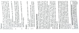
  • 2. Fig. 19.5: Patont duuctus arlerionuns (PDA) ontd. substantial, the neonate becomes short of broath. The additional blood is re-circulated through the lungs and roturned to the left atrium and left ventricle. This causes increased workload on the left side of the heart and increased pulmonary vascular congestion and possibly resistance, and potentialy increased right ventricular pressure and hypertrophy. The additional fluid returning to the lungs increases lung pressure to the pointthat the neonate has greater difficulty inflating the lungs. This uses more calories than normal and often interferes with feeding in infancy. This condition, as a constellation of findings, is called congestive heart failure. In some cases, such as in transposition of the greatvessels (the pulmonary arlery and the aorta), a PDA may need to remain open. In this cardiovascular condition, the PDA is the only way that oxygenated blood can mix with deoxygenated blood. In | these cases, prostaglandins are used to keep the DA open Clinical Features The symptoms of PDA depend on the size of the DA and how much blood flow it carries. Babies with a large PDA might experience symptoms such as a bounding (strong and forceful) pulses result from runoff of bloou from the aorta to the pulmonary artery, fast breathing poor feeding habits, shortness of breath, sweating whie feeding, tiring very easily, poor growth.
  • 3. Patients may be are at risk be asymptom or show sign The N l o n a r y V a s c u l a h r o n i ce x c o Signs of for bacterial endocarditis and ructive disease in later life due ssive pulmonary blood flow. 0eutic Management Therapeut ciatheter-based, ethacin thebased procedures, and surgery. Administration (prostaglane inhibitor) has proved e t r e a t m e n t options tor PDA are medication, sing PDA in premature infants ful in clo newborns. It is usually given orally or l indomethaci stuessti i n s o m e svenously in dose ot0.2 mg/kg of body weight and int the ageof ten days. The dose may be repeated up ree times, at an interval of 12 to 24 hours. based procedure: A catheter (a thin, flexible through their veins using special X-rays a c r o s s thePDAto reaches the heart.A special plug is then inserted remain in position wi ithin the ductus. This is called 1PDAs to prevent the risk of infective endocarditis, C a t h e t e r is fed until it toblockblood tlow to the heart. The plug scatheter device closure.' Sometimes it is done on smallPE e procedure will be able to go home the same day. ction of the lining of the heart, valves, or arteries. lasts approximately two hours and child Surgery Gurgery of PDA is done if the size of the opening is large enough that the lungs could become overloaded with hlood, a condition that can lead to an enlarged heart. A PDAalsomightbe closed to reducethe riskofdeveloping a heart infection known as endocarditis, which affects the tissue lining the heart and blood vessels. Atrioventricular Septal Defect An atrioventricular septal defect (AVSD) is a birth defect of the heart in which important parts at the center of the heart are not fully formed. There are holes between chambers of the heart, and the valves that control the flow of blood between these chambers may not be Tormed correctly. This means that blood flows where it OTmally should not be able to, and extra blood flows to e lungs. This defect is also known as atrioventricular Lanal (AV canal) defect or endocardial cushion deteet. In A septal defect, there is a hole in the wall the prum between the right and left ventricles. nave just one largervalve openingin the middle, instead between the right and left atria. There is also a hole in Inaddition, the tormed correctly. wo a t r i o v e n t r i c u l a r valves are not tone on eachside oftheheart. A baby with AV septal detect may
  • 4. Ogether these problems may ereate a hole in the enter ot the baby's heart. As a result, blood does not tlow the wav it should between the chambers. So the neart has to work harder to pump blood to the lungs and the rest of the body. This condition is also known as atrioventricular canal detect or endocardial cushion delect. Clinical Features Usually these patients have moderate to severe Ci Ihere is a characteristic murmur. Mild cyanoSIS 1s observed, which increases in crying. These children are at high risk for developing pulmonary vascular obstructive disease. Treatments All AVSDs, both partial and complete types, usually require surgery. Surgery depends on how sick the child IS and the specific structure of the AVSD. If possible, surgery should be done before there is pernmanent damage to the lungs. As short-term method medication may be used to treat congestive heart failure, but it is only a short-term measure until the infant is strong enough for surgery. During surgery, any hole in the septa is closed using patches. Sometimes the mitral valve does not close completely, allows blood to flow backwards and make the heart work harder to get enough blood to the rest of the body. This leaky mitral valves need to be repaired or replaced. For a complete AVSD, the common valve is separated into two distinct valves- one on the right side and one on the left. With proper treatment, most babies with AVSD grow up to lead healthy, productive lives. OBSTRUCTIVE HEART DISEASES In an obstructive disorder, the blood flow is restricted or completely blocked. This blockage or narrowing can occur in any of the tour heart valves or above or below the valve. The blockage (atresia) or narrowing (stenosis) can occur in vessels returning blood to the heart (veins) or in vessels pumping blood out of the heart (arteries). Unfortunately, even the most impressive structures, such as the heart, can have problems. Valves do not always work the way they should. If a valve is not formed properly from birth (congenital valve disease) or if it is damaged at some point after birth from age or disease (acquired valve disease), then vital organs, such as the brain and kidneys, may not get the oxygen-rich blood they need to function.
  • 5. B0 Pediatric Nursing Heart valve disease (sometimes called valvular heart disease) can strain the heart, too. But when the valves are defective or do not work the way they should, it can put the heart and other organs at risk. Valve disease can affect one or more of the four valves in the heart (mitral, aortic, tricuspid, and pulmonary). Most often it causes one or both of the following problems: Regurgitation: The valves tissue flaps, or leaflets, that control the flow and direction of the blood, do not fully close, which causes blood to leak back into the heart. Stenosis:The leaflets cannotopen fully to allow enough blood to flow through. The heart has to work harder to compensate for the faulty valve, which can weaken the heart and increase the risk of heart failure (a condition where the heart does not fill up with enough blood or pump enough blood to supply the body with the oxygen and nutrients that it needs) or sudden cardiac arrest (when the heart stops beating). A heart valve problem can also increase the risk of blood clots, which can cause stroke. Valve defects Defective valves may be caused by: Stenosis (narrowing): The valve is not able to open completely. As a result, the heart has to work harder to pump blood through it. Regurgitation: The valve does not close correctly and allows blood to leak backward. Atresia: The valve is missing a hole for the blood to pass through. This is considered a more complex defect. Pulmonary Stenosis Pulmonary stenosis accounts for approximately 5 to 8% of all CHD. Pulmonary stenosis is a narrowing at the entrance to the pulmonary artery (pulmonary valve), and the pulmonary blood flow is decreased. The pulmonary valve is within the heare's right ventricle. Because of the narrowing of the valve, the right ventricle needs to work harder to get blood past the blockage and get hypertrophied. Pulmonary atresia is the extreme form of PS in which the pulmonary valve orifice fails to develop. The valve is completely closed thereby obstructing the outflow of blood from the heart to the lungs. The right ventricle may be hypoplastic. Pathophysiology and Altered Hemodynamics The main pathophysiological consequence of PS is RV strain and an increase in RV pressure. The cellular effect on the RV depends on the timing of obstruction and on the size of Contd..
  • 6. obstruction. If the obstruction is present in a fo. the myocardial response is hyperplasia and a eona vascularity. There is an increase in myocyte siza without an increase in capillary network develops within mature myocardium. Generally children with mild to moderate obstrs increase in ypertophyy when the obstruction are hemodynamically well tolerated and are not a iated on a with cyanosis or cardiac symptoms. In the severe to critica form of PS, right ventricular pressure increases a and causes thickening of right ventricular wall. Right atrial pressure increases i hypertrophy is severe, resulting to right to left shuntiheR foramen of ovale. Cyanosis becomes apparent when re is a right-to-left shunt at the atrial level via an atria septal defectand exaggerated by limited blood flow to the pulmonary vascu bed. If there is no atrial or ventricular level shunt, sudt ncular death may ensue due to compromised cardiac output Clinical Manifestations The most obvious symptom 1s blue, or cyanotic skin in a newborn wh can be noted shortly after bith or several weeks later as the ductus arteriosus closec Clinical symptoms such as dyspnea, heart murmur exercise intolerance, cyanosis, and syncope may occur. In severe PS, the RV will eventually fail as the myocardium becomes unable to support the enhanced work load imposed by the stenosis, and patients develop jugular venous distension, peripheral edema, pleural effusion, ascites, and hepatomegaly. Severe and critical PS may be differentiated clinically by the presence of cyanosis (more pronounced with critical PS) and symptoms of profound heart failure (present with critical PS). Diagnosis Cardiac examination: A systolic ejection mumur is heard best at upper sterna border. Electrocardiogram: The ECG may be normal or show right ventricular hypertrophy with mild to moderate PS. In severe PS, the ECG shows right ventricular hypertrophy and enlargement of right atrium. Chest radiograph: Chest X-ray shows right ventricular hypertrophy and post-stenotic pulmonary artery dialatation. Echocardiogram: The structure of the pulmonary valve the location and severity of the narrowing (stenosis),an the size of the RV and its out flow tract can be visualized d Other imaging tests: Magnetic resonance imaginga CT scans are sometimes used to confirm the diagnosi pulmonary valve stenosis. Cardiac catheterization: During the procedure the .pressure and oxygen measurements are taken the
  • 7. ambers theiheart, as well as the pulmonary isualize the ructures inside the heart. This u r a r t e r ya n d nd aorta. Contrast dye is also injected to more h a r l y generally only done whern child will need balloon pulmonary valve stenosis because be done at the same time as cardiac s t valvuloplasBy to treat that catheterization. M a n a g e m e n t cases of pulmona stenosis are mild and do not S o m e uire. treatment exceptfor routine checkups. However, is more serious child will most likely be admitted once symptoms are noted. Initially, child may be placed ifcasei intensive care unit (ICU) or special care nursery the Oxygen, and possib even on a ventilator, to assist his or her breathing. V medicati may be given to ofpulmonary atresia, as it stops the ductus arteriosus from closing, wing mixing of the pulmonary and systemic asit can cause apnea. Another example of preliminary on lungs function more efficiently. An IV helpthe heart and. m e d i c a t i ation called prostaglandin E1 is used for treatment circulations But prostaglandin E1 can be dangerous atment is heartcatheterization to evaluate the defect or efects of the heart; this procedure is much more invasive. Surgery Surgical repair may be needed for child. The procedure aims at relieving the obstruction of blood flow through the pulmonary valve. This procedure requires heart- lung bypass. Postoperative hospital stay averages 5 to 7days. Child may need either balloon valvuloplasty or open-heart surgery. The decision to perform a balloon vavuloplasty or open-heart surgery depends on the extent to which the pulmonary valve is obstructed. Pulmonary stenosis is classified as mild, moderate 0r severe, depending on a measurement of the blood Presure difference between the right ventricle and pulmonary artery. nav aseries of surgeries to improve the blood flow Ultimately, however, the patient will needto ently. The first surgery will likely be pertormed hortly after birth. A aorta and the pulmonary How to the lungs. As the child grows, so does the heart shunt can be created between the artery to help increaseblood andthe shunt body's requirements. The type of surgery recommended depends on the Size of the rightventricleand the pulmonaryartery may need revised in order to meet the
  • 8. If they are norm in size 1s able to pump blood, open heart surgery can performed to make blood flow through the heart "d normal pattern. and the right ventricle be IT the right ventricle is small and unable to act as a PLmp, doctors may perform another type of operatIO Caled the Fontan procedure. In this three-stag8 procedure, the right atrium is disconnected from tne Puimonary circulation. The systemic venous return gOes directly to the lungs, by-passing the heart. Another treatment option involves the electrosurgical puncture of the atretic valve usinga wire introduced into the patient percutaneously. The puncture is then dilatea using a balloon catheter to allow blood flow from the right ventricle into the pulmonary artery. The outcome varies for every child. If the condition is left uncorrected it may be fatal, but the prognosis has greatly improved over the years for babies with pulmonary atresia. Some factors that affect, how wel the child does include how well the heart is beating the condition of the blood vessels that supply the heart, and how leaky the other heart valves are. Most cases of pulmonary atresia can be helped with surgery. If the patient's right ventricle is exceptionally small, many Surgeries will be needed in order to help stimulate normal circulation of blood to the heart. Aortic Stenosis Aortic stenosis acounts for approximately 5% of all CHD. It is more common in males than females. The aorta is the large artery that supplies oxygen-rich blood to the body. The aortic valve is within the heart's left ventricle and acts as the entrance to the aorta. Narrowing of the aortic valve, which controls blood flow between left ventricle and aorta, is known as aortic stenosis. Depending on the severity of the stenosis, the symptoms at birth can vary from none to decreased blood flow and decreased oxygenation to the body. As the PDA closes, the symptoms usually become more acute. Aortic sternosis is three types: 1. The commonest valvular type is the stricture of aortic valve 2. In subvalvular type, the narrowing remains below the aortic valve 3. In supravalvular, the stenosis remains above the aortic valve.
  • 9. Pathophysiology and Altered Hemodynamics e human aortic valve normally consists of three leatlets neaflefs) and has an orifice of 3.0-4.0 square centimeters. aortic stenosis, the opening of the aortic valve becomes narrowed or constricted (stenotic) S8 Consequence of this stenosis, the left ventricle must generate a higher pressure with each contraction to ettectively move blood forward into the aorta. Increased pressure in tne LV has direct correlation with the degree of obstruction in aortic valve. Ihitially, the LV generates this increased pressure by thickening its muscular walls (myocardial hypertropny). he type of hypertrophy usually seen in aortic stenosIS IS Known as concentric hypertrophy, in which the walls of the Lv are (approximately) equally thickened. In the later stages, the eft ventricle dilates, the wall thins, and the systolic function deteriorates (resulting in impaired ability to pump blood forwara) Heart failure can develop due to this heavy workload. In severe cases, pulmonary edema may occur as a result of increased ert ventricular pressure that may cause a backflow of blood into the lungs. Sometimes, a percutaneous balloon valvuloplasty (opening of a valve) can effectively relieve the valve obstruction. This procedure involves a special catheter (tube) containing a balloon being passed through the aortic valve. The surgeon inflates the balloon to stretch the valve open. The surgical repair aims to relieve the obstruction of blood flow through the aortic valve. This procedure requires heart-lung bypass. Postoperative hospital stay averages 7 to 10 days. Clinical Manifestation Less severe forms of the disease may be 'silent - causing no visible symptoms-until later in life. As the child grows older, increased physical growth requires additional cardiac output. Children may present the following symptoms in severe obstruction. Fatigue and exercise intolerance Having pain, pressure or tightness in their chest Exertional dyspnea Fainting or feeling weak or dizzy when active Having palpitations Babies born with severe aortic stenosis may have to work hard to breathe, have poor appetite or trouble feeding, and failure to thrive. They may also show signs of shock because their heart cannot pump enough blood to the rest of their body. Signs of cardiogenic shock include less frequent urination, cool limbs, increased heart rate, fussiness, poor feeding, rapid breathing, lethargy, mottled skin. They have decreased perfusion and pulmonary congestion. Diagnosis Cardiac examination: A harsh systolic murmur, click, or other abnormal sound is almost always heard at the upper right sternal border, radiating to the upper left sternal
  • 10. border and neck. A vibration or moue can when placing a hand over the hcart. T re ht may car be faint the pulse in the nee pulse or changes in the quality of the pule neck. be Electrocardiogram: The ECG may normal demonstrate left ventriculan hypertrophy. Tmal heart svze is seen. Chest radiograph: Usually normal heart. In severe case of sternosis, dilatation of may be seen. n of ascending aorta well as presence Echocardiogram: Type of stenosis as well of other cardiac defects can be seen. The function of left ventricle and thickness ot its wall is also assessed. Management Regular checkups by a health provider m may be all no symptoms or only mild should ask about that is needed if there is symptoms. The health care provider child's health history, do a physical exam, and perform an echocardiogram. People with severe aortic stenosis may be told not not to play competitive sports, even if they have no sympto If symptoms do occur, strenuous activity must ofteni limited. Medicines are used to treat symptoms of heart failure or abnormal heart rhythms (most commonlv atrial fibrillation). These include diuretics, nitrates, and beta-blockers. High blood pressure should also be treated. If aortic stenosis is severe, this treatment must be done carefully so blood pressure does not drop to dangerously low levels. Treatment may be done in the cardiac catheterization lab using a balloon procedure. A balloon is inserted across the valve. When the balloon is inflated, the valve is stretched open. Then the balloon is removed. This is called balloon valvuloplasty. Some children need surgery to replace their aortic valve with an artificial valve. In some cases the child's n De own pulmonary valve can be used to replace the damaged aortic valve. Aortic Coarctation Coarctation of the aorta is a narrowing of some porto of the aorta (Fig. 19.6A). This narrowing is usuay found just past the arch of the aorta, opposite area of the PDA. Coarctation of the aorta accounts nly approximately 8% of all CHD, it is more com 2ortic tound in male and may accompany other enital h defects like VSD, PDA, tubular hypoplasia or tne isthmus and bicuspid aortic valve.
  • 11. C l i n i c a l M a n i f e s t a t i o n a n d usually become evident after the closure of PDA. The c l i n i c a l features depend upon the type of obstruction The p r e s e n t i r features of neonates are severe CCF, poor rfusion, tachypnea, acidosis and absence of femoral pulse. Theolder e.The older children'sgrowth and development may normal wit normal without symptoms, but overgrowth of upper may be suggestive of CoA. The symptoms may include: extremities and hypertension, bsence of femoral pulse Increased BP in the upper part of the body, resulting in headache, dizziness, fainting, epistaxis and later CVA, encephalopathy. Child may occasionally complaints of weakness and pain in legs after exercise. The femoral and pedal pulse may be absent or diminished, and legs may be cooler than arms. Other manifestations are fatigue, cramps, exertional dyspnea, feeding problem, poor weight gain, irritability and tachycardia. Diagnosis Diagnosis is confirmed by cardiac examination, chest X-ray, barium swallow, electrocardiogram, M cardiac examination, mode echocardiography, cardiac catheterization and angiocardiography. Specific ' E' sign in barium swallow very suggestive of CoA. The first arch ofthe 'E' is Tormed due to dilatation of aorta before the coarctation, ne second arch due to poststenotic dilatation, and middle notch due to the coarctation. Management Medical m a n a g e m e n t of CoA is done with PGE1 nfusion, a n t i b i o t i c s and prevention and t r e a t m e n t of Complications.
  • 12. 483 Cardiovascular System 999 Figs 19.6A and B: A. Coarctation of the aorta, B.Correction of coarctation of the aorta Surgical correction of CoA (Fig. 19.6B) Individualizedto anatomy of CoA lan to include treatment of any additional cardiac defects. Child, Adolescent: Repair is done at 2 to 3 years of age upon diagnosis. Surgery: Four common types of repair-regardless or Technique, usually performed via a left thoracotomy incision. End-to-end anastomosis was done in 1954 by Crawford and Nylin. Excision of CoA area, circumferential anastomosis Is Completed with interrupted sutures anteriorly NURSING CARE OF THE FAMILY AND CHILD WITH CONGENITAL CARDIAC DISEASES The diagnosis of a chronic condition in any family member causes much distress. When an infant is born with a heart defect, the parents may grieve over the loss of the healthy newborn they had anticipated and experience shock, denial, guilt, anger, despair or confusion on learning that their infant has a cardiac defect. Some parents may be unable at first to respond to their newborn. Even greater stress may occur if the condition is one that requires surgical intervention. The nurse should become familiar with parents reaction and nursing intervention require to: Help family to adjust to the disorder: A birth of a baby with severe cardiac anomaly is a shock to the family, parents always suffer with high anxiety and fear that their child will not survive. Here role of nurse is to support the family in their stress, assessing their level of understanding and to provide information as needed (thorough but simple in language). She also helps the other members of the health team to understand the parents' reaction. The nurse needs to support the family in their great pain and emotional investment. She can foster parent intant attachment and encourage parents to hold, touch and look at their child.
  • 13. Teach the parent to care the child which can minimize their anxiety and fear. Introduce parents to other families with similarly affected children. It can help them to adjust to their daily stress. Counseling of family about the disorder and its care: Before describing the defect a review of the basic structure and function of heart is important. Picture, diagram, models are to be used for better understanding of CHD. Increasingly, families are collecting information from different sources. It is important to inform the families about authentic internet sites and medical literature. Parents must know that information from general sources or other families might not be applicable to their own situation. Prepare Child and Family for Surgery Some heart defects need repair soon after birth. For others, it is better to wait months or years. Certain heart defects may not need to be repaired. The surgery is needed for the child's well-being. In general, symptoms that indicate that surgery is needed are blue or gray skin, lips, and nail beds (cyanosis). These symptoms mean there is not enough oxygen in the blood (hypoxia). Difficulty breathing because the lungs are 'wet, congested, or filled with fluid (heart failure). Child suffers with problems of heart rate or heart rhythm (arrhythmias). Child shows feeding or sleeping problem and lack of growth and development. The three most common clinical presentations are: 1. A Murmur 2. Cyanosis 3. Respiratory difficulty.
  • 14. N u r s i n g A s s e s s m e n t Cardiovascular assessment is an important n . in the assessment and managementof and children with chronic conditions. Asse cardiac problems is described in Table 19,1 sing skill nent facutely ill children . Assessment for Look for signs of cardiovascular problems: Inspection Palpation Percussion Auscultation. The general objectives of nursing care for the child with CHD include prevention of physical and emotional fatigue, provision of adequate fluid and nutrition, prevention of intection and care and support to the parents and child to cope with the problems that precipitates before and after surgery. Nursing diagnosis: Impaired gas exchange related to disturbed pulmonary blood flow or pulmonary hypertension. Expected outcome: Patient maintains optimal gas exchange as evidenced by normal ABGs and alert responsive mental condition or no further reduction in mental status. Nursing Intervention Patient is to be placed in semi-upright position which facilitates breathing. Position with proper body alignment tor optimal respiratory functioning (if tolerated, head of bed at 45 degrees). This promotes lung expansio1 au improves air exchange. Routinely check the patient's position so he o she does not slide down in bed. This would cause tn
  • 15. bdomen to co aUSe Tespirator to compress the diaphrag: embarrassment. which would effectively clear the airway. Periodic oral and nasal Patient may eed help if he/she is unable to tions isto be suctioned if needed retovgen saturation and administer oxygen Monitor oxYgen saturation eded P r e ing of respiratory pattern inisterdrugs like diureticaand bronchodialators i f r e q u i r e d 1 re n of aspiration, with continuousmonitor A d m i n i s t e r Analysisofblood gas analysis. ow cardiac output related to Nursing diagnosis: Low reducedmyocardial functions. outcome: Cardiac e c t e d lerance of child is improved. output and activity Nursingintervention Bed ed rest, minimum exercise (care in the bed, indoor play, and other activities of daily living) Administer prescribed drugs like digoxin, diuretics, antihypertensives which improve cardiac output , Organize medication and nursing schedule to provide periods of uninterrupted sleep . Monitor child's condition (heart sound, vital signs). Nursing diagnosis: Activity intolerance related to hypoxia. Fxpected outcome: The child is able to do age appropri- ate activity. Nursing Intervention Oxygen therapy and continuous monitoring of oxygen saturation by pulse oximetry. Pulse oximetry is a useful tool to detect changes in oxygenation. O, saturation should be maintained at 90% or greater. Ahigher liter flow of oxygen is generally required for activity versus rest (e.g. 2 L at rest, and 4 L with activity). Uninterrupted period ofrestand sleep Advice parents to do activities of daily living for the child so that child's energy can be conserved Providing timely feeding, changing diaper and tactile stimulation prevents cry of the child and Saves energy expenditure. Prevent excessive crying provide diversional activities, prevent constipation. Nursi diagnosis: Altered nutrition, less than body rements related to excessive energy demarnds ded by increased cardiac workload. Expected Outcome: Normal nutritional status of child.
  • 16. CordiovgsCular ysterri Nursing Intervention Feed in semi erect position sastric feeding may be started if the ci nable to take oral fecds or get cvanosed while feeai6 Provide small frequent feedings TOvide fomds with high nutritional value (24 Ka o formula) Determine child's likes and dislikes Monitor input and output Daly weight monitoring: Maintaining a healtny Weight is important for cardiac health. Restricting intake of salt can also help lessen fluid retention ana mprove symptoms related to heart valve disease. Assess the child for developmental milestones. VUSIng diagnosis: Increased potential for infection related to poor nutritional status. Expected outcome: No infection. Nursing Intervention revent exposure to communicable diseases. Early detection and treatment of upper respiratory and ui infection. Immunizations shouldbe up-to-date Handwashing should be observed. Maintaining general cleanliness and hygienic measure are important. Be certain that the child receives prophylactic medi- cation for infective endocarditis. Congestive Cardiac Failure (CCF) Every cardiac patient has a potential for developing CCF. Congestive cardiac failure (CCF) by itself is not a diagnosis. It is a clinical syndrome caused by different natomical and or pathological conditions, which is the primary diagnosis. CCF is a term defined as 'inability of the heart to pump enough blood out to the rest of the body, at rest or during stress, necessary for the metabolic needs of the body (systolic failure) and inability to receive blood into the ventricular cavities at low pressure during diastole (diastolic pressure)'. Etiology The etiology of pediatric heart failure may be cardiac or noncardiac and can occur at any age. The predominant etiology during infancy is congenital heart disease. VSD is the commonest defect presenting with heart failure. Myocardial disease especially myocarditis is commonest cause HF in children under 5 years. Rheumatic fever and rheumatic heart disease continue to be an important
  • 17. cause of suffering and HF among children above 5 years. Among noncardiac disease, severe anemia can be a condition to cardiac decompensation at any age. In children, cardiac failure is most often caused by congenital heart disease (left to right shunts) and cardiomyopathy.
  • 18. Clinical Features CCF may become suddenly dyspneic, cyanotic. A young child may develop abdominal pain, The child with tever, anorexia, dyspnea, cough suddenly. Dyspnea, be orthopnea or paroxysmal nocturnal dyspnea can be reported by parent. Sometimes a mother may complaint of palpitation, tachycardia and profuse sweating, pallor or peripheral cyanosis and cold extremities of child. Symptoms are different for children of different age, in babies regardless of the cause of CCF the end result of significant heart failure is poor growth. Slow weight gain is related to two factors. Because of easy fatigability baby takes small feed and there is an excessive loss of calories from increased work of breathing associated
  • 19. with CCF. In addition, as the lungs fill with becomes more difficult for babies to hro fluid, it to breathe and they will use more of the muscles of their chest and belly they to and become very sweaty as of the extra work needed compensate. The baby breathes too fast dri. ing feeding to eat. Uncommonly, there may be an unusual weight gain due to collection of water, manifesting s facial puffiness or as rarely as edema on the feet may be brought with the complaints of persistent hoa crying, breathes too fast with wheezing and excessive the feet. The bal perspiration, restlessness. Diagnosis Physical examination: A child with CCF may presen tachycardia, tachypnea, Gallop rhythm, dyspnea, decreased peripheral pulse and mottling of the extremities, delayed capillary refil, failure to thrive, decreased activity tolerance, sweating, etc. (Table19.2) ChestX-ray: Very commonly X-ray shows cardiomegaly. The heart dialates or hypertrophy occurs both in presence of volume and pressure overload, cardiomyopathy or dysrhythmias. It may gives clues to certain structural heart diseases, and also to right versus left sided heart involvement. Pulmonary markings are often increased, showing pulmonary congestion. ECG: Usually abnormal, and although not useful in assessing HF, may give diagnostic cues for the underlying pathology of heart failure. Urine test: In chronic HF, proteinuria and high specific gravity of urine are common. Blood test: As renal function decreased due to decreased perfusion in CCF, an increase in blood urea nitrogen and creatinine levels may be present. CBC, differentna may give clues to anemia and infection causing complicating HF. Atrial blood gas analysis isimpoTLa Echocardiogram: This test is valuable for evaluau cardiac function and ruling out structural heart disea tests Other tests: Thyroid, renal and hepatic functio are also valuable.
  • 20. rsing Management of CCF utic and NN 7 cardiovascular medicines e October W H O E s s e n t i Therapeutc r n t a i n s d i g o x i n , n t h e complem.. u s e n h a t n a g e m e n t a r e to: rdiac work educingcard sential Medicines List for children (EML section of frusemide, spironolactone an entary list, and, dopamine. The goals of enting myocardial contractility Removeaccumulated fluid and sodium ve tissue oxygenation and decreased oxygen consumption. This is accomplished by on) and using medication to sedate an irritable educing cardiac work: The workload on the heart is needs are kept to a minimum limiting physical activities body temperature, treating hen metabolic bedrest) preserving reducing the effort of breathing (semi Fowler's any p o s i t i o r child Supplement cool humidified oxygen is usuallv amount of oxygen during provided to increase th Baby with CCF is kept in bed rest with minimal degree is maintain It may help the inspiration. handling. ing, The propped up position with an incline about 30 of work of eathing by pooling the edema fluid in the Aanendent areas and to reduce the collection of fluid in lungs. Administration of humidified oxygen (40- 50% concentration) improves impaired oxygenation Cecondary to pulmonary congestion, thus reducing the work of heart by reducing requirements of cardiac output. Administration of Morphine sulfate (0.05 mg/kg SC) or other sedative (Diazepam) may be needed if the child is restless or dyspneic. These drugs reduce anxiety and lower the catecholamine secretion, thus reducing physical activity, the respiratory rate and the heart rate. The workload of heart comes down as the oxygen demand of the body tissues goes down. The sedative also helps in keeping the child in bed. Management of feve, anemia and infection necessary to reduce the workload of heart. At a nperature of 36 to 37 °C, the overall circulatory and eTabolic needs are minimal, thus reducing work or eart. Anemia causes tachycardia and hyperkine culatory state to meetupOxygen demandofthe body. orrection of anemia results in decreased cardiac work. ansfusion is indicated,,3 to 5mL packed red cells/kg reight can be givens every 12 hours. Worseningof transfusion can be prevented, the patient can be Corre body CCF by given frusemide.
  • 21. n be managed by use of vasodialators. Cauces the arteriolarand venous vasoconstrictoy Cuuce the work of heart. "The use of ACE inhibitors is now (captopril well-established in infants and children aptopril dose is 1 mg/kg 8 hourly and can be give creased up to 6 1mg/kg/day). ACE inhibitors Suppre Enin angiotensin/aldosterone system, thus reducin8 soconstriction as well as sodium and water retentio reduce They prevent potassium loss and hence d arrhythmias. These drugs suppress catecholamines al thus its ill effect on myocardium as well as arrhythmias. Augmenting myocardial contractility: Iontropic drugs mprove cardiac output. In infants and children digitalis 8ycosides [digoxin (lanoxin)] is used which decreases heart rate and increases mvocardial contractility. It has rapid onset of action and also eliminated quickly. Besides digitalis, catecholamine ionotropic agents like dopamine has been found to be useful. In a patient with CCF if the blood pressure is low, dopamine may be used. At a dose of less than 5 ug/kg/min of Dopamine causes peripheral vasodilation, increases myocardial contractility and renal blood flow resulting in natriuresis (Table 19.3). Remove accumulated fluid and sodium: Treatment consists of diuretics, possible fluid restriction and possible sodium restriction. Diuretic is used to eliminate excess water and salt to prevent re-accumulation. It reduces total body sodium, thus reducing the blood pressure and peripheral vascular resistance. The potent oral diuretics frusemide is administered which starts its action within 20 minutes. Frusemide interferes with the sodium reabsorption mechanism in descending limb of loop of Henle. Patient on frusemide should be given potassium supplement. Studies suggest that it is preferable to combine frusemide with potassium sparing diuretic. Thee combination is more useful in preventing potassium and magnesium loss, thus reducing arrhythmias as against the combination of frusemide and potassium supplement. ACE inhibitor I drug can be combined with frusemide, if indicated. The patient on ACE I should neither be given potassium supplements or potassium The
  • 22. sparing diuretics like triamterene, amiloride or aldactone even in combination with frusemide, Nursing Diagnoses mpaired gas exchange related to altered pulmonary blood flow or oxygen deprivation Activity intolerance related to decreased cardiac Output due to structural defect and myocardial dysfunction Altered nutrition: Less than body requirements related to the excessive energy demands required by increased cardiac workload Fluid volume excess related to fluid accumulation (edema) Increased potential for respiratory infection secondary to pulmonary vasculature overloading and other infection related to poor nutritional status Anxiety related to diagnostic procedures and hospitalization Developmental delay related to decreased energy inadequate nutrition, physical limitations and social isolation Alteration in parenting related to parental perception of the child as vulnerable. Nursing Intervention and Outcome Impaired gas exchange related to altered pulmonary blood flow or oxygen deprivation. Goal of nursing action: The patient will exhibit improved respiratory function. Nursing intervention includes placing the child in inclined posture of 30 to 45 degree. Tilt mattress support of incubator, place older infant in sent (Fowler's position). Avoid any constricting clothing or restraints around abdomen and chest. Administer humidified O, as prescribed. Expected Outcome: Respirations remain with normal limits; color is good and child rest quietly. Activity intolerance related to decreased cardiac output due to structural defect and myocardial dysfunction (Table 19.4). Goal of nursing action: The patient will exhibit improved cardiac output. Nursing intervention includes administration of digoxin (lanoxin) at regular interval (usually every 12 hours). Nursing established precaution to prevent toxicity. Often an ECG rhythm strip is taken to assess
  • 23. cardiac status before administration. Ensure adequate els (decrease intake of K. Monitor serum potassium levels (dece to enhances digoxin toxicity). Administer medicatione decrease over load as ordered. Check blood pressur Observe for signs of hypotension. Monitor electrolvh e levels. Expected outcome: Heartbeat is strong, regular and within normal limits for age. Peripheral perfusion is adequate. Goal (2): The patient will experience reduction of anxiety. Nursing intervention: Employ flexible feeding schedule. Handle child gently. Hold and comfort the infant. Employ comfort measures found effective in individual cases. Encourage family to provide comfort. Expected outcome: Infant rests quietly and breath easily. Altered nutrition: Less than body requirements related to the excessive energy demands required by increased caráuac workload. Goal of nursing action: The patient will able to conserve energy arnd increase total intake. Nursing Intervention: A child with CCF needs small frequent feeds,asn she has problem in sucking and swallowing a breathing simultaneously.