Cleft lip and palate /certified fixed orthodontic courses by Indian dental academy
•
8 likes•2,111 views

The Indian Dental Academy is the Leader in continuing dental education , training dentists in all aspects of dentistry and offering a wide range of dental certified courses in different formats. Indian dental academy provides dental crown & Bridge,rotary endodontics,fixed orthodontics, Dental implants courses.for details pls visit www.indiandentalacademy.com ,or call 0091-9248678078

Recommended
Recommended
More Related Content
What's hot
What's hot (20)
Viewers also liked
Viewers also liked (10)
Similar to Cleft lip and palate /certified fixed orthodontic courses by Indian dental academy
Similar to Cleft lip and palate /certified fixed orthodontic courses by Indian dental academy (20)
More from Indian dental academy
More from Indian dental academy (20)
Recently uploaded
Recently uploaded (20)
Cleft lip and palate /certified fixed orthodontic courses by Indian dental academy
- 2. INDIAN DENTAL ACADEMY Leader in continuing dental education www.indiandentalacademy.com www.indiandentalacademy.com
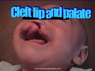
- 3. INTRODUCTION Cleft lip and palate is one of the most common congenital anomalies afflicting humans. The potential problems that may beset the unfortunate victims of the condition include such severe handicaps as impaired suckling and resultant failure to thrive, speech impediment, deafness, malocclusion, gross facial deformity, and severe psychological problems. www.indiandentalacademy.com
- 4. The clefting of lip and/or palate occurs at such a strategic place in the orofacial region and at such a crucial time (before birth) that it becomes a complex congenital deformity and a major therapeutic challenge incapable of resolution by any individual clinician and it warrants the coordinated skills of many modern specialists for its correction. Plastic surgeons, pediatricians, orthodontists, prosthodontists, speech pathologists, otolaryngologists, anesthesiologists, and audiologists form a less than comprehensive list of those who may be required to provide a high degree of interdisciplinary communication and understanding in the treatment of cleft lip and palate deformities. www.indiandentalacademy.com
- 5. HISTORY In ancient times many congenital deformities including cleft lip and palate, were considered to be an evidence of evil spirit in the afflicted child. These children were often removed from the tribe or cultural unit and left to die in the surrounding wilderness. The repair of cleft lip is noted in an ancient monograph during the Chin Dynasty (255-206 BC) and is the earliest report of such surgery any where in the world. Jehan Yipperman, a Flemish surgeon (1295-1351), LeMonnier, a French dentist (1753) described in detail the repair of a cleft lip and palate patient. In the United States, Josiah Flagg, surgeon- dentist, advertised in a handbill in Boston in 1786, ………. that among the many other things he does, he also sews up hare lip. www.indiandentalacademy.com
- 6. Stevens performed the first cleft palate repair in the United States in 1827. Fauchard, in 1786, made valuable contributions and innovation in prosthodontics, particularly in the area of obturators for cleft lip and palate patients. Kingsley who shares with others the claim to be the Father of Orthodontia has written over 100 articles on cleft palate rehabilitation, the inadequacies of cleft palate surgery, obturators, orthodontic diagnosis and orthodontic appliances bear his name. His first book, a treatise on Oral Deformities, as a Branch of Mechanical Surgery, in 1880, featured the orthodontic appliances of the period, including jack screws, retainers and arches with ligatures. www.indiandentalacademy.com
- 7. Calvin Case published a Prosthetic Treatise on the Techniques and Principles of Dental Orthopedia and Prosthetic Correction of the Cleft Palate in 1921. The appliances described were all of the fixed type. It was only a few years ago that orthodontists demonstrated that palatal surgery interfered with maxillary growth. Surgeons were asked not to repair the palate until maxillary growth was complete so as to avoid midfacial deformity. At the same time speech and language pathologists were requesting for early operation on palate to prevent the development of compensatory mechanisms of articulations. www.indiandentalacademy.com
- 8. Taking note of the suggestions made by both orthodontists and speech pathologists the surgeons have modified their techniques, the timing of surgery, and the number of palatal procedures that were performed. Along with surgical refinements, advances have been made in orthodontics and other fields concerned with rehabilitation of cleft lip and palate to such an extent that, the treatment of children with clefts have resulted in a cleft population that is distinctly different from the facially crippled stereo-types of previous generations. www.indiandentalacademy.com
- 10. EMBRYOLOGY OF CLEFT LIP AND PALATE Ø Ralph Millard Jr. once said, “Though various theories have been put forth to enlighten the panorama of the embryological basis, no theory enjoys universal acceptance as no one has been an eye witness to the entire 'in utero show' as yet and thus it is impossible to propose and prove what exactly is happening.” Ø Wilhelm His popularized the theory that separate facial processes fuse to form the mid portion of the face. Ø It was Richard B. Stark of St. Luke Hospital, New York, in 1954, who wrote extensively explaining mesodermal migration. www.indiandentalacademy.com
- 11. There are five principal stages in craniofacial development: Ø Stage – I Germ layer formation and initial organization of craniofacial structures. Ø Stage – II Neural tube formation and initial formation of the oropharynx. Ø Stage – II Origins, migration and interactions of cell populations, especially neural crest cells and their derivatives. Ø Stage – IV formation of organ systems, especially the pharyngeal arches and the primary and secondary palates, and Ø Stage – V Final differentiation of tissues (skeletal, muscular, and nervous elements). www.indiandentalacademy.com
- 12. EMBRYOLOGY OF FACE, NOSE AND PALATE Ø After the formation of the head fold, the developing brain and the pericardium form two prominent bulging on the ventral aspect of the embryo. Ø These bulging are separated by the stomatodaeum. Ø The floor of the stomatodaeum is formed by the buccopharyngeal membrane, which separates it from the foregut. Ø Soon, mesoderm covering the developing forebrain proliferates, and forms a downward projection frontonasal process that overlaps the upper part of the stomatodaeum. www.indiandentalacademy.com
- 13. Ø The pharyngeal arches are laid down in the lateral and ventral walls of the cranial most part of the foregut. These are also, therefore, in very close relationship to the stomatodaeum. Ø It will now be readily appreciated that the face is derived from the following structures that lie around the stomatodaeum: (a) The frontonasal process and (b) The first pharyngeal (or mandibular) arch of each side. Fig.1-Head end of the embryo just before formation of the frontonasal www.indiandentalacademy.com process
- 26. At this stage each mandibular arch forms the lateral wall of th stomatodaeum. This arch gives off a bud from its dorsal end called th maxillary process. It grows ventro-medially cranial to the main part of the arc which is now called the mandibular process. www.indiandentalacademy.com
- 27. The ectoderm overlaying the frontonasal process soon shows bilateral localized thickenings that are situated a little above the stomatodaeum called the nasal placodes. These placodes soon sink below the surface to form nasal pits. The pits are continuous below with the stomatodaeum. The edges of each pit are raised above the surface; the medial raised edge is called the medial nasal process and the lateral edge is called the lateral nasal process. www.indiandentalacademy.com
- 31. Lower lip The mandibular processes of the two sides grow towards each other and fuse in the midline. They now form the lower margin of the stomatodaeum. The mouth develops from the stomatodaeum The fused mandibular processes give rise to the lower lip, and the lower jaw. www.indiandentalacademy.com
- 35. Upper lip Each maxillary process now grows medially and fuses, first with the lateral nasal process, and then with the medial nasal process. The medial and lateral nasal processes also fuse with each other. In this way the nasal pits (now called anterior nares) are cut off from the stomatodaeum. The maxillary processes undergo considerable growth. The frontonasal process becomes much narrower from side to side, so that the two anterior nares come closer together. www.indiandentalacademy.com
- 36. The stomatodaeum is now bounded above by the upper lip which is derived as follows: (i) The mesodermal basis of the lateral part of the lip is formed the maxillary process. (ii) The overlaying skin is derived from ectoderm covering this process. (iii) The mesodermal basis of the lip (called philtrum) is formed from the frontonasal process. (iv) The ectoderm of the maxillary process, however, overgrows this mesoderm to meet that of the opposite maxillary process in the midline. (v) As a result, the skin of the entire upper lip is innervated by the maxillary nerves. www.indiandentalacademy.com
- 37. The muscles of the face (including the lips) are derived from mesoderm of the second branchial arch and are, therefore, supplied by the facial nerve. www.indiandentalacademy.com
- 38. Nose The anterior nares are formed when the nasal pits are cut off from the stomatodaeum by the fusion of the maxillary process with the medial nasal process. The anterior nares gradually approach each other. This is a result of the fact that the frontonasal process becomes progressively narrower and its deeper part ultimately forms the nasal septum. Mesoderm becomes heaped up in the median plane to form the prominence of the nose. Simultaneously, a groove appears between the region of the nose and the bulging forebrain (which may now be called forehead). As the nose becomes prominent the anterior nares come to open downwards instead of forwards. The external form of www.indiandentalacademy.com the nose is thus established.
- 39. Cheeks After formation of the upper and lower lips the stomatodaeum (which can now be called the mouth) is very broad. In its lateral part, it is bound above by the maxillary process and below by the mandibular process. These processes undergo progressive fusion with each other to form the cheeks. During the formation of upper lip the maxillary process fuses with the lateral nasal process. www.indiandentalacademy.com
- 40. This fusion occurs, not only in the region of the lip, but also extends from the stomatodaeum to the medial angle of the developing eye. For some time this line of fusion is marked by a groove called the naso-optic furrow or nasolacrimal sulcus. A strip of ectoderm becomes buried along this furrow and gives rise to the nasolacrimal duct. www.indiandentalacademy.com
- 41. Development anomalies of the face It has been that the formation of various parts of the face involves fusion of diverse components. This fusion is occasionally incomplete and gives rise to various anomalies. www.indiandentalacademy.com
- 42. Harelip The upper lip of the hare normally has a cleft. Hence the term hare-lip is used for clefts of the lips. When one or both maxillary processes do not fuse with the medial nasal process, this gives rise to defects in the upper lip. These may vary in degree and may be unilateral or bilateral. Defective development of the lowermost part of the frontonasal process may give rise to a midline defect of the upper lip. When the two mandibular processes do not fuse with each other the lower lip shows a defect in the midline. The defect usually extends into the jaw. www.indiandentalacademy.com
- 43. Oblique facial cleft Non-fusion of the maxillary and lateral nasal processes gives rise to cleft running from the medial angle of the eye to the mouth. The nasolacrimal duct is not formed. www.indiandentalacademy.com
- 44. INTRODUCTION Cleft lip and palate is one of the most common congenital anomalies afflicting humans. The potential problems that may beast the unfortunate victims of the condition include such severe handicaps as impaired suckling and resultant failure to thrive, speech impediment, deafness, malocclusion, gross facial deformity, and severe psychological problems. www.indiandentalacademy.com
- 45. The clefting of lip and/or palate occurs at such a strategic place in the orofacial region and at such a crucial time (before birth) that it becomes a complex congenital deformity and a major therapeutic challenge incapable of resolution by any individual clinician and it warrants the co-ordinated skills of many modern specialists for its correction. Plastic surgeons, pediatricians, orthodontists, prosthodontists, speech pathologists, otolaryngologists, anesthesiologists, and audiologists form a less than comprehensive list of those who may be required to provide a high degree of interdisciplinary communication and understanding in the treatment of cleft lip and palate deformities. www.indiandentalacademy.com
- 46. The abundance of published material, however, indicates a remarkable lack of consensus in the treatment of these deformities. Each multidisciplinary team develops its own protocol for management of children with cleft lip and palate and those ideas are held tenaciously. What is the effect of surgery as a factor in facial growth. As contrasted with the effect of the inherent growth disturbance associated with the deformity? What is the optimal timing for surgical procedures? By what standards can results be judged? Is there value in using different methods of investigation? These questions have been much discussed over the years and still await resolution. www.indiandentalacademy.com
- 47. One reason for lack of consensus is the apparent inability to design and complete adequate prospective studies that produce unequivocal results and enable comparison with the work of others. The reasons for this failure are apparent. Adequate studies take years- almost the professional lifetime of any one clinician. Often patients move and are lost to follow-up. Professionals move, and others take their place and introduce changes in the original protocol. When a research design must be applied to a complex biological problem, the number of variables may take meaningful standardization almost impossible. www.indiandentalacademy.com
- 48. HISTORY In ancient times many congenital deformities including cleft lip and palate, were considered to be an evidence of evil spirit in the afflicted child. These children were often removed from the tribe or cultural unit and left to die in the surrounding wilderness. www.indiandentalacademy.com
- 49. The repair of cleft lip is noted in an ancient monograph during the Chin Dynasty (255-206 BC) and is the earliest report of such surgery any where in the world. Jehan Yipperman, a Flemish surgeon (1295-1351), LeMonnier, a French dentist (1753) described in detail the repair of a cleft lip and palate patient. In the United States, Josiah Flagg, surgeon- dentist, advertised in a handbill in Boston in 1786, ………. that among the many other things he does, he also sews up hare lip. The first cleft palate repair in the United States was performed by Stevens in 1827. Fauchard, in 1786, made valuable contributions and innovation in prosthodontics, particularly in the area of obturators for cleft lip and palate patients. www.indiandentalacademy.com
- 50. Two men stand out for their early contribution to the orthodontic and prosthodontic treatment of these deformities. Kingsley who shares with others the claim to be the Father of Orthodontia has written over 100 articles on cleft palate rehabilitation, the inadequacies of cleft palate surgery, obturators, orthodontic diagnosis and orthodontic appliances bear his name. His first book, A treatise on Oral Deformities, as a Branch of Mechanical Surgery, in 1880, featured the orthodontic appliances of the period, including jack screws, retainers and arches with ligatures. Calvin Case published A Prosthetic Treatise on the Techniques and Principles of Dental Orthopedia and Prosthetic Correction of the Cleft Palate in 1921. The appliances described were all of the fixed type. www.indiandentalacademy.com
- 51. It was only a few years ago that orthodontists demonstrated that palatal surgery interfered with maxillary growth. Surgeons were asked not to repair the palate until maxillary growth was complete so as to avoid midfacial deformity. At the same time speech and language pathologists were requesting for early operation on palate to prevent the development of compensatory mechanisms of articulations. www.indiandentalacademy.com
- 52. TTaking note of the suggestions made by both orthodontists and speech pathologists the surgeons have modified their techniques, the timing of surgery, and the number of palatal procedures that were performed. Along with surgical refinements, advances have been made in orthodontics and other fields concerned with rehabilitation of cleft lip and palate to such an extent that, the treatment of children with clefts have resulted in a cleft population that is distinctly different from the facially crippled stereo-types of previous generations. www.indiandentalacademy.com
- 53. INCIDENCE OF CLEFT LIP AND PALATE Incidence of major malformations in total births at 26 large hospitals from big cities and towns from different parts of the country is estimated to be 14.82 per 1000 births (2695 total malformations from amongst 1,81,798 births). Of these malformations, the incidence of CL+P was 1.25 and that of cleft palate alone was 0.46 per 1000 births. In other parts of the world, the incidence of this anomaly is reported to be the highest in Afghans as 4.9 and lowest in the Negroid population as 0.4 per 1000 live births. www.indiandentalacademy.com
- 54. EMBRYOLOGY OF CLEFT LIP AND PALATE Ralph Millard Jr. once said Though various theories have been put forth to enlighten the panorama of the embryological basis, no theory enjoys universal acceptance as no one has been an eye witness to the entire 'in utero show' as yet and thus it is impossible to propose and prove what exactly is happening. www.indiandentalacademy.com
- 55. The theory that separate facial processes fuse to form the mid portion of the face was popularized by Wilthelm His. It was Richard B Stark of St. Luke Hospital, New York, in 1954, who wrote extensively explaining mesodermal migration. www.indiandentalacademy.com
- 56. There are five principal stages in craniofacial development:Stage – I Germ layer formation and initial organization of craniofacial structures. Stage – II Neural tube formation and initial formation of the oropharynx. www.indiandentalacademy.com
- 57. Stage – III Origins, migration and interactions of cell populations, especially neural crest cells and their derivatives. Stage – IV Formation of organ systems, especially the pharyngeal arches and the primary and secondary palates, and Stage – V Final differentiation of tissues (skeletal, muscular, and nervous elements). www.indiandentalacademy.com
- 58. EMBRYOLOGY OF FACE, NOSE AND PALATE After the formation of the head fold, the developing brain and the pericardium form two prominent bulgings on the ventral aspect of the embryo. These bulgings are separated by the stometodaeum. The floor of the stomatodaeum is formed by the buccopharyngeal membrane, which separates it from the foregut. Soon, mesoderm covering the developing forebrain proliferates, and forms a downward projection that overlaps the upper part of the stomatodaeum. www.indiandentalacademy.com
- 59. This downward projection is called the frontonasal process. The pharyngeal arches are laid down in the lateral and ventral walls of the cranialmost part of the foregut. These are also, therefore, in very close relationship to the stomatodaeum. It will now be readily appreciated that the face is derived from the following structures that lie around the stomatodaeum: the frontonasal process and the first pharyngeal (or mandibular) arch of each side. www.indiandentalacademy.com
- 60. At this stage each mandibular arch forms the lateral wall of the stomatodaeum. This arch gives off a bud from its dorsal end. This bud is called the maxillary process. It grows ventromedially cranial to the main part of the arch which is now called the mandibular process. www.indiandentalacademy.com
- 61. The ectoderm overlaying the frontonasal process soon shows bilateral localized thickenings, that are situated a little above the stomatadaeum. These are called the nasal placodes. These placodes soon sink below the surface to form nasal pits. The pits are continuous below with the stomatodaeum. The edges of each pit are raised above the surface; the medial raised edge is called the medial nasal process and the lateral edge is called the lateral nasal process. We are now in a position to study the formation of the various parts of the face. www.indiandentalacademy.com
- 62. Lower lip The mandibular processes of the two sides grow towards each other and fuse in the midline. They now form the lower margin of the stomatodaeum. If it is remembered that the mouth develops from the stomatodaeum. It will be readily understood that the fused mandibular processes give rise to the lower lip, and the lower jaw. www.indiandentalacademy.com
- 63. Upper lip Each maxillary process now grows medially and fuses, first with the lateral nasal process, and then with the medial nasal process. The medial and lateral nasal processes also fuse with each other. In this way the nasal pits (now called anterior nares) are cut off from the stomatodaeum. The maxillary processes undergo considerable growth. At the same time the frontonasal process becomes much narrower from side to side, so that the two anterior nares come closer together. www.indiandentalacademy.com
- 64. The stomayodaeum is now bounded above by the upper lip which is derived as follows: ♦ The mesodermal basis of the lateral part of the lip is formed the maxillary process. The overlaying skin is derived from ectoderm covering this process. ♦ The mesodermal basis of the lip (called philtrum) is formed from the frontonasal process. The ectoderm of the maxillary process, however, overgrows this mesoderm to meet that of the opposite maxillary process in the midline. As a result, the skin of the www.indiandentalacademy.com
- 65. TThe muscles of the face (including the lips) are derived from mesoderm of the second branchial arch and are, therefore, supplied by the facial nerve. www.indiandentalacademy.com
- 66. Nose We have seen that the anterior nares are formed when the nasal pits are cut off from the stomatodaeum by the fusion of the maxillary process with the medial nasal process. We have also noted that the anterior nares gradually approach each other. This is a result of the fact that the frontonasal process becomes progressively narrower and its deeper part ultimately forms the nasal septum. Mesoderm becomes heaped up in the median plane to form the prominence of the nose. Simultaneously, a groove appears between the region of the nose and the bulging forebrain (which may now be called forehead). As the nose becomes prominent the anterior nares come to open downwards instead of forwards. The external form of the nose is thus www.indiandentalacademy.com established.
- 67. Cheeks After formation of the upper and lower lips the stomatodaeum (which can now be called the mouth) is very broad. In its lateral part, it is bound above by the maxillary process and below by the mandibular process. These processes undergo progressive fusion with each other to form the cheeks. www.indiandentalacademy.com
- 68. We have already seen that (during the formation of upper lip) the maxillary process fuses with the lateral nasal process. This fusion occurs, not only in the region of the lip, but also extends from the stomatodaeum to the medial angle of the developing eye. For some time this line of fusion is marked by a groove called the naso-optic furrow or nasolacrimal sulcus. A strip of ectoderm becomes buried along this furrow and gives rise to the nasolacrimal duct. www.indiandentalacademy.com
- 69. Development anomalies of the face It has been that the formation of various parts of the face involves fusion of diverse components. This fusion is occasionally incomplete and gives rise to various anomalies. www.indiandentalacademy.com
- 70. Hare-lip: The upper lip of the hare normally has a cleft. Hence the term hare-lip is used for clefts of the lips. ♦ When one or both maxillary processes do not fuse with the medial nasal process, this gives rise to defects in the upper lip. These may vary in degree and may be unilateral or bilateral. ♦ Detective development of the lowermost part of the frontonasal process may give rise to a midline defect of the upper lip. ♦ When the two mandibular processes do not fuse with each other the lower lip shows a defect in the midline. The defect usually extends into the jaw. www.indiandentalacademy.com
- 71. Oblique facial cleft: Non-fusion of the maxillary and lateral nasal processes gives rise to cleft running from the medial angle of the eye to the mouth. The nasolacrimal duct is not formed. www.indiandentalacademy.com
- 72. DEVELOPMENT OF NASAL CAVITIES The nasal cavities are formed by extension of the nasal pits. We have seen that these pits are at first in open communication with the stomatodaeum. Soon the medial and lateral nasal processes fuse, and from a partition between the pit and the stomatodaeum. This is called the primitive palate, and is derived from the frontonasal process. www.indiandentalacademy.com
- 73. The nasal pits now deepen to from the nasal sacs which expand both dorsally and caudally. The dorsal part of the sac is, at first, separated from the stomatodaeum by a thin membrane called the bucconasal membrane (or nasal fin). This soon breaks down. The nasal sac now has a ventral orifice that opens on the face (anterior nares), and a dorsal orifice that opens into the stomatodaeum (primitive posterior nares). www.indiandentalacademy.com
- 74. The two nasal sacs are at first widely separated form one another by the frontonasal process. We have seen, however, that the frontonasal process becomes progressively narrower. This narrowing of the frontonasal process, and the enlargement of the nasal cavities themselves, brings them closer together. The intervening tissue becomes much thinned to from the nasal septum. The ventral part of the nasal septum is attached below to the primitive palate. More posteriorly, the septum is at first attached to the bucconasal membrane, but on disappearance of this membrane it has a free lower edge. The nasal cavities are separated from the mouth by the development of the palate, as described below. www.indiandentalacademy.com
- 75. The lateral wall of the nose is derived on each side from the lateral nasal process. The nasal conchae appear as elevations on the lateral wall of each nasal cavity. The original olfactory placodes form the olfactory epithelium that lies in the roof and adjoining parts of the walls, of the nasal cavity. www.indiandentalacademy.com
- 76. Development of the palate To understand the development of the palate let us have another look at the maxillary processes. From ------- it will be seen that these processes not only from the upper lip but also extend backwards on either side of the stomatodaeum. They can, therefore, be diagrammatically illustrated as in fig…. If we cut a coronal section through the region (along the line X-Y) the maxillary processes will be seen as in……… Finally, if we now correlate …. with …… , the relationship of the maxillary processes to, the developing nasal cavity and mouth is easily understood. www.indiandentalacademy.com
- 77. From each maxillary process a palate like shelf grows medially. This is called the palatal process. We now have three components from which the palate will be formed. These are: (a) the two palatal processes and (b) the primitive palate formed from the frontonasal process. The definitive palate is formed by the fusion of these three parts as follows: (i) Each palatal process fuses with the posterior margin of the primitive palate. (ii) The two palatal processes fuse with each other in midline. Their fusion begins anteriorly and proceeds backwards. (iii) The medial edges of the palatal processes fuse with the free lower edge of the nasal septum, thus separating the two nasal cavities from each other, and from the mouth. www.indiandentalacademy.com
- 78. At a later stage the mesoderm in the palate undergoes intramembranous ossification to form the hard palate. However, ossification does not extend into the posterior-most portion, which remains as the soft palate. The part of the palate derived from the frontonasal process the premaxilla, which carries the incisor teeth. www.indiandentalacademy.com
- 79. Cleft palate Defective fusion of the various components of the palate gives rise to clefts in the palate. These vary considerably in degree as illustrated. Clefts on the palate that extend to its anterior end are associated with cleft-lip, as both the upper lip and the palate are formed by fusion of the maxillary processes with the frontonasal process. Clefts of the palate result in communications between the mouth and the nose. These may be unilateral, or bilateral. www.indiandentalacademy.com
- 80. ETIOLOGICAL FACTORS IN CLEFT LIP AND PALATE Following are the probable etiological factors causing cleft lip and palate: Monogenic or single gene disorder Polygenic or multifactorial inheritance Teratogenic Chromosomal abnormalities Familial Heterogeneity Sex predominance www.indiandentalacademy.com
- 81. Monogenic or Single Gene Disorders Approximately half of the recognized syndromes associated with cleft lip and palate are due to single gene disorders with equal distribution autosomal dominant and autosomal recessive. Single gene defects may give rise to Mendelian patterns of inheritance, either of isolated cleft lip (palate) or in multiple malformations associated with cleft lip with or without cleft palate. www.indiandentalacademy.com
- 82. Teratogenic Drugs Anticonvulsants (a) Diphenylhydantoin: CL (CP) is part of the fetal hydantoin syndrome related to the drug and its analogues. The association between diazepam and cleft lip (palate) has also been reported. Material use of alcohol is potentially teratogenic. Rubella in the first few weeks of gestation is associated with clefting. www.indiandentalacademy.com
- 83. Chromosomal Abnormalities Chromosomal abnormalities account for 18% of the clefting syndromes and would invariably be associated with other malformations. Chromosomal abnormalities notably trisomy D and also less frequently trisomy E, may cause multiple malformations including CL (P). There are multiple deletions and translocations where individual case reports included clefting. Clefting syndromes are three times more frequent in Down's syndrome. www.indiandentalacademy.com
- 84. Familial Familial aggregation has been observed in CL (P). Thus it is inferred that someone with a relative affected by one of these disorders is more likely to be affected than if that person had no affected relatives. The relatives of the index patient shares genes in common in direct proportion to the closeness of their relationship. According to the multifactorial model, the relatives will share some of the disease predisposing genes and hence will be shifted towards the threshold for disease. www.indiandentalacademy.com
- 85. Analysis of family studies indicate degrees of susceptibility. Fogh-Anderson's family studies shows that siblings of patients with cleft lip had increased frequency of cleft lip and cleft palate, but no increased frequency of cleft palate alone. Siblings of patients with cleft palate had increased frequency of cleft palate, but not CL and CP. A cleft uvula, submucous cleft of the palate, and velopharyngeal insufficiency (microforms of clefts) when noted in a parent, is a guideline in discerning autosomal dominant varieties of genetically heterogenous clefting syndromes. www.indiandentalacademy.com
- 86. Of first degree relatives, the incidence in the sibling of index cases of CL (P) is approximately 40 times the incidence in the general population. The incidence in 2nd degree relatives is approximately seven times and that of the 3rd degree relatives is three times that of general. www.indiandentalacademy.com
- 87. Heterogeneity Genetic heterogeneity means what appears first to be one disease turns out to be many diseases with different genetic and non-genetic etiologics. Thus differing genotypes and/or environmental causes may produce a single or similar phenotypes. www.indiandentalacademy.com
- 88. Sex Predominance More males are born with cleft lip and cleft palate than females and more females than males have cleft palate alone. www.indiandentalacademy.com
- 89. CLASSIFICATION Any disease or anomaly has to be defined and classified for the sake of understanding and treating the anomaly. Classification also as a language for communicating with other professionals colleagues. Symbolic representations of cleft lip and palate anomalies serve the clinician as a convenient short hand method for recording conditions. Surgeons, in particular, find it appealing to see at a visual summary of anatomic malformations, particularly congenital anomalies. www.indiandentalacademy.com
- 90. Numerous classifications have been proposed by various authors. Each has its own advantages and disadvantages. Only those classification systems which have found wide clinical acceptance will be described. They are: Davis and Ritchie classification (1922) Veau's classification (1931) Keranhan's striped 'Y' classification (1971) Spina's classification (1974) Millard's classification (1977). www.indiandentalacademy.com
- 91. Davis & Ritchie Classification (1922) They classified the congenital clefts into three groups according to the position of the cleft in relation to the alveolar process. Group – I Prealveolar clefts: ♦ Unilateral ♦ Median ♦ Bilateral www.indiandentalacademy.com
- 92. Group – II Postalveolar clefts: ♦ Soft palate only ♦ Soft & hard palate ♦ Submucous cleft Group – III Alveolar clefts: ♦ Unilateral ♦ Bilateral ♦ Median. www.indiandentalacademy.com
- 93. Veau's Classification (1931) He divided clefts into four groups: Group – I Cleft of soft palate only. Group – II Cleft of the hard and soft palate extending no further than the incisive foramen, thus involving the secondary palate alone. www.indiandentalacademy.com
- 94. Group – III Complete unilateral cleft, extending from the uvula to incisive foramen in the midline, then deviating to one side and usually extending through the alveolus at the position of the future lateral incisor tooth. www.indiandentalacademy.com
- 95. Group – IV Complete bilateral cleft, resembling group –III, with two clefts extending forward from the incisive foramen through the alveolus. When both clefts involve the alveolus, the small anterior element of the palate commonly referred to as the premaxilla, remains suspended from the nasal septum. www.indiandentalacademy.com
- 96. Kernahan's Striped 'Y' Classification (1971) In this classification incisive foramen is the reference point and it is based on embryology rather than morphology. The two arms of the stripes Y Logo are each divided into three sections, representing the lip, the alveolus, and the hard palate as far back as the incisive foramen. The stem of Y is also divided into three parts, representing varying degrees of clefting of the hard and soft palates. Over clefting is represented by stippling and submucous clefting by cross-hatching. www.indiandentalacademy.com
- 97. The advantage of the diagrammatic representation of clefting is that it gives as immediate and readily recognized account of the original extent of the cleft at any time in the long progress of the patient's treatment. www.indiandentalacademy.com
- 98. Spina's Classification (1974) He divided clefts into four groups: Group – I Preincisive foramen clefts (clefts lying anterior to the incisive foramen). Clefts of the lip with or without an alveolar cleft. www.indiandentalacademy.com
- 99. Unilateral: ♦ Right or left: Total when they reach the alveolar arcade or partial. Bilateral: ♦ Total: (On one or both sides). ♦ Partial Median: ♦ Total ♦ Partial www.indiandentalacademy.com
- 100. Group – II Transincisive foramen clefts. (clefts of the lip, alveolus and palate). Unilateral : Bilateral Right or Left. www.indiandentalacademy.com
- 101. Group – III Postinisive foramen clefts Total Partial Group – IV Rare facial clefts. www.indiandentalacademy.com
- 102. Millard's Classification (1977) Millard's symbolic representation is basically a modification of Kernahan's striped 'Y' classification. He has added inverted triangles atop the upright triangular segment which in turn stand on the 'Y' proposed by Kernahan. The inverted triangles represent the nasal arch and the upright triangles represent the nasal floor. www.indiandentalacademy.com
- 103. www.indiandentalacademy.com Leader in continuing dental education www.indiandentalacademy.com