Peripheral nervous system
•
1 like•100 views

For description check YouTube channel : St.Wilfred Institute of Pharmacy,Panvel Contact Details : vijayikale1@gmail.com

Recommended
Recommended
More Related Content
What's hot
What's hot (20)
Similar to Peripheral nervous system
Similar to Peripheral nervous system (20)
Recently uploaded
Recently uploaded (20)
Peripheral nervous system
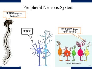
- 1. Peripheral Nervous System ये हम है ये हमारा Nervous System है और ये हमारी Pawri (पार्टी) हो रही है
- 2. Nervous System • A highly complex part of an animal which is made up of nerves and highly specialized cells called as neurons that transmit signals between different parts of the body
- 3. • Neuron : Structural and functional unit of nervous system. Neurons are specialized cells that transmit chemical and electrical signals to facilitate communication between the brain and the body. • Classification of neurons : a) Sensory Neuron b) Motor Neuron c) Interneuron Nervous System
- 4. a) Sensory Neuron : Sensory neurons are neurons responsible for converting external stimuli from the environment into corresponding internal stimuli. They are activated by sensory input, and send projections to other elements of the nervous system, ultimately conveying sensory information to the brain or spinal cord. Nervous System Sensory Neurons Stimuli Brain
- 5. b) Motor Neuron : Motor neurons are neurons located in the central nervous system, and they project their axons outside of the CNS to directly or indirectly control muscles. The interface between a motor neuron and muscle fiber is a specialized synapse called the neuromuscular junction Nervous System Motor Neurons Brain Motor impulse
- 6. c) Interneuron : Interneurons are neither sensory nor motor; rather, they act as the “middle men” that form connections between the other two types. Located in the CNS, they operate locally, meaning their axons connect only with nearby sensory or motor neurons. Interneurons can save time and therefore prevent injury by sending messages to the spinal cord and back instead of all the way to the brain. Nervous System
- 10. Sodium channel Potassium Channel CalciumChannel Chloride channel
- 11. Location of ion channel : Dendrites Acts as a sensor for impulses
- 13. Na+ K+ Cl- Ca++
- 14. Sodium channel Potassium Channel Action Potential : Transfer of information across the length of axon Chloride channel Hyperpolarization of neuron Calcium Channel Helps in neurotransmission
- 17. Peripheral Nervous System • The peripheral nervous system connects the central nervous system to environmental stimuli to gather sensory input and create motor output.
- 18. • The peripheral nervous system (PNS) provides the connection between internal or external stimuli and the central nervous system to allow the body to respond to its environment. • The PNS is made up of different kinds of neurons, or nerve cells, which communicate with each other through electric signaling and neurotransmitters. • The PNS can be broken down into two systems: the autonomic nervous system, which regulates involuntary actions such as breathing and digestion, and the somatic nervous system, which governs voluntary action and body reflexes. • The autonomic nervous system has two complementary parts: the sympathetic nervous system, which activates the “fight-or-flight-or-freeze” stress response, and the parasympathetic nervous system, which reacts with the “rest-and-digest” response after stress. • The somatic nervous system coordinates voluntary physical action. It is also responsible for our reflexes, which do not require brain input. Peripheral Nervous System
- 19. Autonomic Nervous System • The autonomic nervous system regulates involuntary and unconscious actions, such as internal-organ function, breathing, digestion, and heartbeat. • This system consists of two complementary parts: • The sympathetic and parasympathetic systems. • Both divisions work without conscious effort and have similar nerve pathways, but they generally have opposite effects on target tissues.
- 20. • The sympathetic nervous system activates the “fight or flight” response under sudden or stressful circumstances, such as taking an exam or seeing an examiner during class test. • It increases physical arousal levels, raising the heart and breathing rates and dilating the pupils, as it prepares the body to run or confront danger. These are not the only two options; “fight or flight” is perhaps better phrased as “fight or flight or freeze,” where in the third option the body stiffens and action cannot be taken. • This is an autonomic response that occurs in animals and humans; it is a survival mechanism thought to be related to playing dead when attacked by a predator. Autonomic Nervous System (Sympathetic System)
- 21. Khatam Bye Bye …Ta Ta…Goodbye Freeze response
- 22. Autonomic Nervous System (Sympathetic System) Hypothalamus Coordinate with Brain/Spinal Cord Stimulate Preganglionic Neuron/Fiber Release Neurotransmitter – I at Autonomic Ganglion Stimulate Postganglionic Neuron/Fiber Release Neurotransmitter – II at Neuron Effector Junction It stimulate various receptors of respective organs Produce various autonomic action
- 25. Anatomy of sympathetic nervous system (Adrenergic System) Superior control by posterior and lateral part of hypothalamus Preganglionic fibers originate from Thoracic 1 to Lumber 3 segments Activate Preganglionic neuron/fiber (Short) Release neurotransmitter-I (Ach) in Autonomic Ganglion(Junction-I) (Stimulate (NN) or (M1) receptor) Activate postganglionic neuron/fiber (Long) (Stimulate (α1), (α 2), (β1), (β2) or (β3) receptor Release) neurotransmitter-II (Adr) in Neuron Effector Junction (Junction-II) Produce various action
- 26. (Nn and M1) Tissue α1 α2 β1 β2 β3 Pre-Synaptic neurons Post-Synaptic neurons
- 27. Posterior and lateral part of the hypothalamus have the superior control on sympathetic nervous system Preganglionic neuron/fiber of sympathetic nervous systems originate from the thoracic 1 to lumber 3 segment of spinal cord. Preganglionic fiber of sympathetic nervous systems are usually short post ganglionic fiber which is longer than the preganglionic fiber. Here, autonomic ganglion –Junction-I consist neurotransmitter-I that is acetylcholine. Which helps to stimulate various receptors like Nicotinic Neuronal (NN) or Muscarinic (M1) in autonomic ganglion/junction. Stimulation of NN and M1 receptors activate the of post ganglionic neurons/fibers. Post ganglionic neuron/fiber end into the neuron effector junction near the various organ/tissue/cell. Neuron effector junction (Junction-II) consist neurotransmitter II that is noradrenalin (NA) means sympathetic systems consist neurotransmitter-I is acetylcholine (Ach) in autonomic ganglion (Junction-I) and neurotransmitter – II that is adrenalin in neuron effector junction (Junction-II). These neurotransmitter (Noradrenalin - NA) stimulate the various α and β receptors like α1, α2, β1, β2 and β3 Stimulation of various sympathetic receptors gives various kind of autonomic functions. To know these functions it is essential to identify the location of various receptors
- 28. Diagrammatic Representation of Sympathetic transmission
- 30. Adrenergic Receptors 1.Alpha adrenergic receptor 2.Beta adrenergic receptors (Gq) (Gi) (Gs)
- 31. Alpha-1 Receptor • α1 receptors: These are G-protein coupled receptors, it activate phospholipase C and stimulate Inositol Triphosphate (IP3) and Diacylglycerol (DAG) and increase intracellular calcium level and produce below autonomic functions according to their location. Location Function Blood vessels Produce vasoconstriction Iris It contract radial muscles and dilate the pupil known as mydriasis GI tract Contract the GI sphincter and relax the the GI muscle Urinary bladder Contract the trigon and relax the urinary bladder Glands Increase the secretion of glands Uterus It produce contraction in uterus
- 32. Alpha-2 Receptor • α2 receptors: These receptors decrease the cAMPlevel and alter the Ca+ channel conduction Location Function Presynaptic nerve ending It reduce release of noradrenaline Blood vessels Produce constriction of blood vessels(mechanism is unknown) CNS Reduction in central sympathetic flow due to decrease of Noradrenalin level Pancreas Reduce insulin level so increase blood sugar level Platelets Aggregate platelets GI muscle Relaxation of GI muscle
- 33. Beta Receptors • These are G-protein coupled receptors, it act by activation of adenylyl cyclase which increase cyclic AMP (cAMP) level and produce below autonomic functions according to their location. • β1 receptors Location Function Heart Increase force of contraction (Positive Inotropic) Increase heart rate (Positive Chronotropic) Kidney Release of renin, so renin activate angiotensinogen I which convert in angiotensinogen II by the help of angiotensinogen converting enzyme (ACE) and activate the aldosterone. Which retain the Na+ and water and increase the blood volume as well as angiotensinogen act on AT-I and AT-II receptor and contract the blood vessels.
- 34. Beta Receptors • β2 receptors (Gs): Synthesizes special protein which causes relaxation Location Function Blood vessels Dilation of blood vessels Lungs Dilation of bronchial smooth muscles and lungs GI muscle Relaxation of GI muscle Bladder Relaxation of urinary bladder Liver Produce glycogenolysis means conversion of glycogen to glucose and increase blood sugar level Pancreas Increase glucagon secretion which increase blood sugar level Adipose tissue Lipolysis (Break down of fats) Uterus Produce relaxation of uterus
- 35. β3 receptors Located in fat cells, Helps in lipolysis Adipose tissue कस एकदम गोलिगत……. ि िा िी िािा ि िा िी िािा
- 36. Autonomic Nervous System (Parasympathetic System) कभी कभी लगता है अपुन ही भगवान है अहम् ब्रह्मास्मि The parasympathetic nervous system activates a “rest and digest” or “feed and breed” response after these stressful events, which conserves energy and replenishes the system. It reduces bodily arousal, slowing the heartbeat and breathing rate. Together, these two systems maintain homeostasis of body
- 37. • Anatomy of Parasympathetic Nervous System (Cholinergic Nervous System) Superior control by anterior and middle part of hypothalamus Preganglionic neuron/fiber (Long) Release neurotransmitter-I (Ach) in Autonomic Ganglion/Junction (Junction-I) (Stimulate (NN) or (M1) receptor) Activate postganglionic neuron/fiber (Short) Stimulate (M1), (M2), (M3) or (NN) receptor Release neurotransmitter-II (Ach) in Neuron Effector Junction (Junction-II) Produce various action after that Ach is destruct by Acetylcholine Esterase (AchE) Autonomic Nervous System (Parasympathetic System)
- 38. Autonomic Nervous System (Parasympathetic System) Anterior and middle part of the hypothalamus have the superior control on parasympathetic nervous system. It coordinate with the midbrain and spinal cord. Preganglionic fiber of the parasympathetic nervous system originate from the midbrain and sacral part of the spinal cord. Mainly cranial nerve III, VIII, IX, X and gray matter of sacral part of spinal cord emerge out or rise to preganglionic fiber. Preganglionic fibers travel some distance and end in to the junction-I that is autonomic ganglion from which post ganglionic fibers emerge out. Here, neurotransmitter-I that is acetylcholine is released. Which helps to stimulate various receptors like Nicotinic Neuronal (NN) or Muscarinic (M1) in autonomic ganglion/junction. Stimulation of NN and M1 receptors activate the of post ganglionic neurons/fibers. Post ganglionic neuron/fiber end into the neuron effector junction near the various organ/tissue/cell. Neuron effector junction (Junction-II) consist neurotransmitter II that is acetylcholine.
- 39. Autonomic Nervous System (Parasympathetic System) 1. Parasympathetic systems contains same neurotransmitter that is acetylcholine (Ach) in both the junctions. 2. These neurotransmitter (Ach) stimulate the various muscarinic and nicotinic receptors like M1, M2, M3 and Nn. 3. Here the nicotinic muscular receptor (Nm) is not included because it is the part of somatic nervous systems. 4. Stimulation of various parasympathetic receptors gives various kind of autonomic functions. 5. To know these functions it is essential to identify the location of various receptors
- 40. Parasympathetic Receptors Also known as Cholinergic receptors • A. Muscarinic Receptors • B. Nicotinic Receptors(Nn/Nm)
- 41. Muscarinic receptor M1 Receptor (Gq): These are G-protein coupled receptors, it activate phospholipase C and stimulate Inositol Triphosphate (IP3) and Diacylglycerol (DAG) and increase intracellular calcium level and produce below autonomic functions according to their location. Location Function Autonomic ganglion/junction Activation of post ganglionic neuron/fiber Neuron 1 Neuron 2 M1 Ach
- 42. Muscarinic receptor M2 Receptor (Gi): These are G-protein coupled receptors, it act by reducing cyclic AMP (cAMP) level and produce below autonomic functions according to their location.. Heart 1. Decrease force of contraction (Negative Ionotropic) 2. Decrease heart rate (Negative Chronotropic) 3. Decrease conduction (Negative dromotropic)
- 43. Muscarinic receptor M3 receptors (Gq): These are G-protein coupled receptors, it activate phospholipase C and stimulate Inositol Triphosphate (IP3) and Diacylglycerol (DAG) and increase intracellular calcium level and produce below autonomic functions according to their location. Location Function GI smooth muscle Contraction of GI smooth muscle Bronchial smooth muscle Contraction of bronchial smooth muscle (Lungs contraction) Urinary tract Contract detrusor – urinary bladder muscle and produce micturition. Salivary secretion Increase secretion of saliva Lacrimal secretion Increase secretion of tear/lachrymal fluid Gastric secretion Increase secretion of HCl in GI tract Eye Produce meiosis (Contraction of pupils)
- 44. Parasympathetic Receptors • A. Muscarinic Receptors • B. Nicotinic Receptors(Nn/Nm)
- 45. Nicotinic receptor NN receptors: These are the intrinsic ion channel receptors, it act by opening various ion channels like Na+, K+ and Ca+ and produce below autonomic functions according to their location. Location Function Autonomic ganglion/junction (Junction – I) Activation of post ganglionic neuron/fiber NN Receptors
- 46. Overall ANS
- 47. Peripheral Nervous System • The peripheral nervous system refers to parts of the nervous system outside the brain and spinal cord. • It includes the 12 pairs of cranial nerves and 31 pairs of spinal nerves
- 48. When Olfactory nerve is activated इक्तीिा करना तेरे खुशबुको तेरे आणेसे मेरा लदि ओर लदमाग तेजीसे चिने िगता है बवाल चीज है बे साला ससस्टम सहल जाता है”
- 49. Cranial nerves are direct extensions of the brain. • Only Nerve I (olfactory) originates from the cerebrum, the remaining 11 pairs originate from the brain stem. • Nerve I (Olfactory)- for the sense of smell (sensory).
- 50. When Optic nerve is activated तुझे देखा तो ये जाना सनम प्यार होता है दीवाना सनम तुझे देखा तो ये जाना सनम Yee Wrong number hai……..Iska bhi katega
- 51. Cranial nerves are direct extensions of the brain. • Nerve II (Optic)- for the sense of vision (sensory). Jai Hind Dostooo
- 52. Cranial nerves are direct extensions of the brain. • Nerve III (Occulomotor)- for controlling muscles and accessory for controlling muscles and accessory structures of the eyes (motor). • Nerve IV (Trochlear)- for controlling muscles of the eyes (motor). • Nerve VI (Abducens)- for controlling muscles that move the eye (motor)
- 53. Cranial nerves V • Nerve V (Trigeminal)- for controlling muscles of the eyes, upper and lower jaws and tear glands (mixed).
- 54. Cranial nerves VII • Nerve VII (Facial) – for the sense of taste and controlling facial muscles, tear glands and salivary glands (mixed).
- 55. Cranial nerves VIII • Nerve VIII (Vestibulocochlear/Auditory)- for the senses of hearing and equilibrium (sensory).
- 56. Cranial nerves IX • Nerve IX (Glossopharyngeal)- for controlling muscles in the pharynx and to control salivary glands (mixed).
- 57. Cranial nerves X • Nerve X (Vagus)- for controlling muscles used in speech, for controlling muscles used in speech, swallowing, and the digestive tract, and controls cardiac and smooth muscles (mixed).
- 58. Cranial nerves XI • Nerve XI (Accessory)- for controlling muscles of soft palate, pharynx and larynx (motor).
- 59. Cranial nerves XII • Nerve XII (Hypoglossal) for controlling muscles that move the tongue (motor).
- 60. Overview of Cranial Nerves • Nerve I (Olfactory)- for the sense of smell (sensory). • Nerve II (Optic)- for the sense of vision (sensory). • Nerve III (Occulomotor)- for controlling muscles and accessory for controlling muscles and accessory structures of the eyes (motor). • Nerve IV (Trochlear)- for controlling muscles of the eyes (motor). • Nerve V (Trigeminal)- for controlling muscles of the eyes, upper and lower jaws and tear glands (mixed). • Nerve VI (Abducens)- for controlling muscles that move the eye (motor)
- 61. • Nerve VII (Facial) – for the sense of taste and controlling facial muscles, tear glands and salivary glands (mixed). • Nerve VIII (Vestibulocochlear/Auditory)- for the senses of hearing and equilibrium (sensory). • Nerve IX (Glossopharyngeal)- for controlling muscles in the pharynx and to control salivary glands (mixed). • Nerve X (Vagus)- for controlling muscles used in speech, for controlling muscles used in speech, swallowing, and the digestive tract, and controls cardiac and smooth muscles (mixed). • Nerve XI (Accessory)- for controlling muscles of soft palate, pharynx and larynx (primarily motor). • Nerve XII (Hypoglossal) for controlling muscles that move the tongue ( primarily motor).
- 62. Spinal Nerves
- 63. Spinal Nerves • There are 31 pairs of spinal nerves including: • Eight cervical spinal nerves on each side of the spine called C1 to C8 • Twelve thoracic spinal nerves in each side of the body called T1 to T12 • Five lumbar spinal nerves on each side calleed L1 to L5 • Five sacral spinal nerves in each side called S1 to S5 • One coccygeal nerve on each side, Co1
- 64. • Cervical spinal nerves : The cervical spinal nerves innervate the muscles and provide sensation for the head, neck, and diaphragm, as well as the upper limbs and back. • Thoracic spinal nerves : They are involved in the functions of organs and glands in the head, neck, thorax, and abdomen. • Lumbar spinal nerves : Provides sensation to the groin and genital regions and may contribute to the movement of the hip muscles, front part of the thigh and inner side of the lower leg. • Sacral spinal nerves : They are responsible for part of the sensory perception and the movements of the lower extremities of the human body. • Coccygeal nerve : Distributed in skin that send signals to the brain through the spinal nerves. These signals give rise to sensations involving temperature, pressure, and pain
- 68. Neurotransmitters Neurotransmitters are endogenous chemicals acting as signaling molecules that enable transmission of signals across a synapse from one neuron to another neuron, to a muscle cell, or to a gland cell. Excitatory Neurotransmitters Inhibitory Neurotransmitters Inhibitory + Excitatory Neurotransmitters
- 69. Excitatory Neurotransmitters Glutamate Aspartate Learning and Memory NMDA-Glutamate receptor Na,K,Ca
- 70. Inhibitory Neurotransmitters 1. GABA (gamma-Aminobutyric acid) 1. Glycine
- 71. 1. Adrenaline 2. Acetylcholine 3. Dopamine 4. Serotonin 5. Histamine Inhibitory + Excitatory Neurotransmitters
- 72. 1. Adrenaline 2. Acetylcholine 3. Dopamine 4. Serotonin 5. Histamine Inhibitory + Excitatory Neurotransmitters Gs Gq Gi
- 73. Neurotransmitters and Mood Regulation Happy ….Happy Happiness
- 74. Neurotransmitters and Mood Regulation Stay away brooo, Leave me alone Anger
- 75. Neurotransmitters and Mood Regulation Let’s Dance Fun and Enjoyment
- 76. Neurotransmitters and Mood Regulation Why i am crying for no reason Feeling sad
- 77. Neurotransmitters and Mood Regulation Feeling Lonely Feeling sad
- 78. Neurotransmitters and Mood Regulation Love
- 79. Neurotransmitters and Mood Regulation
- 80. Neurotransmitters Regulates your mood What do you think, is it true ?
- 81. Neurotransmitters Balanced All is well
- 83. Mental Disorders Depression Anxiety Alzheimer’s disease Parkinson's disease Mai pagal nahi hu bas thodi tabiyat kharab hai
- 84. Can you define these disorders ? Schizophrenia Manic Disorder Confusion hee confusion hai solution kuchh pata nahi
- 85. What is depression ? Mental state which is characterized by persistent sadness and a lack of interest or pleasure in previously rewarding or enjoyable activities
- 86. WHO report about Depression ● Depression is a common illness worldwide, with more than 264 million people affected (246,000,000 i.e 2460 Lakhs) ● 8 Lakh people die due to suicide every year i.e 1 death every 40 second ● Suicide is the second leading cause of death in 15- 29-year-olds.
- 87. WHO report about India on suicide 1. 800000 people die due to suicide every year (i.e 1 death every 40 second) 1. 1,35000 people are resident of india (17%) 1. According to WHO data, the age standardized suicide rate in India is 16.4 per 100,000 for women (6th highest in the world) and 25.8 for men (ranking 22nd) Statistics
- 88. ● Depressed mood ● Loss of interest and enjoyment ● Reduced energy leading to diminished activities ● Many people with depression also suffer from anxiety symptoms ● Disturbed sleep and appetite ● may have feelings of guilt or low self-worth ● poor concentration. Symptoms
- 89. What is Anxiety ? The American Psychological Association (APA) defines anxiety as “an emotion characterized by feelings of tension, worried thoughts and physical changes like increased blood pressure.”
- 90. Symptoms ● Restlessness ● Uncontrollable feelings of worry ● Increased irritability ● Concentration difficulties ● Sleep difficulties, such as problems in falling or staying asleep
- 91. What is Alzheimer's Disease? Alzheimer’s disease is an irreversible, progressive brain disorder that slowly destroys memory and thinking skills and, eventually, the ability to carry out the simplest tasks
- 92. Early stage: 1. Forgetfulness 2. Losing track of the time 3. Becoming lost in familiar places. Middle stage: 1. Becoming forgetful of recent events and people's names 2. Becoming lost at home 3. Having increasing difficulty with communication 4. Needing help with personal care 5. Experiencing behaviour changes, including wandering and repeated questioning. Late stage: 1. Becoming unaware of the time and place 2. Having difficulty recognizing relatives and friends 3. Having an increasing need for assisted self-care 4. Having difficulty walking 5. Experiencing behaviour changes that may escalate and include aggression. Symptoms
- 93. India has the second highest number of individuals suffering from dementia, at an estimated 4.1 million(41 Lakhs) The figure is expected to double by 2035, as per the ‘Dementia India’ report published by the Alzheimer’s and Related Disorders Society of India Dementia India report on Dementia Statistics
- 94. What is Parkinson’s Disease ? Parkinson's disease (PD) belongs to a group of conditions called motor system disorders, which cause unintended or uncontrollable movements of the body.
- 95. Symptoms The four primary symptoms of PD are: ● Tremor : shaking that has a characteristic rhythmic back and forth motion ● Rigidity : muscle stiffness or a resistance to movement, where muscles remain constantly tense and contracted ● Bradykinesia : slowing of spontaneous and automatic movement that can make it difficult to perform simple tasks or rapidly perform routine movements ● Postural instability : impaired balance and changes in posture that can increase the risk of falls. Other symptoms may include difficulty swallowing, chewing, or speaking; emotional changes; urinary problems or constipation; dementia or other cognitive problems; fatigue; and problems sleeping.
- 96. Dopamine Epinephrine Norepinephrine Serotonin Kuchh toh gadbad hai dayaa
- 97. Dopamine Functions : 1. Reward ( Motivation ) 2. Pleasure 3. Motor functions 4. Compulsion
- 98. Dopamine deficiency Parkinson disease 1. Low motivation 2. Lack of interest 3. Low energy 4. An inability to focus moving or speaking more slowly than usual 5. Feeling fatigued 6. Feeling demotivated 7. Feeling inexplicably sad or tearful mood swings 8. Feeling hopeless 9. Having low self-esteem
- 99. Dopamine Hypersecretion Addiction 1. Alcohol consumption 1. Tea / Coffee consumption 1. Nicotine Consumption 1. Sexual Desire
- 100. Dopamine Hypersecretion Mania ( Bipolar Disorder ) A mental health condition that causes extreme mood swings that include emotional highs (mania or hypomania) and lows (depression).
- 102. Dopamine Hypersecretion Hallucinations There are five types of hallucinations, including: 1 Auditory: Hearing voices or sounds that no one else can (most common type of hallucination) 2 Visual: Seeing people, colors, shapes, or items that aren't real (second most common type of hallucination) 3 Tactile: Feeling sensations (like bugs crawling under your skin) or as if you're being touched when you're not 4 Olfactory: Smelling something that has no physical source (less common than visual and auditory hallucinations) 5 Gustatory: Having a taste in your mouth that has no source (rarest type of hallucination)
- 103. Dopamine Hypersecretion Schizophrenia ( Discussed separately )
- 104. Norepinephrine and Epinephrine Storage site : Adrenergic neurons, Adrenal Gland Together with adrenaline, norepinephrine increases heart rate and blood pumping from the heart, Pupil Dilation. It also increases blood pressure and helps break down fat,Proteins and increase blood sugar levels to provide more energy to the body. In the brain, norepinephrine plays a role in the sleep-wake cycle, helping you in increasing attention and focusing on performing a task , memory storage. It is also important for emotions
- 105. Norepinephrine / Epinephrine Hypersecretion Anxiety, Excess Sweating, Palpitations
- 106. What is Schizophrenia ? Schizophrenia is complex, chronic, mental health disorder characterized by an array of symptoms, including 1. Delusions( fixed, false beliefs that conflict with reality) 2. Hallucinations (Sensations that appear real but are created by your mind) 3. Disorganised speech or behaviour 4. Impaired cognitive ability 5. Impaired working memory Psychoses
- 107. Schizophrenia ( Psychoses )
- 108. Theories Abnormalities in neurotransmission have provided the basis for theories on the pathophysiology of schizophrenia. 1. Most of these theories center on either an excess or a deficiency of neurotransmitters, including dopamine, serotonin, and glutamate. 1. Other theories implicate aspartate, glycine, and gamma- aminobutyric acid (GABA) as part of the neurochemical imbalance of schizophrenia