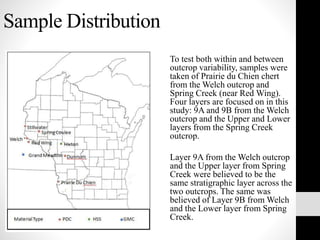
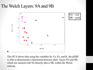
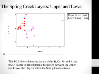
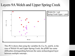
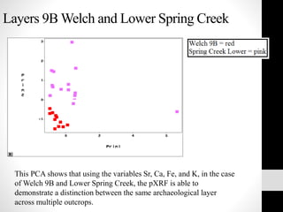
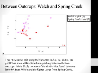
Hand-held Portable X-ray
Fluorescence Spectrometry
(HHpXRF or pXRF) for Sourcing
and Distinguishing Chert
Kia Catina Atsales
Graduate Student: Anthropology
University of Minnesota, Twin Cities
Content
Part I: Background Information
Part II: Research Question and Methodology
Part III: Data and Results
Part IV: Conclusions and Future Work
Part V: Works Cited
Part I: Background Information
What is “chert”?
“Chert” is a general term used for sedimentary rocks that are composed
primarily of microcrystalline quartz (SiO2) and includes materials
referred to as flint, chalcedony, agate, jasper, hornstone, and novaculite.
How does chert form?
Chert formation often begins when silica-secreting marine organisms extract
silica from sea water to produce skeletal or protective material and bind it
within their organic matrix as opal to keep it from re-dissolving into the sea
(Blatt and Robert, 1995). When these organisms die, their bodies’ protective
organic coatings decay and their silica-bearing tissues are deposited on the
ocean/sea/lake floor. A portion of their silica may re-dissolve back into sea
water during transport to the sediment-water interface, but the remainder will
be incorporated into the sedimentary record (Calvert, 1974).
Name Type of
Organism
Habitat Time Range Siliceous
portion of
organism
Radiolarians Single-celled
organisms
Ocean Precambrian to
recent
Skeleton
Sponges Multi-celled
animals
Ocean Early Cambrian
to recent
Spicules
Diatoms Algae Ocean
Freshwater
Cretaceous to
recent
Tertiary to recent
Outer cell wall
Silicoflagellates Single-celled
organisms
Ocean Late Cretaceous
to recent
Skeleton
Major Aquatic Silica-Secreting Organisms (from Table 3.1 in Luedtke, 1992: 23)
How does chert form?
Chert usually forms during burial
diagenesis which is defined as “‘low
temperature/low pressure changes that
frequently take place in sediments prior to,
and often contributing to their lithification’”
(Berry, Mason and Dietrich, 1983: 181,
cited in Luedtke, 1992: 26). The term
“diagenesis” refers to a complex series of
inter-related sedimentary processes
involving compaction, cementation,
chemical alteration, replacement, and
recrystallization (Calvert, 1974). As
sediment containing Opal A is compressed
by overlying material, pore waters become
saturated with respect to dissolved silica
and opaline forms of cristobalite and
tridymite (CT), which begin to
inorganically precipitate (Calvert, 1974).
Additional burial from continuing
sedimentation causes ambient temperatures
to continue to rise and ultimately CT is
converted to quartz (Blatt and Robert, 1995;
Calvert, 1974).
"Haeckel Diatomea" by Ernst Haeckel - Kunstformen der Natur
(1904), plate 84: Diatomeae (see here, here, here and here). Licensed
under Public Domain via Wikimedia Commons -
http://commons.wikimedia.org/wiki/File:Haeckel_Diatomea.jpg#/medi
a/File:Haeckel_Diatomea.jpg
How does chert form?
Bedded Cherts
This style of chert generally occurs
when the ratio of opaline material to
water is high. Bedded cherts form
predominantly in deep, elongated
sedimentary basins (or more rarely in
small basins with restricted
circulation that generate localized
areas of highly concentrated opaline
silica).
Nodular Cherts
This style of chert deposit occurs
as nearly spherical to irregular
shaped concretions within
carbonate sediments that formed
as secondary features during the
migration of silica rich waters
with very high alkalinity (Blatt
and Robert, 1995; Prothero and
Schwab, 2004).
The Children's Museum of Indianapolis [CC BY-SA 3.0
(http://creativecommons.org/licenses/by-sa/3.0)], via Wikimedia Commons
Chert Impurities
The majority of impurities in cherts are minerals that were present in the
original sedimentary environment. Additionally, they can come from the
environment in which re-dissolved silica was precipitated and become
incorporated into the chert. Trace elements in the mineral quartz can be
incorporated into the crystal lattice, contained within microinclusions, or
isolated along quartz grain boundaries. These impurities can include:
• Clays
• Iron-bearing non-silicates
• Carbonates
• Organic matter
• Liquid impurities
http://blogs.egu.eu/divisions/sss/tag/clay-2/
Clay Minerals
How can chert be analyzed?
Visually:
• Color
• Translucency
• Luster
• Texture
Geochemically and Geophysically:
• Neutron Activation Analysis (NAA)
• X-ray Fluorescence Spectrometry (XRF)
• Laser Ablasion Inductively Coupled Plasma Mass Spectrometry (LA-
ICP-MS)
• Magnetic Susceptibility (MS)
http://www.niton.com/Managers/Multimedia/XL3-
presentation/Thermo_20070720_GN.html
What is XRF?
XRF detects both the presence and concentration of an element by
quantifying characteristic x-ray emission wavelength or energy and
then taking this information and quantifying it by measuring the
emitted “characteristic line intensity” (Jenkins, 1999: 101). When
the energy from the X-rays is sufficient, the inner shell electron is
displaced, causing the atom to become unstable and forcing an
outer shell electron into the spot once occupied by the inner shell
electron (Shackley, 2011). This process releases energy which can
be measured and then related to a standard’s known element
concentration.
http://www.elementalcontrols.com/
en/products/products.html
Part II: Research Question and
Methodology
While the portable version of the XRF cannot currently provide
the same level of detail about elemental concentrations in
samples as a lab-based XRF, can the pXRF serve the same
function for archaeology as the lab-based XRF? I.e., can it
provide comparable data for within and between geologic
outcrop comparisons for basic raw material sourcing of chert?
Concern: Can the pXRF work on chert, a sedimentary rock?
E.g. Percent iron in Prairie du Chien chert 0.01% - 0.001%
Niton XL3t GOLDD
The Soils setting was used on the Niton XL3t GOLDD pXRF,
optimized at 80 seconds on the main filter, 40 seconds on the low
filter, and 30 seconds on the high filter.
Main Filter: manganese (MN) through bismuth (Bi) Z= 25-83
Low Filter: potassium (K) through chromium (Cr) Z=19-24
High Filter: silver (Ag) through barium (Ba) Z=47-56
http://genius.com/1894437/Todd-beard-
periodic-table/The-heart-of-chemistry
Sample Distribution
To test both within and between
outcrop variability, samples were
taken of Prairie du Chien chert
from the Welch outcrop and
Spring Creek (near Red Wing).
Four layers are focused on in this
study: 9A and 9B from the Welch
outcrop and the Upper and Lower
layers from the Spring Creek
outcrop.
Layer 9A from the Welch outcrop
and the Upper layer from Spring
Creek were believed to be the
same stratigraphic layer across the
two outcrops. The same was
believed of Layer 9B from Welch
and the Lower layer from Spring
Creek.
Welch Samples
Spring Creek Samples
Welch and Spring Creek Samples
Welch 9A
Welch 9B
Spring Creek Upper
Spring Creek Lower
Principal ComponentAnalysis
Principal component analysis (PCA) was conducted on the pXRF
readings; PCA does a rigid rotation of the data to produce a new set of
axes that summarizes the greatest amount of sample variance (PC1)
and the second most variance that is orthogonal to the first matrix
(PC2). This study focuses on the elemental variation of strontium (Sr),
calcium, (Ca) iron (Fe), and potassium (K). While other elements
provided usable data, these four elements were selected for these
analyses because (1) they are elements that are typically found within
micro-inclusion impurities in cherts; and (2) they affected the
variation of the eigenvectors the most.
Part III: Data and Results
The following are the PCAs
conducted as a comparison of both
intra- and inter-site variability of
Prairie du Chien chert from the Welch
and Spring Creek outcrops. Five
comparisons were conducted on the
variation between:
1) Layers 9A and 9B at the Welch
outcrop
2) The Upper and Lower layers at the
Spring Creek outcrop
3) Layers 9A Welch and Upper Spring
Creek (the same stratigraphic layer
across two outcrops)
4) Layers 9B Welch and Lower Spring
Creek (the same stratigraphic layer
across two outcrops)
3) Between outcrops (Welch and Spring
Creek)
KEY TERMS:
• OUTCROP:
1=Welch
2=Spring Creek
• LEVEL:
A=9A Welch
B=9B Welch
C=Upper Spring Creek
D=Lower Spring Creek
The Welch Layers: 9Aand 9B
This PCA shows that using the variables Sr, Ca, Fe, and K, the pXRF
is able to demonstrate a distinction between chert layers 9A and 9B,
which are stacked with 9A directly above 9B, within the Welch
outcrop.
The Spring Creek Layers: Upper and Lower
This PCA shows that using the variables Sr, Ca, Fe, and K, the
pXRF is able to demonstrate a distinction between the Upper
and Lower chert layers within the Spring Creek outcrop.
Layers 9AWelch and Upper Spring Creek
This PCA shows that using the variables Sr, Ca, Fe, and K, in the
case of Welch 9A and Upper Spring Creek, the pXRF has some
difficulties distinguishing between the same archaeological layer
between multiple outcrops .
Layers 9B Welch and Lower Spring Creek
This PCA shows that using the variables Sr, Ca, Fe, and K, in the case
of Welch 9B and Lower Spring Creek, the pXRF is able to
demonstrate a distinction between the same archaeological layer
across multiple outcrops.
Between Outcrops: Welch and Spring Creek
This PCA shows that using the variables Sr, Ca, Fe, and K, the
pXRF has some difficulties distinguishing between the two
outcrops; this is likely because of the similarities found between
layer 9A from Welch and the Upper Layer from Spring Creek.
Part IV: Conclusions and Future Work
In this study, I attempted to determine whether elemental data from chert
samples analyzed with the pXRF would be able to distinguish (1) between
stratigraphic layers within the same outcrop; (2) between the same
stratigraphic layers across multiple outcrops; and (3) between outcrops when
all data was included. Despite having a small data set, PCAs using the
elemental variable Sr, Fe, Ca and K, demonstrated that the pXRF could be a
helpful tool for distinguishing between stratigraphic layers within a single
outcrop (seen for both the Welch and Spring Creek outcrops). When looking
at the same stratigraphic layer across two outcrops, the results varied, with
Welch 9B and Lower Spring Creek showing clear distinctions (likely because
of differences in environmental conditions under which they formed),
whereas the data for Welch 9A and Upper Spring Creek were less conclusive.
Those inconclusive results were likely why the final PCA, which included the
data from all of the levels, did not demonstrate a clear distinction between the
outcrops.
Future research will include a between outcrop analysis where the
stratigraphic profiles contain differing chert types to determine whether the
pXRF will be able to distinguish between outcrops in less related chert
deposits.
Part V: Works Cited
Blatt, Harvey and Tracy J. Robert.
1995 Petrology : igneous, sedimentary, and metamorphic. New York: W.H. Freeman.
Calvert, S.E.
1974 Deposition and diagenesis of silica in marine sediments. Spec. Publs int. Ass.
Sediment 1: 273-299.
Jenkins, Ron.
1999 X-ray Fluorescence Spectrometry. A Wiley-Interscience Publication: John Wiley and
Sons, Inc. Vol. 152.
Luedtke, Barbara.
1992 An Archaeologist’s Guide to Chert and Flint. Cotsen Institute of Archaeology Press.
Prothero, Donald R. and Fred Schwab.
2004 Sedimentary Geology: An Introduction to Sedimentary Rocks and Stratigraphy. W. H.
Freeman; Second Edition.
Shackley, M. Steven.
2011 “An Introduction to X-ray Fluorescence (XRF) Analysis in Archaeology.” X-ray
Fluorecence Spectrometry (XRF) in Geoarchaeology. Ed. M.S. Shackley. Springer
Science+Business Media, LLC: 7-44.
Acknowledgements
Spring break sample collection crew: Dr. Gilbert Tostevin, Dr.
Joshua Feinberg, and Dan Wendt
My grad committee: Dr. Gilbert Tostevin, Dr. Gilliane Monnier, Dr.
Katherine Hayes, and Dr. Joshua Feinberg
Statistics assistance: Dr. Kieran McNulty and Ryan Knigge
PXRF assistance: Dr. Katherine Hayes and Dr. Ellery Frahm
Funding: Sigma Xi’s Dorothy and Andrew Bird Award and the
UMN Anthropology Department
Special thanks to Dan Wendt and the Minnesota Historical Society
at Fort Snelling

Hand-held Portable X-ray Fluorescence Spectrometry (HHpXRF or pXRF) for Sourcing and Distinguishing Chert

  • 1.
    Hand-held Portable X-ray FluorescenceSpectrometry (HHpXRF or pXRF) for Sourcing and Distinguishing Chert Kia Catina Atsales Graduate Student: Anthropology University of Minnesota, Twin Cities
  • 2.
    Content Part I: BackgroundInformation Part II: Research Question and Methodology Part III: Data and Results Part IV: Conclusions and Future Work Part V: Works Cited
  • 3.
    Part I: BackgroundInformation What is “chert”? “Chert” is a general term used for sedimentary rocks that are composed primarily of microcrystalline quartz (SiO2) and includes materials referred to as flint, chalcedony, agate, jasper, hornstone, and novaculite.
  • 4.
    How does chertform? Chert formation often begins when silica-secreting marine organisms extract silica from sea water to produce skeletal or protective material and bind it within their organic matrix as opal to keep it from re-dissolving into the sea (Blatt and Robert, 1995). When these organisms die, their bodies’ protective organic coatings decay and their silica-bearing tissues are deposited on the ocean/sea/lake floor. A portion of their silica may re-dissolve back into sea water during transport to the sediment-water interface, but the remainder will be incorporated into the sedimentary record (Calvert, 1974). Name Type of Organism Habitat Time Range Siliceous portion of organism Radiolarians Single-celled organisms Ocean Precambrian to recent Skeleton Sponges Multi-celled animals Ocean Early Cambrian to recent Spicules Diatoms Algae Ocean Freshwater Cretaceous to recent Tertiary to recent Outer cell wall Silicoflagellates Single-celled organisms Ocean Late Cretaceous to recent Skeleton Major Aquatic Silica-Secreting Organisms (from Table 3.1 in Luedtke, 1992: 23)
  • 5.
    How does chertform? Chert usually forms during burial diagenesis which is defined as “‘low temperature/low pressure changes that frequently take place in sediments prior to, and often contributing to their lithification’” (Berry, Mason and Dietrich, 1983: 181, cited in Luedtke, 1992: 26). The term “diagenesis” refers to a complex series of inter-related sedimentary processes involving compaction, cementation, chemical alteration, replacement, and recrystallization (Calvert, 1974). As sediment containing Opal A is compressed by overlying material, pore waters become saturated with respect to dissolved silica and opaline forms of cristobalite and tridymite (CT), which begin to inorganically precipitate (Calvert, 1974). Additional burial from continuing sedimentation causes ambient temperatures to continue to rise and ultimately CT is converted to quartz (Blatt and Robert, 1995; Calvert, 1974). "Haeckel Diatomea" by Ernst Haeckel - Kunstformen der Natur (1904), plate 84: Diatomeae (see here, here, here and here). Licensed under Public Domain via Wikimedia Commons - http://commons.wikimedia.org/wiki/File:Haeckel_Diatomea.jpg#/medi a/File:Haeckel_Diatomea.jpg
  • 6.
    How does chertform? Bedded Cherts This style of chert generally occurs when the ratio of opaline material to water is high. Bedded cherts form predominantly in deep, elongated sedimentary basins (or more rarely in small basins with restricted circulation that generate localized areas of highly concentrated opaline silica). Nodular Cherts This style of chert deposit occurs as nearly spherical to irregular shaped concretions within carbonate sediments that formed as secondary features during the migration of silica rich waters with very high alkalinity (Blatt and Robert, 1995; Prothero and Schwab, 2004). The Children's Museum of Indianapolis [CC BY-SA 3.0 (http://creativecommons.org/licenses/by-sa/3.0)], via Wikimedia Commons
  • 7.
    Chert Impurities The majorityof impurities in cherts are minerals that were present in the original sedimentary environment. Additionally, they can come from the environment in which re-dissolved silica was precipitated and become incorporated into the chert. Trace elements in the mineral quartz can be incorporated into the crystal lattice, contained within microinclusions, or isolated along quartz grain boundaries. These impurities can include: • Clays • Iron-bearing non-silicates • Carbonates • Organic matter • Liquid impurities http://blogs.egu.eu/divisions/sss/tag/clay-2/ Clay Minerals
  • 8.
    How can chertbe analyzed? Visually: • Color • Translucency • Luster • Texture Geochemically and Geophysically: • Neutron Activation Analysis (NAA) • X-ray Fluorescence Spectrometry (XRF) • Laser Ablasion Inductively Coupled Plasma Mass Spectrometry (LA- ICP-MS) • Magnetic Susceptibility (MS) http://www.niton.com/Managers/Multimedia/XL3- presentation/Thermo_20070720_GN.html
  • 9.
    What is XRF? XRFdetects both the presence and concentration of an element by quantifying characteristic x-ray emission wavelength or energy and then taking this information and quantifying it by measuring the emitted “characteristic line intensity” (Jenkins, 1999: 101). When the energy from the X-rays is sufficient, the inner shell electron is displaced, causing the atom to become unstable and forcing an outer shell electron into the spot once occupied by the inner shell electron (Shackley, 2011). This process releases energy which can be measured and then related to a standard’s known element concentration. http://www.elementalcontrols.com/ en/products/products.html
  • 10.
    Part II: ResearchQuestion and Methodology While the portable version of the XRF cannot currently provide the same level of detail about elemental concentrations in samples as a lab-based XRF, can the pXRF serve the same function for archaeology as the lab-based XRF? I.e., can it provide comparable data for within and between geologic outcrop comparisons for basic raw material sourcing of chert? Concern: Can the pXRF work on chert, a sedimentary rock? E.g. Percent iron in Prairie du Chien chert 0.01% - 0.001%
  • 11.
    Niton XL3t GOLDD TheSoils setting was used on the Niton XL3t GOLDD pXRF, optimized at 80 seconds on the main filter, 40 seconds on the low filter, and 30 seconds on the high filter. Main Filter: manganese (MN) through bismuth (Bi) Z= 25-83 Low Filter: potassium (K) through chromium (Cr) Z=19-24 High Filter: silver (Ag) through barium (Ba) Z=47-56 http://genius.com/1894437/Todd-beard- periodic-table/The-heart-of-chemistry
  • 12.
    Sample Distribution To testboth within and between outcrop variability, samples were taken of Prairie du Chien chert from the Welch outcrop and Spring Creek (near Red Wing). Four layers are focused on in this study: 9A and 9B from the Welch outcrop and the Upper and Lower layers from the Spring Creek outcrop. Layer 9A from the Welch outcrop and the Upper layer from Spring Creek were believed to be the same stratigraphic layer across the two outcrops. The same was believed of Layer 9B from Welch and the Lower layer from Spring Creek.
  • 13.
  • 14.
  • 15.
    Welch and SpringCreek Samples Welch 9A Welch 9B Spring Creek Upper Spring Creek Lower
  • 16.
    Principal ComponentAnalysis Principal componentanalysis (PCA) was conducted on the pXRF readings; PCA does a rigid rotation of the data to produce a new set of axes that summarizes the greatest amount of sample variance (PC1) and the second most variance that is orthogonal to the first matrix (PC2). This study focuses on the elemental variation of strontium (Sr), calcium, (Ca) iron (Fe), and potassium (K). While other elements provided usable data, these four elements were selected for these analyses because (1) they are elements that are typically found within micro-inclusion impurities in cherts; and (2) they affected the variation of the eigenvectors the most.
  • 17.
    Part III: Dataand Results The following are the PCAs conducted as a comparison of both intra- and inter-site variability of Prairie du Chien chert from the Welch and Spring Creek outcrops. Five comparisons were conducted on the variation between: 1) Layers 9A and 9B at the Welch outcrop 2) The Upper and Lower layers at the Spring Creek outcrop 3) Layers 9A Welch and Upper Spring Creek (the same stratigraphic layer across two outcrops) 4) Layers 9B Welch and Lower Spring Creek (the same stratigraphic layer across two outcrops) 3) Between outcrops (Welch and Spring Creek) KEY TERMS: • OUTCROP: 1=Welch 2=Spring Creek • LEVEL: A=9A Welch B=9B Welch C=Upper Spring Creek D=Lower Spring Creek
  • 18.
    The Welch Layers:9Aand 9B This PCA shows that using the variables Sr, Ca, Fe, and K, the pXRF is able to demonstrate a distinction between chert layers 9A and 9B, which are stacked with 9A directly above 9B, within the Welch outcrop.
  • 19.
    The Spring CreekLayers: Upper and Lower This PCA shows that using the variables Sr, Ca, Fe, and K, the pXRF is able to demonstrate a distinction between the Upper and Lower chert layers within the Spring Creek outcrop.
  • 20.
    Layers 9AWelch andUpper Spring Creek This PCA shows that using the variables Sr, Ca, Fe, and K, in the case of Welch 9A and Upper Spring Creek, the pXRF has some difficulties distinguishing between the same archaeological layer between multiple outcrops .
  • 21.
    Layers 9B Welchand Lower Spring Creek This PCA shows that using the variables Sr, Ca, Fe, and K, in the case of Welch 9B and Lower Spring Creek, the pXRF is able to demonstrate a distinction between the same archaeological layer across multiple outcrops.
  • 22.
    Between Outcrops: Welchand Spring Creek This PCA shows that using the variables Sr, Ca, Fe, and K, the pXRF has some difficulties distinguishing between the two outcrops; this is likely because of the similarities found between layer 9A from Welch and the Upper Layer from Spring Creek.
  • 23.
    Part IV: Conclusionsand Future Work In this study, I attempted to determine whether elemental data from chert samples analyzed with the pXRF would be able to distinguish (1) between stratigraphic layers within the same outcrop; (2) between the same stratigraphic layers across multiple outcrops; and (3) between outcrops when all data was included. Despite having a small data set, PCAs using the elemental variable Sr, Fe, Ca and K, demonstrated that the pXRF could be a helpful tool for distinguishing between stratigraphic layers within a single outcrop (seen for both the Welch and Spring Creek outcrops). When looking at the same stratigraphic layer across two outcrops, the results varied, with Welch 9B and Lower Spring Creek showing clear distinctions (likely because of differences in environmental conditions under which they formed), whereas the data for Welch 9A and Upper Spring Creek were less conclusive. Those inconclusive results were likely why the final PCA, which included the data from all of the levels, did not demonstrate a clear distinction between the outcrops. Future research will include a between outcrop analysis where the stratigraphic profiles contain differing chert types to determine whether the pXRF will be able to distinguish between outcrops in less related chert deposits.
  • 24.
    Part V: WorksCited Blatt, Harvey and Tracy J. Robert. 1995 Petrology : igneous, sedimentary, and metamorphic. New York: W.H. Freeman. Calvert, S.E. 1974 Deposition and diagenesis of silica in marine sediments. Spec. Publs int. Ass. Sediment 1: 273-299. Jenkins, Ron. 1999 X-ray Fluorescence Spectrometry. A Wiley-Interscience Publication: John Wiley and Sons, Inc. Vol. 152. Luedtke, Barbara. 1992 An Archaeologist’s Guide to Chert and Flint. Cotsen Institute of Archaeology Press. Prothero, Donald R. and Fred Schwab. 2004 Sedimentary Geology: An Introduction to Sedimentary Rocks and Stratigraphy. W. H. Freeman; Second Edition. Shackley, M. Steven. 2011 “An Introduction to X-ray Fluorescence (XRF) Analysis in Archaeology.” X-ray Fluorecence Spectrometry (XRF) in Geoarchaeology. Ed. M.S. Shackley. Springer Science+Business Media, LLC: 7-44.
  • 25.
    Acknowledgements Spring break samplecollection crew: Dr. Gilbert Tostevin, Dr. Joshua Feinberg, and Dan Wendt My grad committee: Dr. Gilbert Tostevin, Dr. Gilliane Monnier, Dr. Katherine Hayes, and Dr. Joshua Feinberg Statistics assistance: Dr. Kieran McNulty and Ryan Knigge PXRF assistance: Dr. Katherine Hayes and Dr. Ellery Frahm Funding: Sigma Xi’s Dorothy and Andrew Bird Award and the UMN Anthropology Department Special thanks to Dan Wendt and the Minnesota Historical Society at Fort Snelling