This document summarizes research on how shear stress regulates SIRT1 and its role in vascular homeostasis. The key points are:
1) Applying laminar flow to endothelial cells in vitro increased SIRT1 levels and activity as well as mitochondrial biogenesis. Higher SIRT1 levels were observed under physiologically relevant pulsatile flow compared to pathophysiological oscillatory flow.
2) Laminar flow activated both AMPK and SIRT1 independently in endothelial cells. Knockdown and genetic ablation studies showed they do not regulate each other.
3) Laminar flow increased the association between SIRT1 and eNOS and decreased eNOS acetylation. AMPK
Il 17 signaling pathway a new therapeutic targetsafoora pordel
Interleukin 17 (IL-17) and its closest relative, IL-17F, have recently drawn much attention in the field of immunology. IL-17 and IL-17F are expressed by a distinct type of T cells, T helper 17 cells and certain other lymphocytes. These cytokines play key regulatory roles in host defense and inflammatory diseases.
"Mechanisms of nitric oxide synthase uncoupling in endotoxin-induced acute lung injury: role of asymmetric dimethylarginine" appeared in a 2010 issue of Vascular Pharmacology and summarized Stephen M. Black's research into acute lung injury/sepsis.
"Bioluminescent Imaging of Histidyl-Transfer RNA Synthetase-Induced Myositis ...Nicholas Young
My talk from the American College of Rheumatology Meeting in Boston, MA, November 14-19, 2014: • "Bioluminescent Imaging of Histidyl-Transfer RNA Synthetase-Induced Myositis Reveals Early-Phase Involvement of NF-kB-Mediated Inflammation".
Cardiac inflammation plays a major role during myocardial infarction, reduction of inflammation decreases the severity of disease progression. SNRK is a AMPK family member protein which is highly involved in decreasing the inflammation in heart and protects cardiac function.
Crimson Publishers-Noncoding RNA Modulation for Cardiovascular Therapeutics CrimsonPublishers-SBB
Noncoding RNA Modulation for Cardiovascular Therapeutics by Ki-Chul Hwang* in Significances of Bioengineering & Biosciences
Cardiovascular diseases are multifactorial diseases that involve alteration of multiple genes and subsequent phenotypic changes. Non-coding RNAs regulate gene expression, affecting many physiological and pathophysiological processes in humans. Accumulating evidence indicates that noncoding RNA, such as microRNAs, expression profile changes can lead to cardiovascular diseases. This article reviews the current findings regarding the roles of noncoding RNAs in cardiovascular diseases, and strategy to modulate them for therapeutics.
A typical Realization of the process with linear recovery of AldosteroneIJERA Editor
Hypercortisolism as a sign of hypothamamus-pituitary-adrenocortical (HPA) axis overactivity and sleep EEG
changes are frequently observed in depression. Closely related to the
HPA axis is the renin-angiotensin-aldosterone system (RAAS) as 1. adrenocorticotropic hormone (ACTH) is a
common stimulus for cortisol and aldosterone, 2. cortisol release is suppressed by mineralocorticoid receptor
(MR) agonists 3. angiotensin II (ATII) releases CRH and vasopressin from the hypothalamus. The first passage
time and the bounds of the survival functions for the application are also obtained
Alcohol and e-cigarette damage alveolar-epithelial barrier by activation of ...
Chen et al PNAS-2010
1. Shear stress, SIRT1, and vascular homeostasis
Zhen Chena,1
, I-Chen Penga,b,1
, Xiaopei Cuia
, Yi-Shuan Lic
, Shu Chienc,2
, and John Y-J. Shyya,2
a
Division of Biomedical Sciences, b
Biochemistry and Molecular Biology Graduate Program, University of California, Riverside, CA 92521-0121; and
c
Department of Bioengineering, University of California at San Diego, La Jolla, CA 92093-0412
Contributed by Shu Chien, March 23, 2010 (sent for review February 5, 2010)
Shear stress imposed by blood flow is crucial for maintaining
vascular homeostasis. We examined the role of shear stress in reg-
ulating SIRT1, an NAD+
-dependent deacetylase, and its functional
relevance in vitro and in vivo. The application of laminar flow in-
creased SIRT1 level and activity, mitochondrial biogenesis, and ex-
pression of SIRT1-regulated genes in cultured endothelial cells
(ECs). When the effects of different flow patterns were compared
in vitro, SIRT1 level was significantly higher in ECs exposed to phys-
iologically relevant pulsatile flow than pathophysiologically rele-
vant oscillatory flow. These results are in concert with the finding
that SIRT1 level was higher in the mouse thoracic aorta exposed to
atheroprotective flow than in the aortic arch under atheroprone
flow. Because laminar shear stress activates AMP-activated protein
kinase (AMPK), with subsequent phosphorylation of endothelial
nitric oxide synthase (eNOS) at Ser-633 and Ser-1177, we studied
the interplay of AMPK and SIRT1 on eNOS. Laminar flow increased
SIRT1-eNOS association and eNOS deacetylation. By using the
AMPK inhibitor and eNOS Ser-633 and -1177 mutants, we demon-
strated that AMPK phosphorylation of eNOS is needed to prime
SIRT1-induced deacetylation of eNOS to enhance NO production.
To verify this finding in vivo, we compared the acetylation status of
eNOS in thoracic aortas from AMPKα2−/−
mice and their AMPKα2+/+
littermates. Our finding that AMPKα2−/−
mice had a higher eNOS
acetylation indicates that AMPK phosphorylation of eNOS is re-
quired for the SIRT1 deacetylation of eNOS. These results suggest
that atheroprotective flow, via AMPK and SIRT1, increases NO bio-
availability in endothelium.
NAD+
-dependent deacetylase | AMP-activated protein kinase | endothelial
nitric oxide synthase | NO bioavailability | endothelial homeostasis
SIRT1, also known as Sirtuin 1 (silent mating type information
regulation 2 homolog), contributes to the caloric restriction
(CR)-induced increase in lifespan in species ranging from yeast
to mammals (1–3). Functioning as an NAD+
-dependent class III
histone deacetylase (4), SIRT1 deacetylates multiple targets in
mammalian cells, including tumor suppressor p53, Forkhead box
O1 and 3 (FOXO1 and FOXO3), peroxisome proliferator-acti-
vated receptor γ (PPARγ) coactivator 1α (PGC-1α), liver X re-
ceptor, and hypoxia-inducible factor 2α (5–14). By regulating
these molecules involved in cell survival and in carbohydrate and
lipid metabolism, SIRT1 functions as a master regulator of stress
response and energy homeostasis.
SIRT1 is also an important modulator in cardiovascular functions
in health and disease. The beneficial effects of SIRT1 on endothelial
cell (EC) biology were demonstrated by several previous studies. Ota
et al. (15) showed that overexpression of SIRT1 prevented oxidative
stress-induced endothelial senescence, whereas inhibition of SIRT1
led to premature senescence. Treatment of human coronary arterial
ECs with resveratrol (RSV), an SIRT1 activator, increased the mito-
chondrial mass and key factors mediating mitochondrial biogenesis,
such as PGC-1α and nuclear respiratory factor 1 (NRF1) (16).
Mattagajasingh et al. (17) demonstrated that inhibition of SIRT1
in rat arteries attenuated endothelium-dependent vasodilation, which
might be due to the enhanced acetylation of endothelial nitric oxide
synthase(eNOS).Inmice,RSVadministrationincreasedaorticeNOS
activity (18). Furthermore, EC-specific overexpression of SIRT1 de-
creased atherosclerosis in ApoE-knockout mice (19).
The endothelium forms a bioactive interface between the
circulating blood and the vessel wall. The constant exposure of
ECs to shear stress maintains vascular tone, which is mediated in
part through its regulation of eNOS. Depending on the flow
pattern, the associated shear stress can be atheroprotective or
atheroprone. Steady pulsatile flow present in the straight parts of
the arterial tree is atheroprotective, which increases the eNOS-
derived NO bioavailability and exerts antiinflammatory and
antioxidative effects. In bends and bifurcations, disturbed flow
patterns induce the expression of molecules involved in athero-
genesis and elevate the level of reactive oxygen species (ROS) in
ECs (20). Using the flow channel as a model system, we have
shown that laminar flow causes activation of AMP-activated
protein kinase (AMPK), which in turn phosphorylates eNOS at
Ser-633 and -1177, thus leading to enhanced NO bioavailability
(21, 22).
In hepatocytes, SIRT1 regulates lipid metabolism through
AMPK (23). In skeletal muscles, however, AMPK regulates
SIRT1 by modulating the activity of nicotinamide phosphor-
ibosyl transferase (24). A recent report by Canto et al. (25)
showed that the phosphorylation of PGC-1α by AMPK primes
the PGC-1α deacetylation by SIRT1 in myocytes. These studies
suggest that AMPK may crosstalk with SIRT1 to modulate
downstream targets. Because shear stress regulates the endo-
thelium in health and disease, and both AMPK and SIRT1 play
critical roles in endothelial biology, we investigated the effect of
shear stress on SIRT1 in ECs and its functional consequences.
Our results show that laminar flow elevates SIRT1 in ECs and
that the laminar flow-activated SIRT1 acts synergistically with
AMPK to increase NO bioavailability in vitro and in vivo.
Results
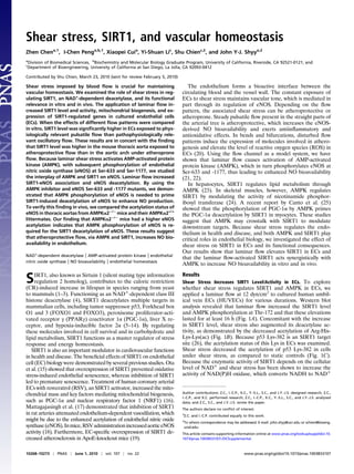
Shear Stress Increases SIRT1 Level/Activity in ECs. To explore
whether shear stress regulates SIRT1 and AMPK in ECs, we
applied a laminar flow at 12 dyn/cm2
to cultured human umbil-
ical vein ECs (HUVECs) for various durations. Western blot
analysis revealed that laminar flow increased the SIRT1 level
and AMPK phosphorylation at Thr-172 and that these elevations
lasted for at least 16 h (Fig. 1A). Concomitant with the increase
in SIRT1 level, shear stress also augmented its deacetylase ac-
tivity, as demonstrated by the decreased acetylation of Arg-His-
Lys-Lys(ac) (Fig. 1B). Because p53 Lys-382 is an SIRT1 target
site (26), the acetylation status of this Lys in ECs was examined.
Shear stress decreased the acetylation of p53 Lys-382 in cells
under shear stress, as compared to static controls (Fig. 1C).
Because the enzymatic activity of SIRT1 depends on the cellular
level of NAD+
and shear stress has been shown to increase the
activity of NAD(P)H oxidase, which converts NADH to NAD+
Author contributions: Z.C., I.-C.P., X.C., Y.-S.L., S.C., and J.Y.-J.S. designed research; Z.C.,
I.-C.P., and X.C. performed research; Z.C., I.-C.P., X.C., Y.-S.L., S.C., and J.Y.-J.S. analyzed
data; and Z.C., S.C., and J.Y.-J.S. wrote the paper.
The authors declare no conflict of interest.
1
Z.C. and I.-C.P. contributed equally to this work.
2
To whom correspondence may be addressed. E-mail: john.shyy@ucr.edu or schien@bioeng.
ucsd.edu.
This article contains supporting information online at www.pnas.org/lookup/suppl/doi:10.
1073/pnas.1003833107/-/DCSupplemental.
10268–10273 | PNAS | June 1, 2010 | vol. 107 | no. 22 www.pnas.org/cgi/doi/10.1073/pnas.1003833107
2. (27), we compared the cellular level of NAD+
in ECs under
shear stress and in controls. Compared with static control con-
ditions, laminar flow increased NAD+
level by ∼50% (Fig. 1D).
Together, Fig. 1 suggests that laminar flow elevates SIRT1 level
with an attendant increase in SIRT1 deacetylation activity.
Shear Stress Increases Mitochondrial Biogenesis in ECs. In several cell
types, the elevation of SIRT1 is tightly correlated with increased
mitochondrial biogenesis (12, 16, 25). In the present study, laminar
flow indeed increased the mitochondrial mass (Fig. 2A), with at-
tendant increase in the activity of mitochondrial reductase (Fig. 2B).
Because PGC-1α and NRF1 are key regulators of mitochondrial
biogenesis and COX4 is involved in the mitochondrial respiratory
chain (28), we examined whether laminar flow increases the ex-
pressionofthesemoleculesinECs.AsshowninFig.2C,laminarflow
increased the mRNA level of PGC-1α, NRF1, and COX4 in a time-
dependent manner. Importantly, SIRT1 knockdown by small in-
terferingRNA (siRNA)abolishedtheflow-inducedPGC-1α,NRF1,
and COX4 (Fig. 2 D and E). These results suggest that laminar flow
increases mitochondrial biogenesis in ECs and that this effect is
mediated through SIRT1.
Shear Stress Induction of SIRT1 Is Independent of AMPK. Depending
on the stimuli, AMPK and SIRT1 can regulate each other recip-
rocally (23, 24). Because laminar flow concurrently activated AMPK
and SIRT1, we investigated whether AMPK regulates SIRT1 or vice
versa in ECs under shear stress. As shown in Fig. 3A, AMPKα knock-
down by siRNA did not affect the laminar flow induction of SIRT1,
indicating that AMPK is not upstream of SIRT1. That the shear-
inducedincreaseinSIRT1levelisindependentofAMPKwasfurther
verified by the finding that ablation of both AMPKα1 and α2 in
murine embryonic fibroblasts (MEFs) had little effect on the shear
induction of SIRT1 (Fig. 3C). Furthermore, the level of SIRT1 was
comparable in wild-type MEFs infected with Ad-null or Ad-AMPK-
CA expressing the constitutively activated form of AMPK (Fig. 3E).
These experiments demonstrate that AMPK is neither necessary nor
sufficient for the laminar flow-induced SIRT1. We also knocked
downSIRT1inECsandusedMEFsisolatedfromSIRT1-nullmouse
embryos to determine whether SIRT1 regulates AMPK. As shown in
Fig. 3 B and D, laminar flow was still able to activate AMPK in ECs
with SIRT1 knocked down or in SIRT1−/−
MEFs, indicating that
SIRT1 is also not upstream to AMPK in our system. The results
presented inFig. 3 suggest that AMPK and SIRT1 are independently
activated by laminar flow; neither is upstream to the other.
AMPK and SIRT1 Synergistically Activate eNOS. Although AMPK
and SIRT1 were independently activated by laminar flow, there
is the possibility that they may act synergistically on eNOS to
increase NO bioavailability. Hence, we examined first whether
laminar flow can increase the association of SIRT1 with eNOS.
Following immunoprecipitation of SIRT1 from ECs, subsequent
anti-eNOS immunoblotting showed an increase in the level of
coimmunoprecipitated eNOS in lysates from ECs that had been
subjected to laminar flow (Fig. 4A). The increased association
between SIRT1 and eNOS in ECs exposed to laminar flow was
further confirmed by double immunostaining (Fig. 4B).
Fig. 1. Laminar shear stress increases SIRT1 level/activity in ECs. Confluent
monolayers of HUVECs were subjected to laminar flow at 12 dyn/cm2
for
various times or kept as static controls represented as time 0. (A and C) Cell
lysates were analyzed by Western blotting with anti-SIRT1, anti-phospho-
AMPK (Thr-172; T172), anti-AMPK, anti-α-tubulin, anti-acetyl-p53 (Lys-382;
K382), and anti-p53 antibodies. The bar graphs below are results of densi-
tometry analyses of the ratios of SIRT1 to α-tubulin, phospho-AMPK to
AMPK, and acetyl-p53 to p53. (B) SIRT1 deacetylase activity in HUVECs under
shear stress or static conditions was assessed by using fluorogenic Arg-His-
Lys-Lys (ac) as the substrate. The fluorescent intensities obtained from dif-
ferent groups were compared, with the value obtained at time 0 set as 1. (D)
NAD+
level in HUVECs under laminar flow or static condition for 16 h was
measured by spectrophotometry. The reading of the static group was set as
1. The bar graphs in all panels are mean ± SD averaged from three in-
dependent experiments. *, P < 0.05 vs. static control.
Fig. 2. Shear stress-increased mitochondrial biogenesis is mediated by
SIRT1. Confluent monolayer of HUVECs were subjected to laminar flow for
16 h or kept under static condition. (A) Mitotracker Green FM staining. (B)
Quantification of mitochondrial reductase activity by MTT assay from ab-
sorbance at 540 nm. (C) Assay of levels of mRNA encoding PGC-1α, NRF1, and
COX4 in HUVECs subjected to laminar flow for various durations by quan-
titative RT-PCR (qRT-PCR). (D and E) HUVECs were transfected with SIRT1
siRNA or control RNA (20 nM) and then exposed to laminar flow for 16 h or
were kept as static controls. SIRT1, PGC-1α, and α-tubulin levels were
assessed by Western blot analysis in (D), and NRF1 and COX4 mRNA levels
were analyzed by qRT-PCR in (E). Bar graphs are mean ± SD from three in-
dependent experiments. *, P < 0.05 between indicated groups.
Chen et al. PNAS | June 1, 2010 | vol. 107 | no. 22 | 10269
PHYSIOLOGY
3. Next, we examined the functional consequence of the SIRT1-
eNOS association. eNOS immunoprecipitation followed by immu-
noblottingwithanantibody recognizingacetylatedLysrevealedthat
acetylation of eNOS was decreased in ECs exposed to laminar flow
(Fig. 4C). We have shown that the application of laminar flow
causes AMPK activation before SIRT1 elevation (22) (Fig. 1), sug-
gesting that eNOS phosphorylation by AMPK may prime eNOS for
deacetylation by SIRT1. This possibility was verified by the finding
that the increase in eNOS phosphorylation and deacetylation in-
duced by laminar flow were blocked by the AMPK-inhibitor Com-
pound C (Fig. 4C). In contrast, SIRT1 inhibition by nicotinamide
(NAM in Fig. 4C) abolished eNOS deacetylation without affecting
eNOS phosphorylation.
To further delineate the requirement of eNOS phosphoryla-
tion for SIRT1 deacetylation, we transfected human embryonic
kidney 293 (HEK293) cells with plasmids overexpressing gain- or
loss-of-function mutants of bovine eNOS (bovine eNOS Ser-635
and -1179 corresponds to human eNOS Ser-633 and -1177).
S635D and S1179D (Asp replacing Ser) are the phosphomi-
metics that simulate the phosphorylation by AMPK, whereas
S635A and S1179A (Ala replacing Ser) are the non-
phosphorylatable mutants. As shown in Fig. 4D, eNOS S635A
and S1179A were more acetylated than the wild-type, S635D, or
S1179D. The transfected cells were also infected with adenovirus
overexpressing the wild-type SIRT1 (Ad-SIRT1) to mimic the
induction by flow (Fig. 4E). In parallel control experiments, cells
were infected with a dominant negative mutant of SIRT1 (Ad-
SIRT1-DN) (Fig. 4F). Overexpression of Ad-SIRT1, but not Ad-
SIRT1-DN, increased NO production by cells overexpressing
S635D or S1179D. In cells transfected with S635A or S1179A,
the NO production was not changed by overexpressing SIRT1,
nor by using SIRT1-DN (Fig. 4 E and F).
Pulsatile and Oscillatory Flows Have Opposite Effects on Regulating
SIRT1. To correlate the results obtained from flow channel
experiments with physiological and pathophysiological flow
conditions in arteries, we compared the SIRT1 level in ECs
subjected to pulsatile flow (12 ± 4 dyn/cm2
) and oscillatory flow
(1 ± 4 dyn/cm2
) representing steady flow that has a distinct di-
rection and disturbed flow with little net direction, respectively.
As shown in Fig. 5A, pulsatile flow, but not oscillatory flow, in-
creased the SIRT1 level in ECs. As a positive control, RSV
treatment increased the SIRT1 level in ECs similar to that with
pulsatile flow. Consistent with the increase in SIRT1 level, the
NAD+
level in ECs was higher under pulsatile than oscillatory
flow (Fig. 5B). Mitochondrial mass, revealed by Mitotracker
Green staining, was also increased by pulsatile, but not oscilla-
tory, flow (Fig. 5C).
Poly(ADP ribose) polymerase-1 (PARP-1), which is highly
sensitive to ROS, consumes NAD+
for catalysis of poly(ADP
ribosyl)ation on target proteins (29, 30). Because oscillatory flow
causes a sustained activation of NAD(P)H oxidase and a high
Fig. 3. Shear stress-increased SIRT1 is AMPK independent. (A and B)
HUVECs were transfected with AMPK α1 and α2 siRNAs (10 nM each), SIRT1
siRNA (20 nM), or with control RNA (20 nM) before being subjected to
laminar flow for 16 h. (C and D) MEFs ablated with AMPKα or SIRT1 (i.e.,
AMPKα−/−
and SIRT1−/−
) were subjected to laminar flow, and cell extracts
were collected at the indicated times. (E) Wild-type MEFs were infected with
Ad-AMPK-CA [100 multiplicities of infection (MOIs)] or Ad-null for 48 h. Total
cell proteins were then resolved by SDS/PAGE and subjected to Western
blot analysis with various antibodies as indicated.
Fig. 4. Synergistic effects of AMPK and SIRT1
on eNOS. (A) HUVECs were exposed to lami-
nar flow for the durations indicated or kept as
static controls. SIRT1 was immunoprecipitated
(IP) with anti-SIRT1, and immunoblotting (IB)
was performed with anti-eNOS. In parallel,
IgG was used as an IP control. The level of
immunoprecipitated SIRT1 was also assessed
by Western blot. (B) HUVECs under laminar
flow or static condition for 4 h were subjected
to immunostaining with anti-SIRT1 and anti-
eNOS, followed by conjugated secondary
antibodies. The cell nuclei were counter-
stained with DAPI. The colocalization of SIRT1
and eNOS is demonstrated by the merging of
pseudocolors. (C) HUVECs were pretreated
with DMSO, Compound C (10 μM), or nico-
tinamide (NAM) (5 mM) for 30 min before
being exposed to laminar flow for 4 h. The
phosphorylation of eNOS Ser-633 and Ser-
1177 was probed in the input lysates. In ad-
dition, eNOS was immunoprecipitated from
whole-cell lysates, and the acetylation of
eNOS and total eNOS were assessed by im-
munoblotting with anti-Ace-Lys and anti-
eNOS. (D–F) HEK293 cells were transfected with plasmids expressing bovine eNOS wild-type (WT), S635A, S635D, S1179A, or S1179D. (D) eNOS was immu-
noprecipitated and then examined by Western blot analysis with anti-eNOS and anti-Ace-Lys. (E and F) Transfected HEK293 cells were infected with Ad-(Flag)-
SIRT1 or Ad-(Flag)-SIRT1-DN with various MOIs, as indicated. Twenty-four hours after infection, the NO bioavailability in the cells was determined by Griess
assay and expressed as NOx. The expressions of SIRT1 and eNOS were examined by Western blot.
10270 | www.pnas.org/cgi/doi/10.1073/pnas.1003833107 Chen et al.
4. level of intracellular ROS (27, 31), we considered the possibility
that PARP-1 may be involved in the differential regulation of
SIRT1 in ECs subjected to different flow patterns. Fig. 5D shows
the level of PARP-1 was indeed higher under oscillatory than
pulsatile flow.
Differential Regulation of SIRT1-eNOS in the Vessel Wall. To de-
termine whether SIRT1 is differentially regulated in atheroprone
vs. atheroprotective areas in the arterial tree, we isolated the
aortic arch and thoracic aorta from C57BL6 mice. These areas
correspond to regions under disturbed and pulsatile flows, re-
spectively (32). Western blot analysis showed that the SIRT1
level was higher in the thoracic aorta (under pulsatile flow) than
in the aortic arch (under disturbed flow) (Fig. 6A). Because the
isolated tissues were a mixture of ECs, vascular smooth muscle
cells, and connective tissues, we performed en face staining on
the mouse arterial tree. Confocal microscopy revealed that the
level of SIRT1 in the endothelium of the thoracic aorta was
significantly higher than that in the inner curvature of the aortic
arch (Fig. 6B). Furthermore, colocalization of SIRT1 and eNOS
was noticeably higher in the thoracic aorta than aortic arch en-
dothelium. To confirm the synergistic effect of AMPK and
SIRT1 on eNOS in vivo, we compared the phosphorylation and
acetylation of eNOS in thoracic aortas from AMPKα2−/−
mice
and their AMPKα2+/+
littermates. As seen in Fig. 6 C and D,
acetyl-CoA carboxylase (ACC), the AMPK canonical target, and
eNOS phosphorylations were lower, whereas eNOS acetylation
level was higher in the thoracic aortas of AMPKα2−/−
mice, as
compared with their AMPKα2+/+
littermates. However, the
SIRT1 level was comparable between the two lines of mice.
These results suggest that eNOS deacetylation by SIRT1 in the
vessel wall requires AMPK.
Discussion
Dysfunctional ECs, signified by enhanced inflammation and ox-
idative stress, prelude atherosclerosis and other vascular im-
pairments. TheeffectsofshearstressonECscanbeatheroprotective
or atheroprone depending on the associated flow patterns. Because
SIRT1 exerts antiinflammatory and antioxidative effects on various
cell types, including ECs, we studied the shear stress regulation of
SIRT1inECs.Ourresultsindicatethatlaminarflowcausesamarked
increase in SIRT1 level, and that AMPK and SIRT1 synergistically
increase the eNOS-derived NO bioavailability in ECs responding to
laminar flow.
Consistent with its effects of elevating the levels of SIRT1 and
NAD+
, laminar flow increased the mitochondria mass and levels
of PGC-1α, NRF1, and COX4. siRNA knockdown experiments
presented in Fig. 2 showed that these key regulators of mito-
chondrial biogenesis are up-regulated by the flow-elevated
SIRT1 (Fig. 2 C–E). Increased mitochondrial biogenesis is im-
plicated in the balance of the endothelial redox state (33). By
scavenging free radicals (e.g., ROS and reactive nitrogen species)
through PGC-1α-induced ROS detoxifying enzymes (34), the
laminar flow-enhanced mitochondrial biogenesis may protect
ECs against oxidative stress. We have previously shown that
laminar flow activates endogenous PPARγ and its target genes in
ECs and that these effects are ligand-dependent (35). Together
with the ligand-activated PPARγ, the elevated PGC-1α, func-
tioning as a transcriptional coactivator of PPARγ, may also
contribute to the positive effects of PPARγ on EC biology, in-
cluding reverse cholesterol transport, antiinflammation, and
antiatherosclerosis (36). In addition, laminar flow is known to
arrest the EC cycle at the G0/G1 phase (37), which may explain
the quiescence of ECs in the straight part of the arterial tree.
Inhibition of SIRT1 by sirtinol or siRNA causes premature se-
nescence-like growth arrest in ECs (15). The implication of this
notion in the current study is that the elevated SIRT1 by laminar
flow may ameliorate aging-related senescence of ECs, even if
they are quiescent.
eNOS-derived NO bioavailability, which is critically important
for EC physiological functions, is regulated by multiple mecha-
nisms at transcriptional and posttranslational levels (38). Among
the various mechanisms, phosphorylations at Ser-633 and Ser-
1177 enhance eNOS activity, and AMPK can phosphorylate both
sites (21, 22, 39). Although the shear stress activation of AMPK
and SIRT1 seems to be independent processes, the activated
AMPK and SIRT1 converge at eNOS in that AMPK phos-
phorylation may prime eNOS for SIRT1 deacetylation (depicted
in Fig. 6E). Such a model is supported by the following obser-
vations: eNOS was no longer deacetylated following inhibition of
AMPK activity by Compound C (Fig. 4C), and eNOS S635A and
S1179A were much more acetylated than eNOS S635D and
S1179D (Fig. 4D). This mode of action is reminiscent of the
synergistic effects of AMPK and SIRT1 on PGC-1α (25). By
using AMPKα2−/−
mice, we showed that this synergism occurs in
vivo. Because shear stress appears to preferentially activate
AMPKα2 in ECs (40), and AMPKα1 ablation caused anemia
and severe splenomegaly in the knockout animals (41),
AMPKα2−/−
instead of AMPKα1−/−
mice were used. AMPKα2
ablation resulted in a lower level of eNOS phosphorylation and
a higher level of eNOS acetylation, even though SIRT1 was as
abundant as in the wild type (Fig. 6 C and D). These results
indicate that AMPK phosphorylation of eNOS is required for the
deacetylation by SIRT1 in the mouse thoracic aorta. SIRT1 in-
hibition by nicotinamide resulted in superphosphorylation of
eNOS (Fig. 4C). It seems that eNOS deacetylation is also cor-
related with its dephosphorylation. Such a mechanism may re-
store the set point for eNOS activation. Despite the fact that
eNOS phosphorylation sites have been extensively studied, the
acetylation sites remain unclear. Two potential residues are Lys-
Fig. 5. SIRT1 is differentially regulated by pulsatile flow vs. oscillatory flow.
HUVECs were exposed to pulsatile flow (PS) (12 ± 4 dyn/cm2
, 1 Hz) or oscillatory
flow (OS) (1 ± 4 dyn/cm2
, 1 Hz), treated with resveratrol (RSV) (100 μM), or kept
under static condition for 16 h. (A) The levels of SIRT1, phospho-AMPK (Thr-
172), AMPK, and α-tubulin were assessed by Western blot analysis. *, P < 0.05
compared to static control; #, P < 0.05 between PS and OS. (B) NAD+
level was
measured spectrophotometrically. *, P < 0.05 compared to static control. (C)
The mitochondria in static cells and those exposed to pulsatile or oscillatory
flow were stained with Mitotracker Green FM. *, P < 0.05 between indicated
groups. (D) PARP-1 expression in HUVECs exposed to pulsatile or oscillatory
flow was revealed by Western blot analysis. *, P < 0.05 compared to PS. Bar
graphs represent densitometry analysis of three independent experiments,
with the static group set as 1 in (A), (B), and (C). In (D), the level of PARP-1/
α-tubulin in ECs exposed to pulsatile flow was set as 1.
Chen et al. PNAS | June 1, 2010 | vol. 107 | no. 22 | 10271
PHYSIOLOGY
5. 494 and Lys-504 (17). The identification of SIRT1-targeted sites
in eNOS and their interplay with phosphorylation sites warrant
future research.
Cellular NAD+
level can modulate SIRT1 activity and ex-
pression (42, 43). Thus, one possible mechanism by which lam-
inar/pulsatile flow augments SIRT1 level and/or activity is
through the elevated NAD+
level. Such an enhanced oxidized
state in ECs may be caused by the increase in NAD+
(Figs. 1D
and 5B) resulting from an increased activity of NAD(P)H oxi-
dase. Indeed, laminar flow induces a transient increase in the
activity of NAD(P)H oxidase (27). Previously, Nisoli et al. (44)
found that the SIRT1 induction by CR involves eNOS-derived
NO, as evidenced by the attenuated SIRT1 in white adipose
tissue of the eNOS-null mice. However, laminar flow was able to
increase SIRT1 level in eNOS-null MEFs and in HUVECs
treated with the NOS inhibitor nitro-L-arginine methyl ester (L-
NAME) (Fig. S1). These results suggest that the flow induction
of SIRT1 does not depend on eNOS. One possible explanation
for this independency is that the moderate and transient increase
in ROS by pulsatile flow is able to induce SIRT1. This is in line
with the recent finding that oxidative stress generated by short-
term H2O2 treatment enhanced SIRT1 (45). In contrast to pul-
satile flow, oscillatory flow causes a sustained activation of NAD
(P)H oxidase with an attendant strong elevation of ROS (27, 31,
46). This pathophysiological ROS level should induce or activate
PARP-1 (29), which in turn depletes cellular NAD+
to diminish
the expression or activity of SIRT1 as seen in Fig. 5 A and B.
The differential effects of pulsatile vs. oscillatory flow on
SIRT1 expression was verified in the mouse arterial tree (Fig. 6).
SIRT1 level was higher, with increased eNOS colocalized in the
atheroprotective areas. It is known that endothelial dysfunction
(which is signified by increased oxidative and inflammatory
responses) predisposes the arteries to atherosclerosis. Hence,
SIRT1 activation by pulsatile flow may prevent EC dysfunction
and counteract the risk factors associated with atherosclerosis.
Compared with therapeutic interventions such as RSV and sev-
eral small molecules developed for SIRT1 activation, shear stress
would be more physiologically relevant. The molecular basis
underlying the marked differences between pulsatile and oscil-
latory flows in the modulation of SIRT1 may provide new
insights into the mechanism of regulation of EC biology by
shear stress.
Materials and Methods
The resources of antibodies and reagents, as well as detailed methods for cell
culture, adenoviral infection, siRNA knockdown, transient transfection,
Western blot analysis, immunoprecipitation, qRT-PCR, NAD+
measurement,
MTT assay, and NO bioavailability assay are described in SI Materials
and Methods.
Fluid Shear Stress Experiments. The shear stress experiments were performed
as previously described (22, 47, 48). Various types of cells were exposed to
laminar flow at 12 dyn/cm2
for different times. In some experiments, a re-
ciprocating syringe pump was connected to the circulating system to in-
troduce a sinusoidal component with a frequency of 1 Hz onto the shear
stress. Pulsatile shear flow or oscillatory shear flow was applied to ECs with
a shear stress of 12 ± 4 dyn/cm2
or 1 ± 4 dyn/cm2
, respectively.
SIRT1 Activity Assay. SIRT1 activity was assessed as previously described (24),
with minor modification. Briefly, 10–20 μg of EC extracts were used in
a deacetylation assay, with Fluor de Lys-SIRT1 as the substrate (Biomol). The
deacetylation of the substrate was measured by use of a microplate-reading
fluorimeter, with excitation set at 380 nm and emission at 460 nm.
Animal Experiments. The animal experimental protocols were approved by
the Institutional Animal Care and Use Committee of University of California,
Riverside. The thoracic aorta and aortic arch from male C57BL6 mice older than
20 weeks were isolated for both Western blot analysis and en face immu-
nostaining as previously described (47, 48). In addition, thoracic aortas were
dissected from 20-week-old AMPKα2−/−
mice and their AMPKα2+/+
litter-
mates. Tissue extracts from aortas were pooled for eNOS immunoprecipita-
tion and immunoblotting.
Mitotracker and Immunofluorescence Staining. Mitotracker Green FM (Invi-
trogen) was used to stain mitochondria in HUVECs as previously described
(49). The cells were washed twice with PBS and then incubated with 20 nM
Mitotracker Green FM for 30 min. After 3 washes with PBS, the cells were
subjected to fluorescence detection (excitation, 490 nm; emission, 516 nm)
by using a Leica SP2 confocal microscope.
For immunostaining, HUVECs were fixed with 4% paraformaldehyde in
PBS (pH 7.4) for 30 min at room temperature. For en face staining, mouse
aortas were perfused with 4% paraformaldehyde in PBS for 15 min, and the
dissected specimens were fixed for another 60 min after the removal of
tunica adventitia. Primary antibodies, i.e., rabbit anti-SIRT1 and mouse anti-
Fig. 6. Differential regulation of SIRT1/eNOS in the vessel wall
in vivo. (A) Tissue extracts of the aortic arch (AA) and thoracic
aorta (TA) from two C57BL6 male mice were pooled and sub-
jected to Western blot analysis with anti-SIRT1 and α-tubulin
antibodies. Data shown represent results from a total of eight
mice. (B) Confocal microscope images of en face immunos-
taining of SIRT1 and eNOS in the endothelium of the aortic
arch and thoracic aorta of C57BL6 mice. The primary anti-
bodies were anti-SIRT1 and anti-eNOS, and secondary anti-
bodies were Alexa Fluor 488-conjugated chicken anti-rabbit or
Alexa Fluor 555-conjugated chicken anti-mouse, respectively.
Nuclei were counterstained with DAPI. Shown are represen-
tative images from six animals. (C and D) Tissue extracts were
isolated from the thoracic aorta of AMPKα2−/−
and their
AMPKα2+/+
littermates. In (C), the level of phospho-eNOS Ser-
633, Ser-1177, AMPKα, phosphorylated ACC, SIRT1, and
α-tubulin was determined by Western blot. In (D), eNOS was
immunoprecipitated and the acetylation of eNOS in the
immunoprecipitates was determined by immunoblotting with
anti-Ace-Lys. After stripping, the blot was reprobed with anti-
eNOS. The presented results on AMPKα2−/−
and AMPKα2+/+
mice were pooled from eight aortas in each group. The
experiments were repeated with another group of seven ani-
mals per mouse line. Bar graphs below are densitometry
analysis of the ratios of acetyl-eNOS to eNOS from the two experiments. (E) Schematic model of the synergistic effects of AMPK and SIRT1 on eNOS activity.
Laminar/pulsatile shear stress increases the NAD+
level in ECs, thus leading to elevation of SIRT1. Concomitantly, AMPK is activated by upstream AMPK kinase
(AMPKK), which in turn phosphorylates eNOS. The phosphorylated eNOS enhances its affinity toward SIRT1 so that eNOS is deacetylated. Such a synergism
between AMPK and SIRT1 augments the eNOS-derived NO bioavailability.
10272 | www.pnas.org/cgi/doi/10.1073/pnas.1003833107 Chen et al.
6. eNOS, were applied at 1:100 dilution to the specimens, which were in-
cubated at 4 °C overnight and then washed off with PBS and 0.1% Tween-20
(PBST), followed by the application of Alexa Fluor 488-conjugated chicken
anti-rabbit or Alexa Fluor 555-conjugated chicken anti-mouse (Invitrogen).
After PBST wash, coverslips were mounted with Prolong Gold Antifade Re-
agent (Invitrogen) and viewed with a Leica SP2 confocal microscope. For all
immunofluorescence experiments, parallel groups of cells or aorta speci-
mens were stained with only primary antibody or secondary antibody as
a control. Multiple images were captured from three to four regions in the
aortic arch and thoracic aorta of every mouse.
Statistical Analysis. The significance of variability was determined by Stu-
dent’s t test or one-way ANOVA. All results are presented as mean ± SD from
at least three independent experiments. Unless otherwise indicated, P < 0.05
was considered statistically significant.
ACKNOWLEDGMENTS. We thank Dr. Michael McBurney (Department of
Medicine, University of Ottawa) for providing SIRT1-null MEF, and Mr. Phu
Nguyen (Department of Bioengineering, University of California at San
Diego) for technical services. This work was supported in part by National
Institutes of Health Grants HL89940 (to J.Y.-J.S.), HL93731 (to J.Y.-J.S.),
HL080518 (to S.C.), and HL085159 (to S.C.).
1. Lin SJ, Defossez PA, Guarente L (2000) Requirement of NAD and SIR2 for life-span
extension by calorie restriction in Saccharomyces cerevisiae. Science 289:2126–2128.
2. Bordone L, et al. (2007) SIRT1 transgenic mice show phenotypes resembling calorie
restriction. Aging Cell 6:759–767.
3. Boily G, et al. (2008) SirT1 regulates energy metabolism and response to caloric
restriction in mice. PLoS One 3:e1759.
4. Imai S, Armstrong CM, Kaeberlein M, Guarente L (2000) Transcriptional silencing and
longevity protein Sir2 is an NAD-dependent histone deacetylase. Nature 403:795–800.
5. Motta MC, et al. (2004) Mammalian SIRT1 represses forkhead transcription factors.
Cell 116:551–563.
6. Brunet A, et al. (2004) Stress-dependent regulation of FOXO transcription factors by
the SIRT1 deacetylase. Science 303:2011–2015.
7. Nemoto S, Fergusson MM, Finkel T (2004) Nutrient availability regulates SIRT1
through a forkhead-dependent pathway. Science 306:2105–2108.
8. Yang Y, Hou H, Haller EM, Nicosia SV, Bai W (2005) Suppression of FOXO1 activity by
FHL2 through SIRT1-mediated deacetylation. EMBO J 24:1021–1032.
9. Luo J, et al. (2001) Negative control of p53 by Sir2α promotes cell survival under stress.
Cell 107:137–148.
10. Vaziri H, et al. (2001) hSIR2(SIRT1) functions as an NAD-dependent p53 deacetylase.
Cell 107:149–159.
11. Rodgers JT, et al. (2005) Nutrient control of glucose homeostasis through a complex
of PGC-1α and SIRT1. Nature 434:113–118.
12. Lagouge M, et al. (2006) Resveratrol improves mitochondrial function and protects
against metabolic disease by activating SIRT1 and PGC-1α. Cell 127:1109–1122.
13. Li X, et al. (2007) SIRT1 deacetylates and positively regulates the nuclear receptor LXR.
Mol Cell 28:91–106.
14. Dioum EM, et al. (2009) Regulation of hypoxia-inducible factor 2α signaling by the
stress-responsive deacetylase sirtuin 1. Science 324:1289–1293.
15. Ota H, et al. (2007) Sirt1 modulates premature senescence-like phenotype in human
endothelial cells. J Mol Cell Cardiol 43:571–579.
16. Csiszar A, et al. (2009) Resveratrol induces mitochondrial biogenesis in endothelial
cells. Am J Physiol Heart Circ Physiol 297:H13–H20.
17. Mattagajasingh I, et al. (2007) SIRT1 promotes endothelium-dependent vascular
relaxation by activating endothelial nitric oxide synthase. Proc Natl Acad Sci USA 104:
14855–14860.
18. Orimo M, et al. (2009) Protective role of SIRT1 in diabetic vascular dysfunction.
Arterioscler Thromb Vasc Biol 29:889–894.
19. Zhang QJ, et al. (2008) Endothelium-specific overexpression of class III deacetylase
SIRT1 decreases atherosclerosis in apolipoprotein E-deficient mice. Cardiovasc Res 80:
191–199.
20. Chien S (2008) Effects of disturbed flow on endothelial cells. Ann Biomed Eng 36:
554–562.
21. Zhang Y, et al. (2006) AMP-activated protein kinase is involved in endothelial NO
synthase activation in response to shear stress. Arterioscler Thromb Vasc Biol 26:
1281–1287.
22. Chen Z, et al. (2009) AMP-activated protein kinase functionally phosphorylates
endothelial nitric oxide synthase Ser633. Circ Res 104:496–505.
23. Hou X, et al. (2008) SIRT1 regulates hepatocyte lipid metabolism through activating
AMP-activated protein kinase. J Biol Chem 283:20015–20026.
24. Fulco M, et al. (2008) Glucose restriction inhibits skeletal myoblast differentiation by
activating SIRT1 through AMPK-mediated regulation of Nampt. Dev Cell 14:661–673.
25. Cantó C, et al. (2009) AMPK regulates energy expenditure by modulating NAD+
metabolism and SIRT1 activity. Nature 458:1056–1060.
26. Langley E, et al. (2002) Human SIR2 deacetylates p53 and antagonizes PML/p53-
induced cellular senescence. EMBO J 21:2383–2396.
27. De Keulenaer GW, et al. (1998) Oscillatory and steady laminar shear stress
differentially affect human endothelial redox state: role of a superoxide-producing
NADH oxidase. Circ Res 82:1094–1101.
28. Hock MB, Kralli A (2009) Transcriptional control of mitochondrial biogenesis and
function. Annu Rev Physiol 71:177–203.
29. Chiarugi A (2002) Poly(ADP-ribose) polymerase: killer or conspirator? The ‘suicide
hypothesis’ revisited. Trends Pharmacol Sci 23:122–129.
30. Ha HC, Snyder SH (1999) Poly(ADP-ribose) polymerase is a mediator of necrotic cell
death by ATP depletion. Proc Natl Acad Sci USA 96:13978–13982.
31. Hwang J, et al. (2003) Pulsatile versus oscillatory shear stress regulates NADPH oxidase
subunit expression: implication for native LDL oxidation. Circ Res 93:1225–1232.
32. Suo J, et al. (2007) Hemodynamic shear stresses in mouse aortas: implications for
atherogenesis. Arterioscler Thromb Vasc Biol 27:346–351.
33. Davidson SM, Duchen MR (2007) Endothelial mitochondria: contributing to vascular
function and disease. Circ Res 100:1128–1141.
34. St-Pierre J, et al. (2006) Suppression of reactive oxygen species and neurodegeneration
by the PGC-1 transcriptional coactivators. Cell 127:397–408.
35. Liu Y, et al. (2004) Laminar flow activates peroxisome proliferator-activated receptor-
gamma in vascular endothelial cells. Circulation 110:1128–1133.
36. Tontonoz P, Spiegelman BM (2008) Fat and beyond: the diverse biology of
PPARgamma. Annu Rev Biochem 77:289–312.
37. Lin K, et al. (2000) Molecular mechanism of endothelial growth arrest by laminar
shear stress. Proc Natl Acad Sci USA 97:9385–9389.
38. Sessa WC (2004) eNOS at a glance. J Cell Sci 117:2427–2429.
39. Boo YC, et al. (2006) Coordinated regulation of endothelial nitric oxide synthase
activity by phosphorylation and subcellular localization. Free Radic Biol Med 41:
144–153.
40. Fisslthaler B, Fleming I (2009) Activation and signaling by the AMP-activated protein
kinase in endothelial cells. Circ Res 105:114–127.
41. Föller M, et al. (2009) Regulation of erythrocyte survival by AMP-activated protein
kinase. FASEB J 23:1072–1080.
42. Lin SJ, Ford E, Haigis M, Liszt G, Guarente L (2004) Calorie restriction extends yeast life
span by lowering the level of NADH. Genes Dev 18:12–16.
43. Zhang Q, et al. (2007) Metabolic regulation of SIRT1 transcription via a HIC1:CtBP
corepressor complex. Proc Natl Acad Sci USA 104:829–833.
44. Nisoli E, et al. (2005) Calorie restriction promotes mitochondrial biogenesis by
inducing the expression of eNOS. Science 310:314–317.
45. Nasrin N, et al. (2009) JNK1 phosphorylates SIRT1 and promotes its enzymatic activity.
PLoS One, 10.1371/journal.pone.0008414.
46. Hwang J, et al. (2003) Oscillatory shear stress stimulates endothelial production of O2
-
from p47phox-dependent NAD(P)H oxidases, leading to monocyte adhesion. J Biol
Chem 278:47291–47298.
47. Guo D, Chien S, Shyy JY (2007) Regulation of endothelial cell cycle by laminar versus
oscillatory flow: distinct modes of interactions of AMP-activated protein kinase and
Akt pathways. Circ Res 100:564–571.
48. Young A, et al. (2009) Flow activation of AMP-activated protein kinase in vascular
endothelium leads to Krüppel-like factor 2 expression. Arterioscler Thromb Vasc Biol
29:1902–1908.
49. Schulz E, et al. (2008) Suppression of the JNK pathway by induction of a metabolic
stress response prevents vascular injury and dysfunction. Circulation 118:1347–1357.
Chen et al. PNAS | June 1, 2010 | vol. 107 | no. 22 | 10273
PHYSIOLOGY
7. Supporting Information
Chen et al. 10.1073/pnas.1003833107
SI Materials and Methods
Antibodies and Reagents. The antibodies used in the present study
wereanti-SIRT1fromMillipore,andanti-pan-AMPKα,anti-phospho-
AMPK Thr-172, anti-phospho-ACC Ser-79, anti-acetyl-p53 Lys-379/
382, anti-p53, anti-PGC-1α, anti-eNOS, anti-acetylated lysine, anti-
PARP-1, anti-α-tubulin, horseradish peroxide-conjugated anti-rabbit,
and anti-mouse antibodies from Cell Signaling Technology. Anti-
phospho-eNOS Ser-633/635, anti-phospho-eNOS Ser-1177/1179, and
anti-eNOS used in immunoprecipitation were from BD Transduction
Laboratory.
Cell Culture. HUVECs were cultured in medium M199 (Gibco)
supplemented with 15% FBS (Omega), 3 ng/mL β-EC growth
factor (Sigma), 4 U/mL heparin (Sigma), and 100 U/mL peni-
cillin-streptomycin. MEFs and HEK293 cells were cultured in
DMEM containing 10% FBS.
Adenoviral Infection, siRNA Knockdown, and Transient Transfection.
Ad-AMPK-CA,arecombinantadenovirusexpressingaconstitutively
active(CA)formof AMPKα2,wasdescribedpreviously (1). MEFsat
70% confluency were seeded on six-well plates, infected with Ad-null
or Ad-AMPK-CA at 100 multiplicities of infection (MOI), and in-
cubated for 24 h before further experimentation.
Transient transfection was performed with Lipofectamine
RNAiMAX (Invitrogen). In brief, HUVECs at 70% confluency
were transfected with AMPKα1 and AMPKα2 at 10 nM each
(Qiagen, SI02622235 and SI02758595), and SIRT1 (Qiagen,
SI00098434) or scramble (ctrl) siRNA, each at 20 nM, in Opti-
MEM (Gibco). Four hours after transfection, the medium was
changed to fresh M199, and cells were kept in culture for 48 h
before shear stress experiments.
HEK293 cells were transiently transfected with 1 μg of re-
spective DNA and 2.5 μL Lipofectamine 2000 (Invitrogen) per
106
cells, following a standard protocol. Twenty-four hours after
transfection, cells were infected with Ad-SIRT1 or Ad-SIRT1-
DN (2) for another 24 h.
Western Blot Analysis and Immunoprecipitation. Lysates from ECs,
MEFs, HEK293 cells, or mouse aortas were resolved on 8% SDS/
PAGE, and proteins were transferred to PVDF membrane. The
immunoblotting with different antibodies followed instructions
provided by various manufacturers. Immunoprecipitation for
SIRT1 and eNOS was performed following the standard protocol
provided by the manufacturers (Millipore for SIRT1 and BD
Biosciences for eNOS).
Quantitative RT-PCR. Total RNA was isolated with TRIzol reagent
(Invitrogen). Reverse transcription was carried out with 3 μg of
total RNA by the SuperScript II reverse transcriptase (In-
vitrogen). The synthesized cDNA was used to perform real-time
quantitative PCR (qPCR) with the iQ SYBR Green supermix
(Bio-Rad) on the iCycler real-time PCR detection system
(Bio-Rad). The sequences of primer sets used were as follows:
for PGC-1α, forward: GGAGCAATAAAGCGAAGA; reverse:
GAGGAGTTGTGGGAGGAG; for NRF1, forward: ACTCT-
ACAGGTCGGGGAAAA; reverse: AGTGAGACAGTGCCA-
TCAGG; for COX4, forward: GCAGTGGCGGCAGAATGT;
reverse: GGCTAAGCCCTGGATGGG.
NAD+
Measurement and MTT Assays. The cellular level of NAD+
and was measured as previously described (3). The cell extract
was neutralized and centrifuged to collect the supernatant. Forty
microliters of supernatant was added to 80 μL of an NAD+
re-
action mixture and incubated for 5 min at 37 °C. After measuring
the basal absorbance at 570 nm, the reaction was initiated by
adding 20 μL of alcohol dehydrogenase solution and then in-
cubated at 37 °C for 20 min. The absorbance of the reaction
mixture was determined again at 570 nm. The NAD+
level was
obtained by subtracting the basal absorbance from the second
reading.
3-(4,5-Dimethylthiazol-2-yl)-2,5-diphenyltetrazolium bromide
(MTT) (Molecular Probes) was used to assess mitochondrial
activity with a protocol adapted from Xia et al (4). HUVECs were
incubated with 150 μg/mL MTT under normal growth conditions
(5% CO2, 37 °C) for 2 h. The medium was removed, the for-
mazan product was dissolved in DMSO, and the solution was
then incubated at 37 °C for 10 min. The absorbance of formazan
product from MTT was measured at 540 nm by using a spectro-
photometer. After subtracting the background, the reading was
normalized to total protein concentration.
NO Bioavailability Assays. The NO production from cells trans-
fected with various plasmids was detected as the accumulated
nitrite (NO2
–
), a stable breakdown product of NO, in cell culture
media by using Griess reagent (Sigma). An aliquot of cell culture
media was mixed with an equal volume of Griess reagent and
then incubated at room temperature for 15 min. The azo dye
production was analyzed by use of a spectrophotometer with
absorbance set at 540 nm.
1. Foretz M, et al. (2005) Short-term overexpression of a constitutively active form of
AMP-activated protein kinase in the liver leads to mild hypoglycemia and fatty liver.
Diabetes 54:1331–1339.
2. Luo J, et al. (2001) Negative control of p53 by Sir2α promotes cell survival under stress.
Cell 107:137–148.
3. Jacobson EL, Jacobson MK (1976) Pyridine nucleotide levels as a function
of growth in normal and transformed 3T3 cells. Arch Biochem Biophys 175:
627–634.
4. Xia S, Laterra J (2006) Hepatocyte growth factor increases mitochondrial mass in
glioblastoma cells. Biochem Biophys Res Commun 345:1358–1364.
Chen et al. www.pnas.org/cgi/content/short/1003833107 1 of 2
8. Fig. S1. Confluent monolayer of the wild-type MEF (A) and eNOS−/−
MEF (B) subjected to a laminar flow for the time as indicated or kept under static
condition (time 0). In (C), HUVECs were treated with L-NAME (100 μM) 30 min before the exposure to the laminar flow for 16 h. In parallel static controls,
HUVECs were incubated with or without L-NAME. The collected lysates were analyzed by Western blot with anti-SIRT1 and anti-α-tubulin.
Chen et al. www.pnas.org/cgi/content/short/1003833107 2 of 2