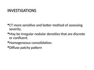
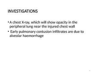
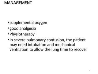
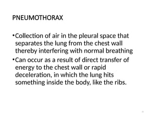
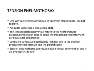
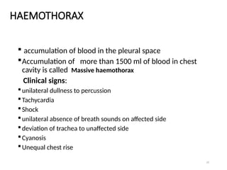
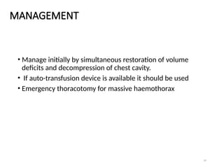
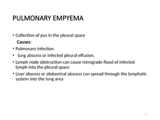
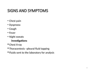
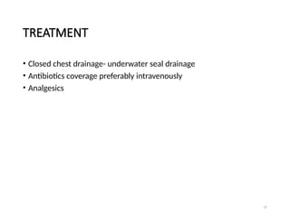
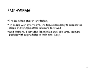
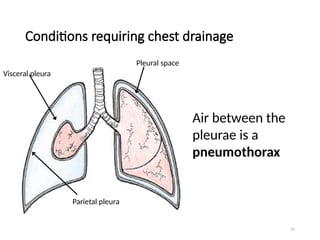
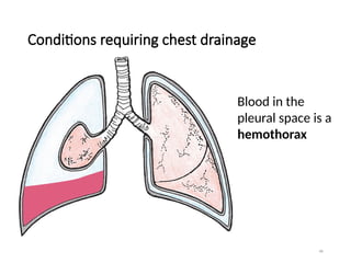
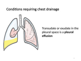
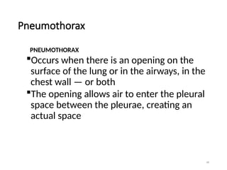
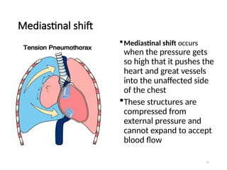
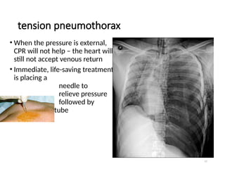
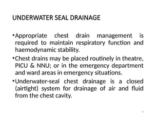
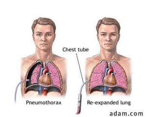
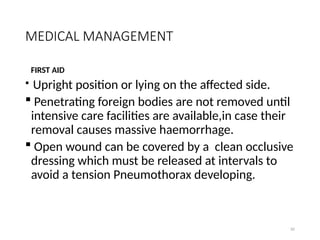
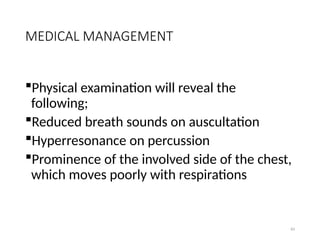
1
Disorders Of the Respiratory
System
Chest injuries
2
INTRODUCTION
•Injuries to the chest and abdomen can be
difficult to recognize and treat, and many
injuries can go unnoticed until they become
very serious.
• The muscle and bones that serve to protect
vital organs can also mask their injuries - or at
worst contribute to them.
• It is important for the rescuer to consider
injuries that lie beneath the skin
3
Intro cont ……….
• Different organs react in different ways when
subjected to trauma.
•Hollow organs (such as the bladder) tend to
rupture, releasing their contents into the
surrounding space.
•Solid organs (such as the liver) tend to tear
instead, often bleeding at a slow enough rate to
be overlooked.
4
CLOSED CHEST INJURIES
•Chest injuries can be inherently serious, as
this area of the body houses many critical
organs, such as the heart, lungs, and many
blood vessels.
• Most chest trauma injuries should receive
professional medical attention.
5
OPEN CHEST WOUNDS
•An open Pneumothorax or sucking chest
wound - the chest wall has been penetrated
(by knife, bullet, falling onto a sharp object)
Recognition
•An open chest wound – escaping air
•Entrance and possible exit wound (exit
wounds are more severe)
6
SIGNS AND SYMPTOMS
•Trouble breathing
•Sucking sound as air passes through opening
in chest wall
•Blood or blood-stained bubbles may be
expelled with each exhalation
•Coughing up blood
7
TREATMENT
•Assess ABCs and intervene as necessary
•Do not remove any embedded objects
•Call for an ambulance
•Flutter valve over wound.
•Lateral positioning: victim's injured side
down
•Treat for shock
•Conduct a secondary survey
•Monitor vitals carefully
8
MAKING A FLUTTER VALVE
•Get some sort of plastic that is bigger than the
wound.
• Tape the plastic patch over the wound on only 3
sides.
• The 4th side is left open, allowing blood to
drain and air to escape.
•This opening should be at the bottom (as
determined by the victim’s position).
•When the casualty inhales, the bag will be
sucked in, but when the casualty exhales, the air
will exit through the untaped side.
9
RIB INJURIES
•A common result of trauma to the chest is
damage to the victim's rib cage.
• The curved shape of the rib cage helps to deflect
the force of some injuries, but damage to
cartilage or the ribs themselves can still result.
• While a single broken rib can be very painful for
the patient, a number of broken ribs can lead to
other complications.
• A victim with broken ribs may take very shallow
breaths without even noticing it, as their body
tries to prevent the pain with taking a full breath.
10
•When many adjoining ribs are broken in
different places, a portion of the rib cage can
move in the opposite direction the chest
should.
•This is known as a "flail" segment, and can
make breathing very painful and less
effective.
11
SIGNS AND SYMPTOMS
 Trouble breathing
•Shallow breathing
•Tenderness at site of injury
•Deformity & bruising of chest
•Pain upon movement/deep
breathing/coughing
•Dusky or blue lips or nail beds
•May cough up blood
•Crackling feeling upon touching victim's skin.
12
TREATMENT
•Assess ABCs and intervene as necessary
•Call for an ambulance
•Assist the victim into a position of comfort
(typically seated upright, to avoid fighting
gravity)
•Conduct a secondary survey
•Monitor patient's condition carefully
•Be vigilant, keep alert for any changes.
•If a flail segment is suspected, tightly secure a
bulky dressing (such as a tightly folded hand
towel) to help stabilize the injury.
13
FLAIL CHEST
•Major physiological insult is contusion of
underlying lung and decreased vital capacity
•Occurs when 3 or more consecutive ribs or
costal cartilages are fractured bifocally.
•These circumscribed segments, having lost
continuity with the rigid thorax, move
inwards with inspiration and push outward
with exhalation, thus moving paradoxically.
14
PRESENTING SYMPTOMS
 Chest pains
 Tachypnoea
 Dyspnoea
 Thoracic splinting, along with chest wall contusions,
 Tenderness,
 Crepitance,
 Palpable rib fractures
N.B : these symptoms are are suggestive, but
paradoxical chest wall motion is the diagnostic sine
qua non.
15
INVESTIGATIONS
may be difficult to diagnose if patient is
already mechanically ventilated, in pain,
obese, or has large breasts or subcutaneous
emphysema.
CXR is helpful in identifying multiple
fractured ribs, but will not reveal
cartilaginous disruptions.
 Major value of the CXR is in detecting
associated injures
16
MANAGEMENT
Half the cases can be managed without ventilation
others require ventilation for 1-3 weeks
chest wall usually stabilises in 1-2 weeks
operative fixation is suggested by some authors.
Main benefit is to prevent deformity.
weaning should not wait till paradoxical movement
improves, rather should be initiated when gas
exchange is adequate.
In absence of systemic hypotension control
administration of IV fluids to prevent over hydration
17
LUNG CONTUSION
Essentially a bruise of the lung.
Direct injury causes pulmonary vascular damage
with secondary alveolar haemorrhage
Initially not much shunt as these alveoli are poorly
perfused
Subsequently tissue inflammation develops.
18
INVESTIGATIONS
CT more sensitive and better method of assessing
severity.
May be irregular nodular densities that are discrete
or confluent.
Homogeneous consolidation.
Diffuse patchy pattern
19
INVESTIGATIONS
•A chest X-ray, which will show opacity in the
peripheral lung near the injured chest wall
• Early pulmonary contusion infiltrates are due to
alveolar haemorrhage
20
MANAGEMENT
•supplemental oxygen
•good analgesia
•Physiotherapy
•In severe pulmonary contusion, the patient
may need intubation and mechanical
ventilation to allow the lung time to recover
21
PNEUMOTHORAX
•Collection of air in the pleural space that
separates the lung from the chest wall
thereby interfering with normal breathing
•Can occur as a result of direct transfer of
energy to the chest wall or rapid
deceleration, in which the lung hits
something inside the body, like the ribs.
22
TENSION PNEUMOTHORAX
 One way valve effect allowing air to enter the pleural space, but not
to leave.
 Air builds up forcing a mediastinal shift.
 This leads to decreased venous return to the heart and lung
collapse/compression causing acute life-threatening respiratory and
cardiovascular compromise.
 Ventilated patients are particularly high risk due to the positive
pressure forcing more air into the pleural space.
 Tension pneumothorax can result in rapid clinical deterioration and is
an emergency situation
23
HAEMOTHORAX
 accumulation of blood in the pleural space
Accumulation of more than 1500 ml of blood in chest
cavity is called Massive haemothorax
Clinical signs:
unilateral dullness to percussion
Tachycardia
Shock
unilateral absence of breath sounds on affected side
deviation of trachea to unaffected side
Cyanosis
Unequal chest rise
24
MANAGEMENT
• Manage initially by simultaneous restoration of volume
deficits and decompression of chest cavity.
• If auto-transfusion device is available it should be used
• Emergency thoracotomy for massive haemothorax
25
PULMONARY EMPYEMA
• Collection of pus in the pleural space
Causes:
• Pulmonary infection
• lung abscess or infected pleural effusion.
• Lymph node obstruction can cause retrograde flood of infected
lymph into the pleural space
• Liver abscess or abdominal abscess can spread through the lymphatic
system into the lung area
26
SIGNS AND SYMPTOMS
• Chest pain
• Dyspnoea
• Cough
• Fever
• Night sweats
investigations
 Chest X-ray
 Thoracentesis –pleural fluid tapping
 Fluids sent to the laboratory for analysis
27
TREATMENT
• Closed chest drainage- underwater seal drainage
• Antibiotics coverage preferably intravenously
• Analgesics
28
EMPHYSEMA
 The collection of air in lung tissue.
 In people with emphysema, the tissues necessary to support the
shape and function of the lungs are destroyed.
 As it worsens, it turns the spherical air sacs into large, irregular
pockets with gaping holes in their inner walls.
29
EMPHYSEMA
 Also destroys the elastic fibers that hold
open the small airways leading to the air
sacs.
This allows them to collapse when breathing
out, so the air in the lungs can't escape.
This reduces the surface area of the lungs
and, in turn, the amount of oxygen that
reaches the bloodstream.
30
CAUSES
Smoking tobacco
exposure to air pollution
Marijuana smoke
 Manufacturing fumes
Coal and silica dust
31
SYMPTOMS
Dyspnea on exertion
 Expanded chest.
 Trouble coughing
 Decreased amounts of sputum.
 Weight loss.
32
INVESTIGATIONS
• Computerized tomography (CT). CT scans combine X-ray images
taken from many different directions to create cross-sectional views
of internal organs.
• Pulmonary function tests or spirometry, can measure the air flow
into and out of the lungs and be used to predict the severity of
emphysema
• They can also measure how well the lungs deliver oxygen to the
bloodstream.
33
INVESTIGATIONS
•Oximetry -to measure the percentage of red
blood cells that have oxygen.
•A plain chest X-ray may show lungs that have
become too inflated and have lost normal
lung markings, consistent with destruction of
alveoli and lung tissue.
34
TREATMENT
Medications
•Bronchodilators
•Corticosteroids
•Antibiotics
•Oxygen
35
CARDIAC TAMPONADE
 The mechanical compression of the heart resulting from large
amounts of fluid collecting in the pericardial space and limiting the
heart’s normal range of motion
 Most commonly results from penetrating injuries but may follow
blunt trauma
 Relatively small amounts of blood (approx. 100 ml) required to
restrict cardiac activity and interfere with cardiac filling.
 Removal of small amounts of blood or fluid (often as little as 15-20
ml) by pericardiocentesis may have enormous beneficial effects
36
MANAGEMENT OF CHEST DISORDERS
• THORACOSTOMY is the creation of a surgical opening in the chest
wall through which a chest tube (also called thoracic catheter) is
placed, which allows air and fluid to flow out of the chest
37
INDICATIONS
Pneumothorax: accumulation of air or gas in the
pleural space
Pleural effusion: accumulation of fluid in the
pleural space
Chylothorax: a collection of lymphatic fluid in
the pleural space
Empyema: a pyogenic infection of the pleural
space
38
Haemothorax: accumulation of blood in
the pleural space
Hydrothorax: accumulation of serous fluid
in the pleural space
Postoperative: for example,thoracotomy,
cardiac surgery
39
THORACOTOMY
Thoracotomy is an operation that is used to enter the
chest to gain access to the thoracic organs, most
commonly the heart, the lungs, or the esophagus, or for
access to the thoracic aorta.
 It is used for lung operations such as lobectomy,
pneumonectomy, removal of deep lung tumors, or
extensive decortication.
It may be required for the removal of mediastinal
tumors, or small tumors that are adherent to large blood
vessels.
40
THORACOTOMY
The intercostal muscle, which is the small, sheet-like
muscle that connects the ribs to each other, is then
separated from the rib, and the chest is entered.
Metal devices called retractors are then placed into
the opening that has been made between the ribs,
and the opening is slowly enlarged.
The ribs are somewhat flexible, like a bow, and can
be "sprung open" with the use of the retractor.
41
THORACOTOMY
At this point, more of the intercostal muscle
is separated from the rib forward and
backward (beneath the skin) to release the
rib so that the opening can be enlarged
without breaking the rib.
After the main operation has been
performed, one or two chest tubes (drains)
are placed, and secured to the skin.
42
The retractors are removed, the ribs are put
back together, and the skin is closed with
absorbable suture.
The chest tubes remain in place for three to
four days, on average.
43
DECORTICATION
 It is a surgical procedure that removes a
restrictive layer of fibrous tissue overlying the
lung, chest wall, and diaphragm.
The aim of decortication is to remove this
layer and allow the lung to re-expand.
 When the peel is removed, compliance in
the chest wall returns, the lung is able to
expand and deflate, and patient symptoms
improve rapidly.
When pressures are disrupted
• If air or fluid enters the pleural
space between the parietal and
visceral pleura, the -4cmH20
pressure gradient that normally
keeps the lung against the chest
wall disappears and the lung
collapses
44
45
Conditions requiring chest drainage
Air between the
pleurae is a
pneumothorax
Pleural space
Visceral pleura
Parietal pleura
46
Conditions requiring chest drainage
Blood in the
pleural space is a
hemothorax
47
Conditions requiring chest drainage
Transudate or exudate in the
pleural space is a pleural
effusion
48
Pneumothorax
PNEUMOTHORAX
Occurs when there is an opening on the
surface of the lung or in the airways, in the
chest wall — or both
The opening allows air to enter the pleural
space between the pleurae, creating an
actual space
49
Open pneumothorax
OPEN PNEUMOTHORAX
Opening in the chest wall
(with or without lung
puncture)
Allows atmospheric air to
enter the pleural space
Penetrating trauma: stab,
gunshot, impalement
Surgery
50
Open pneumothorax
CLOSED
PNEUMOTHORAX
Chest wall is intact
Rupture of the lung
and visceral pleura
(or airway) allows air
into the pleural
space
51
Open pneumothorax
• An open pneumothorax is also called a “sucking chest wound”
• With the pressure changes in the chest that normally occur with
breathing, air moves in and out of the chest through the opening in
the chest wall
• Looks bad and sounds worse, but the opening acts as a vent so
pressure from trapped air cannot build up in the chest
52
Closed pneumothorax
In a closed pneumothorax, a
patient who is breathing
spontaneously can have an
equilibration of pressures
across the collapsed lung
The patient will have
symptoms, but this is not life-
threatening
53
Tension pneumothorax
A tension pneumothorax can kill
Chest wall is intact
Air enters the pleural space from the lung or
airway, and it has no way to leave
There is no vent to the atmosphere as there is
in an open pneumothorax
Most dangerous when patient is receiving
positive pressure ventilation in which air is
forced into the chest under pressure
54
Tension pneumothorax
Tension pneumothorax
occurs when a closed
pneumothorax creates
positive pressure in the
pleural space that
continues to build
That pressure is then
transmitted to the
mediastinum (heart and
great vessels)
55
Mediastinal shift
Mediastinal shift occurs
when the pressure gets
so high that it pushes the
heart and great vessels
into the unaffected side
of the chest
These structures are
compressed from
external pressure and
cannot expand to accept
blood flow
56
Mediastinal shift
• Mediastinal shift can quickly lead to cardiovascular collapse
• The vena cava and the right side of the heart cannot accept venous
return
• With no venous return, there is no cardiac output
• No cardiac output = not able to sustain life
57
tension pneumothorax
• When the pressure is external,
CPR will not help – the heart will
still not accept venous return
• Immediate, life-saving treatment
is placing a
needle to
relieve pressure
followed by
chest tube
58
Hemothorax
• Hemothorax occurs after thoracic surgery and many traumatic
injuries
• As with pneumothorax, the negative pressure between the pleurae is
disrupted, and the lung will collapse to some degree, depending on
the amount of blood
• The risk of mediastinal shift is insignificant, as the amount of blood
needed to cause the shift would result in a life-threatening
intravascular loss
59
Hemothorax is best seen
in an upright chest
radiograph
Any accumulation of
fluid that hides the
costophrenic angle on an
upright CXR is enough to
require drainage
Note air/fluid meniscus
60
Pleural effusion
Fluid in the pleural space is pleural effusion
Transudate is a clear fluid that collects in
the pleural space when there are fluid
shifts in the body from conditions such as
CHF, malnutrition, renal and liver failure
Exudate is a cloudy fluid with cells and
proteins that collects when the pleurae are
affected by malignancy or diseases such as
tuberculosis and pneumonia
61
Treatment for pleural conditions
1. Remove fluid & air as promptly as possible
2. Prevent drained air & fluid from returning to the pleural space
3. Restore negative pressure in the pleural space to re-expand the
lung
62
Remove fluid & air
 Thoracostomy creates an opening in the chest wall through which a
chest tube (also called thoracic catheter) is placed, which allows air
and fluid to flow out of the chest
63
Remove fluid and air
A clamp dissects over the rib to avoid the
nerves and vessels below the rib
Small
incision
The clamp opens
to spread the
muscles
Finger is used
to explore the
space to avoid
sharp
instrument
Clamp holds
chest tube and
guides into place
64
Remove fluid & air
65
Remove fluid & air through chest tube
• Also called “thoracic catheters”
Different sizes
 From infants to adults
 Small for air, larger for
fluid
Different configurations
 Curved or straight
 Types of plastic
 PVC
 Silicone
Coated/Non-Coated
 Heparin
 Decrease friction
66
Remove fluid and air after thoracic
surgery
•At the end of the procedure, the surgeon
makes a stab wound in the chest wall
through which the chest tube is placed into
the pleural space
67
Prevent air & fluid from returning to the
pleural space
Chest tube is attached to a drainage device
Allows air and fluid to leave the chest
Contains a one-way valve to prevent air &
fluid returning to the chest
Designed so that the device is below the
level of the chest tube for gravity drainage
68
Prevent air & fluid from returning to the
pleural space
It’s all
about
bottles
and
straws
How does a chest drainage system work?
69
Prevent air & fluid from returning to the
pleural space
Most basic concept
Straw attached to chest
tube from patient is
placed under 2cm of
fluid (water seal)
Just like a straw in a
drink, air can push
through the straw, but
air can’t be drawn back
up the straw
Tube open
to
atmosphere
vents air Tube
from
patient
70
Prevent air & fluid from returning to the
pleural space
This system works if only air is leaving the
chest
If fluid is draining, it will add to the fluid in
the water seal, and increase the depth
As the depth increases, it becomes harder for
the air to push through a higher level of
water, and could result in air staying in the
chest
71
Prevent air & fluid from returning to the
pleural space
For drainage, a second bottle
was added
The first bottle collects the
drainage
The second bottle is the
water seal
With an extra bottle for
drainage, the water seal will
then remain at 2cm
Fluid drainage
2cm fluid
Tube open to
atmosphere
vents air
Tube
from
patie
nt
72
Prevent air & fluid from returning to the
pleural space
The two-bottle system is the key for chest
drainage
A place for drainage to collect
A one-way valve that prevents air or fluid
from returning to the chest
73
How a chest drainage system works
Expiratory positive pressure from the patient helps
push air and fluid out of the chest (cough, Valsalva)
Gravity helps fluid drainage as long as the chest
drainage system is below the level of the chest
Suction can improve the speed at which air and
fluid are pulled from the chest
74
At the bedside
Keep drain below the chest
for gravity drainage
This will cause a pressure
gradient with relatively
higher pressure in the chest
Fluid, like air, moves from an
area of higher pressure to an
area of lower pressure
Same principle as raising an
IV bottle to increase flow rate
75
Monitoring intrathoracic pressure
The water seal chamber and suction control
chamber provide intrathoracic pressure
monitoring
Gravity drainage without suction: Level of
water in the water seal chamber =
intrathoracic pressure (chamber is calibrated
manometer)
76
Monitoring intrathoracic pressure
Slow, gradual rise in water level over time
means more negative pressure in pleural
space and signals healing
Goal is to return to -8cmH20
With suction: Level of water in suction
control + level of water in water seal
chamber = intrathoracic pressure
77
Monitoring air leak
Water seal is a window into the pleural space
Not only for pressure
If air is leaving the chest, bubbling will be seen here
Air leak meter (1-5) provides a way to “measure”
the leak and monitor over time – getting better or
worse?
78
Disposable chest drains
Collection chamber
Fluids drain directly into chamber, calibrated in
mL fluid, write-on surface to note level and time
Water seal
One way valve, U-tube design, can monitor air
leaks & changes in intrathoracic pressure
Suction control chamber
U-tube, narrow arm is the atmospheric vent,
large arm is the fluid reservoir, system is
regulated, easy to control negative pressure
79
UNDERWATER SEAL DRAINAGE
•Chest drains are inserted to allow draining of
the pleural spaces of air, blood or fluid,
allowing expansion of the lungs and
restoration of negative pressure in the
thoracic cavity.
• The underwater seal also prevents backflow
of air or fluid into the pleural cavity.
80
UNDERWATER SEAL DRAINAGE
•Appropriate chest drain management is
required to maintain respiratory function and
haemodynamic stability.
•Chest drains may be placed routinely in theatre,
PICU & NNU; or in the emergency department
and ward areas in emergency situations.
•Underwater-seal chest drainage is a closed
(airtight) system for drainage of air and fluid
from the chest cavity.
81
82
MEDICAL MANAGEMENT
FIRST AID
 Upright position or lying on the affected side.
 Penetrating foreign bodies are not removed until
intensive care facilities are available,in case their
removal causes massive haemorrhage.
 Open wound can be covered by a clean occlusive
dressing which must be released at intervals to
avoid a tension Pneumothorax developing.
83
MEDICAL MANAGEMENT
Physical examination will reveal the
following;
Reduced breath sounds on auscultation
Hyperresonance on percussion
Prominence of the involved side of the chest,
which moves poorly with respirations
84
MEDICAL MANAGEMENT
Deviation of the trachea away from (closed)
or toward (open) the affected side
Chest x-ray for diagnosis. Chest tubes may be
needed to allow the air to escape and the
lung to re-inflate.
85
MEDICAL MANAGEMENT
An apical intercostal chest drain will be inserted and
connected to under water seal drainage system
Narcotic analgesia are given to manage the patient’s
pain and anxiety
86
NURSING CARE OF A CLIENT ON UWSD
Assisting with insertion of a chest drain
The nurse assisting should offer simple explanations
and reassurance.
 If possible the patient should be sat up so that he
can lean his arms forward on an over bed table.
This will help to expand the thoracic cavity and
provide good support for the patient.
The pressure needed to pierce the chest wall is
unpleasant but should not be painful.
87
The nurse assembles all the equipment. Often a
complete sterile chest drain set is kept prepackaged
in the emergence unit.
 Before the chest drain is inserted, the drainage
system should be opened, and the drainage jar filled
with sterile water to the requisite level.
 The whole system should be carefully checked to
ensure that it is airtight
88
A local anaesthetic is then given at the chosen
site ,a small incision made ,and the chest drain
inserted into the pleural cavity, connected to the
underwater seal drainage system and sutured in
place.
 A chest X-ray must be performed after a chest drain
has been inserted to verify its correct placement,
the degree of re-expansion of the lung and the
residual pleural fluid and/or Pneumothorax.
89
Once a chest drain is inserted, it is important
for the nursing staff to ensure that the
patient and the drain are closely monitored.
90
Mechanism of functioning
The intra pleural drain is attached to the drainage
tube. This should lead to the long tube whose end is
under seal.
The water seal drainage provides for escape of air,
fluids and debris into a drainage bottle .
The water acts as a seal and keeps the air from
being drawn back into the pleural space
 The drainage tubing should be 2.5cm below the
water level.
This level prevents it from emerging above water
line when the bottle is moved.
91
Mechanism of functioning
If the tube is too deep, a higher intrapleural
pressure is required to expel the air.
The other shorter tube is left open to the
atmosphere and attached to the controlled suction
apparatus.
This is to allow gas to escape.
Although drainage of liquids and /or debris relies
on gravity and the mechanics of respiration,
additional controlled suction may be necessary to
accelerate the process.
92
POSITIONING
The patient should be placed in a semi-
recumbent position with regular position changes
in order to encourage drainage and prevent
stiffening of the shoulder joints.
 These might enhance breathing and
expectoration, as well as allowing full lung
expansion and possibly preventing complications
of prolonged immobilization.
93
PAIN MANAGEMENT
The pain could be substantial and might affect
coughing, ventilation, sleep as well as re-expansion
of the lung.
 Nurses should be aware of the potential need for
prescribed on-demand pain killers or inform the
surgeon about the possible requirements.
Proper patient positioning can contribute greatly to
pain relief.
94
PAIN MANAGEMENT
Chest tubes are painful as the parietal pleura is very
sensitive.
 Patients require regular pain relief for comfort, and
to allow them to complete physiotherapy or
mobilise
Pain assessment should be conducted frequently
and documented
95
MARKING FLUID LEVELS
Use a piece of tape for marking or calibrated bottle.
This provides baseline for measurement fluid
drainage.
When recording fluid drainage; note the date and
time.
Mark hourly or daily increments by taping the level
on the drainage bottles.
When using the tape, specify whether the
upper,mid or lower border of the tape is the level to
be measured at.
96
The marking will show the amount of fluid
loss and how fast fluids collecting in the
drainage bottle.
If the fluid is blood, it serves as a basis for
retransfusion or reopening if following
surgery.
Inaccuracies of 100-200ml can occur if the
incorrect border is used.
97
CARE OF THE DRAINAGE TUBING
Secure tubing to the bed clothes by the use of tape
and safety pins.
This helps to prevent kinking, looping or pressure on
the tubing which may cause reflux of the fluid into
the pleural space or impede drainage causing
blocking of the intrapleural drain by debris.
Ensure that artery clamps are in close proximity to
patient .i.e taped to the wall, clamped to the bed
clothes or on the bed side locker.
98
CARE OF THE DRAINAGE TUBING
In an event of accidental disconnection of the
drainage tubing from the intrapleural drain,
the artery clamps should be applied
immediately to the intrapleural drain.
This is to prevent entry of air (on inspiration)
into the pleural space leading to
Pneumothorax.
99
CARE OF THE DRAINAGE TUBING
When moving the patient, the drainage
tubing is more likely to become
disconnected, therefore ,the artery clamps
should be readily available.
There may be medical order to clamp the
tubing such as during instillation of drugs or
radioactive substances to delay the drainage.
100
CARE OF THE DRAINAGE TUBING
Volume
Document hourly the amount of fluid in the
drainage chamber on the Fluid Balance
Chart
•Calculate and document total hourly output
if multiple drains
•Calculate and document cumulative total
output
101
 Notify medical staff if there is a sudden
increase in amount of drainage
 greater than 5mls/kg in 1 hour
 greater than 3mls/kg consistently for 3 hours
 Blocked drains are a major concern for
cardiac surgical patients due to the risk of
cardiac tamponade
102
Notify medical staff if a drain with
ongoing loss suddenly stops draining
If the chamber tips over and blood has
spilt into next chamber, simply tip the
chamber up to allow blood to flow to
original chamber
103
OSCILLATION
The water in the water seal chamber will rise and
fall (swing) with respirations.
 This will diminish as the Pneumothorax resolves.
Watch for unexpected cessation of swing as this
may indicate the tube is blocked or kinked.
Cardiac surgical patients may have some of their
drains in the mediastinum in which case there will
be no swing in the water seal chamber.
Document on Fluid Balance Chart
104
CHEST DRAIN DRESSINGS
Dressings should be changed if:
No longer dry and intact, or signs of infection
e.g. redness, swelling, exudate
Infected drain sites require daily changing, or
when wet or soiled
No evidence for routine dressing change after 3
or 7 days
This procedure is a risk for accidental drain
removal so avoid unnecessary dressing changes
105
SUCTION AND CHEST DRAINS
Suction may be attached to the underwater
seal drain to manage a non-resolving
Pneumothorax following thoracic surgery or
to facilitate drainage of a pleural effusion.
 Ideally, a high volume /low-pressure system
should be used.
106
DRAIN PATENCY
Drainage can be impeded by excessive coiling,
dependent loops, kinked or blocked tubes, and
which potentially might lead to tension
Pneumothorax or surgical emphysema.
 The tubing should be lifted regularly to drain the
fluid into the collection bottle if the coilings cannot
be avoided.
The effects of clamping, milking and striping of
chest tubes are controversial and are usually not
advised.
107
OBSERVATIONS
Start of shift checks:
Patient assessment
Chest drain assessment
Equipment
Other considerations e.g physiotherapy
referral
108
OBSERVATIONS
Drain insertion site
Observe for signs of infection and
inflammation and document findings
Check dressing is clean and intact
Observe sutures remain intact &
secure (particularly long term drains
where sutures may erode over time)
109
OBSERVATIONS
Patient’s vital signs, oxygen saturation as well as the
presence of tidaling and bubbling in chest drainage
system should be closely monitored.
 Any deterioration or distress of the patient should be
reported to the doctors immediately.
 ensure that there is fluctuation(swinging) of the fluid
level in the drainage tube underwater seal.
This shows that there is effective communication btn
the pleural cavity and the drainage bottle.
110
OBSERVATIONS
It provides the valuable indication of the
patency of the drainage system, and is a gauge
of intrapleural pressure.
Ensure that the drainage bottle remains at
floor level, except when the patient is being
helped to move.
The drainage bottle should never be raised
above the level of intrapleural drain to prevent
backflow of fluid into the pleural space.
111
OBSERVATIONS
Caution visitors and ancillary staff against
handling any part of the system or displacing
the drainage bottle.
This is to prevent backflow and also to guard
against accidental disconnection of the
tubing which would allow air entry.
112
OBSERVATIONS
Observations should include breath sounds and equality of
chest movements, respiration rate, pattern, depth and effort
associated with breathing.
 If any deterioration or distress is detected, the
medical team must be notified at once and another
chest X-ray should be ordered.
The patient and the chest drain site should be
assessed at least daily for signs of systemic or local
infection.
113
OBSERVATIONS
The drainage bottle must be kept below chest level
to prevent fluid re-entering the pleural space.
 The activity of the chest drain, including fluctuation
of the water level in the underwater seal chamber
(swinging) and the bubbling of air through the
underwater seal, should be monitored.
114
OBSERVATIONS
The fluid level in the underwater seal drain
should be checked regularly and the level of
drainage marked on the bottle each time, as
therapeutic decisions are based on the
quantity of drainage and its colour and
consistency.
 Immediately after chest surgery the extent
of the drainage must be monitored every 30
minutes.
115
Colour and Consistency
Monitor the colour/type of the drainage.
 If there is a change eg. Haemoserous to
bright red or serous to creamy, notify medical
staff.
116
OBSERVATIONS
 Following thoracic surgery, the patient may
drain up to 100ml/h of blood and
haemoserous fluid for three to four hours.
If drainage suddenly ceases, this may indicate
that the drain is blocked.
 If there is an increase in blood and
haemoserous fluid this may be an indication of
haemorrhage, and the nurse should inform the
surgeon.
117
RECORDING AND OBSERVING DRAINAGE
 Volume, color, tidaling, bubbling of drainage
fluid and level of suction pressure should be
regularly evaluated and recorded on patient’s
chest drain chart.
 The frequency of recording will vary depending
on the condition of the patients and their
underlying disease(s).
118
DRAIN SECURITY AND WOUND MANAGEMENT
The use of transparent, water-proof and secure
tapings might be necessary in a busy and congested
ward environment.
The insertion site should be checked everyday to
ensure that the wound is dry and clean, with no
loosen sutures or visible side hole(s) of chest tube
(i.e. slipping out).
 Presence of or increasing surgical emphysema, pus,
or excessive bleeding around insertion sites should
also be noted.
119
Potentially dangerous conditions that require urgent
attention
Large amount of bubbling in the water seal
chamber, which might signify a large patient
air leak or a leak in a system
 Sudden or unexpected cessation of bubbling,
which may indicate a blockage in the tubing.
 Large amount of bloody discharge might
indicate haemothorax or trauma to
underlying organ(s)
120
Increasing dyspnoea, increased heart rate, lowered
blood pressure and low oxygen saturation.
 These may signify recurrent Pneumothorax (after
drain removal) or insufficient drainage or tube
blockage
 Absence of gentle bubbling in suction control
bottle/ chamber may indicate disconnection of the
suction pressure or inadequate suction force to
counteract the large air leakage.
121
CLAMPING DRAINS
 Clamping a chest drain tube can increase the
risk of a tension pneumothorax.
This occurs when air from the alveoli enters,
but cannot leave, the pleural space.
 The air can build up, causing a mediastinal
shift towards the unaffected lung, leading to
compression of the vena cava, which is
associated with shock and collapse.
The condition can be fatal. If bubbling is
observed in the underwater seal drain, the
chest tube should never be clamped.
122
MILKING AND STRIPPING DRAINAGE TUBING
Routine milking or stripping of tubing to
maintain the patency of the drainage system
should be avoided as this increases the
negative pressure in the intrathoracic cavity.
123
Changing the Chamber
•Indications
•The chest drain chamber needs to be replaced
when it is ¾ full or when the UWSD system sterility
has been compromised eg. Accidental
disconnection.
•Equipment Required
New UWSD
Dressing pack
Gloves
Eye Protection
124
•Procedure
Perform hand hygiene
Use personal protective equipment to protect
from possible body fluid exposure
Using an aseptic technique, remove the unit
from packaging and place adjacent to old
chamber
Prepare the new UWSD as per manufacturers
directions supplied with drain
125
Ensure patients drain is clamped to prevent air
being sucked back into chest
Disconnect old chamber by holding down the clip
on the in line connector to pull the tubing away
from the chamber.
Insert the tubing into the new chamber until you
hear it click Unclamp the chest drain
Check drain is back on suction
Place old chamber into yellow infectious waste bag
& tie
Perform hand hygiene
126
MOBILITY WITH A CHEST DRAIN
Patients should be encouraged to walk
around the ward or exercise around the bed
if their drain is attached to wall suction.
This facilitates drainage and prevents
stiffness of the shoulder joints.
 Deep breathing exercises and coughing
should be encouraged so as to open the
airways and increase intrathoracic pressure
and promote re-expansion of the lungs
127
REMOVAL OF THE CHEST DRAIN
Chest drains are usually removed when the
drainage is less than 100-150ml over 24 hours,
breath sounds have returned to normal, bubbling in
the underwater seal drain has ceased and the chest
X-ray shows that the underlying problem has been
resolved.
Two nurses must perform the procedure: one
removes the drain, the other ties the suture to close
the wound.
If the patient has had a Pneumothorax, the chest
drain should not be clamped when it is removed.
128
Analgesia and a full explanation of the
procedure must be given to patients.
Before any attempt is made to remove the
drain, the position and viability of the suture
used to close the wound should be inspected
to ensure that it will close the site.
If in doubt, the medical staff should be
notified, as a further suture may be required
before removing the drain.
129
In order to reduce the complication of
recurrent pneumothorax, Valsalva’s
manoeuvre can be used.
This requires patients to hold their breath and
to bear down or try to breathe out against a
closed glottis.
This increases the intra-thoracic pressure,
which reduces the possibility of air re-entering
the pleural space through the drain site.
130
 The drain can be removed while the patient is
holding her breath or on expiration.
 As soon as one nurse has briskly removed the
drain, the other nurse immediately ties the suture in
order
to form an airtight seal.
 The patient can then breathe normally.
A chest X-ray should be performed to check that a
Pneumothorax has not recurred, and both the
patient and the drain site should be monitored
closely.
131
COMPLICATIONS
Pneumothorax
Request urgent CXR
Ensure drain system is intact with no leaks, or
blockages such as kinks or clamps
Prepare for insertion/ repositioning of chest drain
132
Bleeding at the drain site
Apply pressure to insertion site
Place occlusive dressing over site
Notify medical staff
Check Coagulation results
Check drain chamber to ensure no excessive
blood loss
133
Infection of insertion site
Notify medical staff
Swab wound site
Consider blood cultures
134
Accidental disconnection of system
Clamp the drain tubing. Clean ends of drain and
reconnect.
Ensure all connections are cable tied.
 If a new drainage system is needed cover the exposed
patent end of the drain with sterile dressing while new
drain is setup.
 Ensure clamp removed when problem resolved
Check vital signs
Alert medical staff
135
Accidental drain removal
•Apply pressure to the exit site and seal with
steri-strips. Place an occlusive dressing over
the top
•Check vital signs
•Alert medical staff.
136
THE END!!!!

Cheat injuries nursing students and.pptx

  • 1.
    1 Disorders Of theRespiratory System Chest injuries
  • 2.
    2 INTRODUCTION •Injuries to thechest and abdomen can be difficult to recognize and treat, and many injuries can go unnoticed until they become very serious. • The muscle and bones that serve to protect vital organs can also mask their injuries - or at worst contribute to them. • It is important for the rescuer to consider injuries that lie beneath the skin
  • 3.
    3 Intro cont ………. •Different organs react in different ways when subjected to trauma. •Hollow organs (such as the bladder) tend to rupture, releasing their contents into the surrounding space. •Solid organs (such as the liver) tend to tear instead, often bleeding at a slow enough rate to be overlooked.
  • 4.
    4 CLOSED CHEST INJURIES •Chestinjuries can be inherently serious, as this area of the body houses many critical organs, such as the heart, lungs, and many blood vessels. • Most chest trauma injuries should receive professional medical attention.
  • 5.
    5 OPEN CHEST WOUNDS •Anopen Pneumothorax or sucking chest wound - the chest wall has been penetrated (by knife, bullet, falling onto a sharp object) Recognition •An open chest wound – escaping air •Entrance and possible exit wound (exit wounds are more severe)
  • 6.
    6 SIGNS AND SYMPTOMS •Troublebreathing •Sucking sound as air passes through opening in chest wall •Blood or blood-stained bubbles may be expelled with each exhalation •Coughing up blood
  • 7.
    7 TREATMENT •Assess ABCs andintervene as necessary •Do not remove any embedded objects •Call for an ambulance •Flutter valve over wound. •Lateral positioning: victim's injured side down •Treat for shock •Conduct a secondary survey •Monitor vitals carefully
  • 8.
    8 MAKING A FLUTTERVALVE •Get some sort of plastic that is bigger than the wound. • Tape the plastic patch over the wound on only 3 sides. • The 4th side is left open, allowing blood to drain and air to escape. •This opening should be at the bottom (as determined by the victim’s position). •When the casualty inhales, the bag will be sucked in, but when the casualty exhales, the air will exit through the untaped side.
  • 9.
    9 RIB INJURIES •A commonresult of trauma to the chest is damage to the victim's rib cage. • The curved shape of the rib cage helps to deflect the force of some injuries, but damage to cartilage or the ribs themselves can still result. • While a single broken rib can be very painful for the patient, a number of broken ribs can lead to other complications. • A victim with broken ribs may take very shallow breaths without even noticing it, as their body tries to prevent the pain with taking a full breath.
  • 10.
    10 •When many adjoiningribs are broken in different places, a portion of the rib cage can move in the opposite direction the chest should. •This is known as a "flail" segment, and can make breathing very painful and less effective.
  • 11.
    11 SIGNS AND SYMPTOMS Trouble breathing •Shallow breathing •Tenderness at site of injury •Deformity & bruising of chest •Pain upon movement/deep breathing/coughing •Dusky or blue lips or nail beds •May cough up blood •Crackling feeling upon touching victim's skin.
  • 12.
    12 TREATMENT •Assess ABCs andintervene as necessary •Call for an ambulance •Assist the victim into a position of comfort (typically seated upright, to avoid fighting gravity) •Conduct a secondary survey •Monitor patient's condition carefully •Be vigilant, keep alert for any changes. •If a flail segment is suspected, tightly secure a bulky dressing (such as a tightly folded hand towel) to help stabilize the injury.
  • 13.
    13 FLAIL CHEST •Major physiologicalinsult is contusion of underlying lung and decreased vital capacity •Occurs when 3 or more consecutive ribs or costal cartilages are fractured bifocally. •These circumscribed segments, having lost continuity with the rigid thorax, move inwards with inspiration and push outward with exhalation, thus moving paradoxically.
  • 14.
    14 PRESENTING SYMPTOMS  Chestpains  Tachypnoea  Dyspnoea  Thoracic splinting, along with chest wall contusions,  Tenderness,  Crepitance,  Palpable rib fractures N.B : these symptoms are are suggestive, but paradoxical chest wall motion is the diagnostic sine qua non.
  • 15.
    15 INVESTIGATIONS may be difficultto diagnose if patient is already mechanically ventilated, in pain, obese, or has large breasts or subcutaneous emphysema. CXR is helpful in identifying multiple fractured ribs, but will not reveal cartilaginous disruptions.  Major value of the CXR is in detecting associated injures
  • 16.
    16 MANAGEMENT Half the casescan be managed without ventilation others require ventilation for 1-3 weeks chest wall usually stabilises in 1-2 weeks operative fixation is suggested by some authors. Main benefit is to prevent deformity. weaning should not wait till paradoxical movement improves, rather should be initiated when gas exchange is adequate. In absence of systemic hypotension control administration of IV fluids to prevent over hydration
  • 17.
    17 LUNG CONTUSION Essentially abruise of the lung. Direct injury causes pulmonary vascular damage with secondary alveolar haemorrhage Initially not much shunt as these alveoli are poorly perfused Subsequently tissue inflammation develops.
  • 18.
    18 INVESTIGATIONS CT more sensitiveand better method of assessing severity. May be irregular nodular densities that are discrete or confluent. Homogeneous consolidation. Diffuse patchy pattern
  • 19.
    19 INVESTIGATIONS •A chest X-ray,which will show opacity in the peripheral lung near the injured chest wall • Early pulmonary contusion infiltrates are due to alveolar haemorrhage
  • 20.
    20 MANAGEMENT •supplemental oxygen •good analgesia •Physiotherapy •Insevere pulmonary contusion, the patient may need intubation and mechanical ventilation to allow the lung time to recover
  • 21.
    21 PNEUMOTHORAX •Collection of airin the pleural space that separates the lung from the chest wall thereby interfering with normal breathing •Can occur as a result of direct transfer of energy to the chest wall or rapid deceleration, in which the lung hits something inside the body, like the ribs.
  • 22.
    22 TENSION PNEUMOTHORAX  Oneway valve effect allowing air to enter the pleural space, but not to leave.  Air builds up forcing a mediastinal shift.  This leads to decreased venous return to the heart and lung collapse/compression causing acute life-threatening respiratory and cardiovascular compromise.  Ventilated patients are particularly high risk due to the positive pressure forcing more air into the pleural space.  Tension pneumothorax can result in rapid clinical deterioration and is an emergency situation
  • 23.
    23 HAEMOTHORAX  accumulation ofblood in the pleural space Accumulation of more than 1500 ml of blood in chest cavity is called Massive haemothorax Clinical signs: unilateral dullness to percussion Tachycardia Shock unilateral absence of breath sounds on affected side deviation of trachea to unaffected side Cyanosis Unequal chest rise
  • 24.
    24 MANAGEMENT • Manage initiallyby simultaneous restoration of volume deficits and decompression of chest cavity. • If auto-transfusion device is available it should be used • Emergency thoracotomy for massive haemothorax
  • 25.
    25 PULMONARY EMPYEMA • Collectionof pus in the pleural space Causes: • Pulmonary infection • lung abscess or infected pleural effusion. • Lymph node obstruction can cause retrograde flood of infected lymph into the pleural space • Liver abscess or abdominal abscess can spread through the lymphatic system into the lung area
  • 26.
    26 SIGNS AND SYMPTOMS •Chest pain • Dyspnoea • Cough • Fever • Night sweats investigations  Chest X-ray  Thoracentesis –pleural fluid tapping  Fluids sent to the laboratory for analysis
  • 27.
    27 TREATMENT • Closed chestdrainage- underwater seal drainage • Antibiotics coverage preferably intravenously • Analgesics
  • 28.
    28 EMPHYSEMA  The collectionof air in lung tissue.  In people with emphysema, the tissues necessary to support the shape and function of the lungs are destroyed.  As it worsens, it turns the spherical air sacs into large, irregular pockets with gaping holes in their inner walls.
  • 29.
    29 EMPHYSEMA  Also destroysthe elastic fibers that hold open the small airways leading to the air sacs. This allows them to collapse when breathing out, so the air in the lungs can't escape. This reduces the surface area of the lungs and, in turn, the amount of oxygen that reaches the bloodstream.
  • 30.
    30 CAUSES Smoking tobacco exposure toair pollution Marijuana smoke  Manufacturing fumes Coal and silica dust
  • 31.
    31 SYMPTOMS Dyspnea on exertion Expanded chest.  Trouble coughing  Decreased amounts of sputum.  Weight loss.
  • 32.
    32 INVESTIGATIONS • Computerized tomography(CT). CT scans combine X-ray images taken from many different directions to create cross-sectional views of internal organs. • Pulmonary function tests or spirometry, can measure the air flow into and out of the lungs and be used to predict the severity of emphysema • They can also measure how well the lungs deliver oxygen to the bloodstream.
  • 33.
    33 INVESTIGATIONS •Oximetry -to measurethe percentage of red blood cells that have oxygen. •A plain chest X-ray may show lungs that have become too inflated and have lost normal lung markings, consistent with destruction of alveoli and lung tissue.
  • 34.
  • 35.
    35 CARDIAC TAMPONADE  Themechanical compression of the heart resulting from large amounts of fluid collecting in the pericardial space and limiting the heart’s normal range of motion  Most commonly results from penetrating injuries but may follow blunt trauma  Relatively small amounts of blood (approx. 100 ml) required to restrict cardiac activity and interfere with cardiac filling.  Removal of small amounts of blood or fluid (often as little as 15-20 ml) by pericardiocentesis may have enormous beneficial effects
  • 36.
    36 MANAGEMENT OF CHESTDISORDERS • THORACOSTOMY is the creation of a surgical opening in the chest wall through which a chest tube (also called thoracic catheter) is placed, which allows air and fluid to flow out of the chest
  • 37.
    37 INDICATIONS Pneumothorax: accumulation ofair or gas in the pleural space Pleural effusion: accumulation of fluid in the pleural space Chylothorax: a collection of lymphatic fluid in the pleural space Empyema: a pyogenic infection of the pleural space
  • 38.
    38 Haemothorax: accumulation ofblood in the pleural space Hydrothorax: accumulation of serous fluid in the pleural space Postoperative: for example,thoracotomy, cardiac surgery
  • 39.
    39 THORACOTOMY Thoracotomy is anoperation that is used to enter the chest to gain access to the thoracic organs, most commonly the heart, the lungs, or the esophagus, or for access to the thoracic aorta.  It is used for lung operations such as lobectomy, pneumonectomy, removal of deep lung tumors, or extensive decortication. It may be required for the removal of mediastinal tumors, or small tumors that are adherent to large blood vessels.
  • 40.
    40 THORACOTOMY The intercostal muscle,which is the small, sheet-like muscle that connects the ribs to each other, is then separated from the rib, and the chest is entered. Metal devices called retractors are then placed into the opening that has been made between the ribs, and the opening is slowly enlarged. The ribs are somewhat flexible, like a bow, and can be "sprung open" with the use of the retractor.
  • 41.
    41 THORACOTOMY At this point,more of the intercostal muscle is separated from the rib forward and backward (beneath the skin) to release the rib so that the opening can be enlarged without breaking the rib. After the main operation has been performed, one or two chest tubes (drains) are placed, and secured to the skin.
  • 42.
    42 The retractors areremoved, the ribs are put back together, and the skin is closed with absorbable suture. The chest tubes remain in place for three to four days, on average.
  • 43.
    43 DECORTICATION  It isa surgical procedure that removes a restrictive layer of fibrous tissue overlying the lung, chest wall, and diaphragm. The aim of decortication is to remove this layer and allow the lung to re-expand.  When the peel is removed, compliance in the chest wall returns, the lung is able to expand and deflate, and patient symptoms improve rapidly.
  • 44.
    When pressures aredisrupted • If air or fluid enters the pleural space between the parietal and visceral pleura, the -4cmH20 pressure gradient that normally keeps the lung against the chest wall disappears and the lung collapses 44
  • 45.
    45 Conditions requiring chestdrainage Air between the pleurae is a pneumothorax Pleural space Visceral pleura Parietal pleura
  • 46.
    46 Conditions requiring chestdrainage Blood in the pleural space is a hemothorax
  • 47.
    47 Conditions requiring chestdrainage Transudate or exudate in the pleural space is a pleural effusion
  • 48.
    48 Pneumothorax PNEUMOTHORAX Occurs when thereis an opening on the surface of the lung or in the airways, in the chest wall — or both The opening allows air to enter the pleural space between the pleurae, creating an actual space
  • 49.
    49 Open pneumothorax OPEN PNEUMOTHORAX Openingin the chest wall (with or without lung puncture) Allows atmospheric air to enter the pleural space Penetrating trauma: stab, gunshot, impalement Surgery
  • 50.
    50 Open pneumothorax CLOSED PNEUMOTHORAX Chest wallis intact Rupture of the lung and visceral pleura (or airway) allows air into the pleural space
  • 51.
    51 Open pneumothorax • Anopen pneumothorax is also called a “sucking chest wound” • With the pressure changes in the chest that normally occur with breathing, air moves in and out of the chest through the opening in the chest wall • Looks bad and sounds worse, but the opening acts as a vent so pressure from trapped air cannot build up in the chest
  • 52.
    52 Closed pneumothorax In aclosed pneumothorax, a patient who is breathing spontaneously can have an equilibration of pressures across the collapsed lung The patient will have symptoms, but this is not life- threatening
  • 53.
    53 Tension pneumothorax A tensionpneumothorax can kill Chest wall is intact Air enters the pleural space from the lung or airway, and it has no way to leave There is no vent to the atmosphere as there is in an open pneumothorax Most dangerous when patient is receiving positive pressure ventilation in which air is forced into the chest under pressure
  • 54.
    54 Tension pneumothorax Tension pneumothorax occurswhen a closed pneumothorax creates positive pressure in the pleural space that continues to build That pressure is then transmitted to the mediastinum (heart and great vessels)
  • 55.
    55 Mediastinal shift Mediastinal shiftoccurs when the pressure gets so high that it pushes the heart and great vessels into the unaffected side of the chest These structures are compressed from external pressure and cannot expand to accept blood flow
  • 56.
    56 Mediastinal shift • Mediastinalshift can quickly lead to cardiovascular collapse • The vena cava and the right side of the heart cannot accept venous return • With no venous return, there is no cardiac output • No cardiac output = not able to sustain life
  • 57.
    57 tension pneumothorax • Whenthe pressure is external, CPR will not help – the heart will still not accept venous return • Immediate, life-saving treatment is placing a needle to relieve pressure followed by chest tube
  • 58.
    58 Hemothorax • Hemothorax occursafter thoracic surgery and many traumatic injuries • As with pneumothorax, the negative pressure between the pleurae is disrupted, and the lung will collapse to some degree, depending on the amount of blood • The risk of mediastinal shift is insignificant, as the amount of blood needed to cause the shift would result in a life-threatening intravascular loss
  • 59.
    59 Hemothorax is bestseen in an upright chest radiograph Any accumulation of fluid that hides the costophrenic angle on an upright CXR is enough to require drainage Note air/fluid meniscus
  • 60.
    60 Pleural effusion Fluid inthe pleural space is pleural effusion Transudate is a clear fluid that collects in the pleural space when there are fluid shifts in the body from conditions such as CHF, malnutrition, renal and liver failure Exudate is a cloudy fluid with cells and proteins that collects when the pleurae are affected by malignancy or diseases such as tuberculosis and pneumonia
  • 61.
    61 Treatment for pleuralconditions 1. Remove fluid & air as promptly as possible 2. Prevent drained air & fluid from returning to the pleural space 3. Restore negative pressure in the pleural space to re-expand the lung
  • 62.
    62 Remove fluid &air  Thoracostomy creates an opening in the chest wall through which a chest tube (also called thoracic catheter) is placed, which allows air and fluid to flow out of the chest
  • 63.
    63 Remove fluid andair A clamp dissects over the rib to avoid the nerves and vessels below the rib Small incision The clamp opens to spread the muscles Finger is used to explore the space to avoid sharp instrument Clamp holds chest tube and guides into place
  • 64.
  • 65.
    65 Remove fluid &air through chest tube • Also called “thoracic catheters” Different sizes  From infants to adults  Small for air, larger for fluid Different configurations  Curved or straight  Types of plastic  PVC  Silicone Coated/Non-Coated  Heparin  Decrease friction
  • 66.
    66 Remove fluid andair after thoracic surgery •At the end of the procedure, the surgeon makes a stab wound in the chest wall through which the chest tube is placed into the pleural space
  • 67.
    67 Prevent air &fluid from returning to the pleural space Chest tube is attached to a drainage device Allows air and fluid to leave the chest Contains a one-way valve to prevent air & fluid returning to the chest Designed so that the device is below the level of the chest tube for gravity drainage
  • 68.
    68 Prevent air &fluid from returning to the pleural space It’s all about bottles and straws How does a chest drainage system work?
  • 69.
    69 Prevent air &fluid from returning to the pleural space Most basic concept Straw attached to chest tube from patient is placed under 2cm of fluid (water seal) Just like a straw in a drink, air can push through the straw, but air can’t be drawn back up the straw Tube open to atmosphere vents air Tube from patient
  • 70.
    70 Prevent air &fluid from returning to the pleural space This system works if only air is leaving the chest If fluid is draining, it will add to the fluid in the water seal, and increase the depth As the depth increases, it becomes harder for the air to push through a higher level of water, and could result in air staying in the chest
  • 71.
    71 Prevent air &fluid from returning to the pleural space For drainage, a second bottle was added The first bottle collects the drainage The second bottle is the water seal With an extra bottle for drainage, the water seal will then remain at 2cm Fluid drainage 2cm fluid Tube open to atmosphere vents air Tube from patie nt
  • 72.
    72 Prevent air &fluid from returning to the pleural space The two-bottle system is the key for chest drainage A place for drainage to collect A one-way valve that prevents air or fluid from returning to the chest
  • 73.
    73 How a chestdrainage system works Expiratory positive pressure from the patient helps push air and fluid out of the chest (cough, Valsalva) Gravity helps fluid drainage as long as the chest drainage system is below the level of the chest Suction can improve the speed at which air and fluid are pulled from the chest
  • 74.
    74 At the bedside Keepdrain below the chest for gravity drainage This will cause a pressure gradient with relatively higher pressure in the chest Fluid, like air, moves from an area of higher pressure to an area of lower pressure Same principle as raising an IV bottle to increase flow rate
  • 75.
    75 Monitoring intrathoracic pressure Thewater seal chamber and suction control chamber provide intrathoracic pressure monitoring Gravity drainage without suction: Level of water in the water seal chamber = intrathoracic pressure (chamber is calibrated manometer)
  • 76.
    76 Monitoring intrathoracic pressure Slow,gradual rise in water level over time means more negative pressure in pleural space and signals healing Goal is to return to -8cmH20 With suction: Level of water in suction control + level of water in water seal chamber = intrathoracic pressure
  • 77.
    77 Monitoring air leak Waterseal is a window into the pleural space Not only for pressure If air is leaving the chest, bubbling will be seen here Air leak meter (1-5) provides a way to “measure” the leak and monitor over time – getting better or worse?
  • 78.
    78 Disposable chest drains Collectionchamber Fluids drain directly into chamber, calibrated in mL fluid, write-on surface to note level and time Water seal One way valve, U-tube design, can monitor air leaks & changes in intrathoracic pressure Suction control chamber U-tube, narrow arm is the atmospheric vent, large arm is the fluid reservoir, system is regulated, easy to control negative pressure
  • 79.
    79 UNDERWATER SEAL DRAINAGE •Chestdrains are inserted to allow draining of the pleural spaces of air, blood or fluid, allowing expansion of the lungs and restoration of negative pressure in the thoracic cavity. • The underwater seal also prevents backflow of air or fluid into the pleural cavity.
  • 80.
    80 UNDERWATER SEAL DRAINAGE •Appropriatechest drain management is required to maintain respiratory function and haemodynamic stability. •Chest drains may be placed routinely in theatre, PICU & NNU; or in the emergency department and ward areas in emergency situations. •Underwater-seal chest drainage is a closed (airtight) system for drainage of air and fluid from the chest cavity.
  • 81.
  • 82.
    82 MEDICAL MANAGEMENT FIRST AID Upright position or lying on the affected side.  Penetrating foreign bodies are not removed until intensive care facilities are available,in case their removal causes massive haemorrhage.  Open wound can be covered by a clean occlusive dressing which must be released at intervals to avoid a tension Pneumothorax developing.
  • 83.
    83 MEDICAL MANAGEMENT Physical examinationwill reveal the following; Reduced breath sounds on auscultation Hyperresonance on percussion Prominence of the involved side of the chest, which moves poorly with respirations
  • 84.
    84 MEDICAL MANAGEMENT Deviation ofthe trachea away from (closed) or toward (open) the affected side Chest x-ray for diagnosis. Chest tubes may be needed to allow the air to escape and the lung to re-inflate.
  • 85.
    85 MEDICAL MANAGEMENT An apicalintercostal chest drain will be inserted and connected to under water seal drainage system Narcotic analgesia are given to manage the patient’s pain and anxiety
  • 86.
    86 NURSING CARE OFA CLIENT ON UWSD Assisting with insertion of a chest drain The nurse assisting should offer simple explanations and reassurance.  If possible the patient should be sat up so that he can lean his arms forward on an over bed table. This will help to expand the thoracic cavity and provide good support for the patient. The pressure needed to pierce the chest wall is unpleasant but should not be painful.
  • 87.
    87 The nurse assemblesall the equipment. Often a complete sterile chest drain set is kept prepackaged in the emergence unit.  Before the chest drain is inserted, the drainage system should be opened, and the drainage jar filled with sterile water to the requisite level.  The whole system should be carefully checked to ensure that it is airtight
  • 88.
    88 A local anaestheticis then given at the chosen site ,a small incision made ,and the chest drain inserted into the pleural cavity, connected to the underwater seal drainage system and sutured in place.  A chest X-ray must be performed after a chest drain has been inserted to verify its correct placement, the degree of re-expansion of the lung and the residual pleural fluid and/or Pneumothorax.
  • 89.
    89 Once a chestdrain is inserted, it is important for the nursing staff to ensure that the patient and the drain are closely monitored.
  • 90.
    90 Mechanism of functioning Theintra pleural drain is attached to the drainage tube. This should lead to the long tube whose end is under seal. The water seal drainage provides for escape of air, fluids and debris into a drainage bottle . The water acts as a seal and keeps the air from being drawn back into the pleural space  The drainage tubing should be 2.5cm below the water level. This level prevents it from emerging above water line when the bottle is moved.
  • 91.
    91 Mechanism of functioning Ifthe tube is too deep, a higher intrapleural pressure is required to expel the air. The other shorter tube is left open to the atmosphere and attached to the controlled suction apparatus. This is to allow gas to escape. Although drainage of liquids and /or debris relies on gravity and the mechanics of respiration, additional controlled suction may be necessary to accelerate the process.
  • 92.
    92 POSITIONING The patient shouldbe placed in a semi- recumbent position with regular position changes in order to encourage drainage and prevent stiffening of the shoulder joints.  These might enhance breathing and expectoration, as well as allowing full lung expansion and possibly preventing complications of prolonged immobilization.
  • 93.
    93 PAIN MANAGEMENT The paincould be substantial and might affect coughing, ventilation, sleep as well as re-expansion of the lung.  Nurses should be aware of the potential need for prescribed on-demand pain killers or inform the surgeon about the possible requirements. Proper patient positioning can contribute greatly to pain relief.
  • 94.
    94 PAIN MANAGEMENT Chest tubesare painful as the parietal pleura is very sensitive.  Patients require regular pain relief for comfort, and to allow them to complete physiotherapy or mobilise Pain assessment should be conducted frequently and documented
  • 95.
    95 MARKING FLUID LEVELS Usea piece of tape for marking or calibrated bottle. This provides baseline for measurement fluid drainage. When recording fluid drainage; note the date and time. Mark hourly or daily increments by taping the level on the drainage bottles. When using the tape, specify whether the upper,mid or lower border of the tape is the level to be measured at.
  • 96.
    96 The marking willshow the amount of fluid loss and how fast fluids collecting in the drainage bottle. If the fluid is blood, it serves as a basis for retransfusion or reopening if following surgery. Inaccuracies of 100-200ml can occur if the incorrect border is used.
  • 97.
    97 CARE OF THEDRAINAGE TUBING Secure tubing to the bed clothes by the use of tape and safety pins. This helps to prevent kinking, looping or pressure on the tubing which may cause reflux of the fluid into the pleural space or impede drainage causing blocking of the intrapleural drain by debris. Ensure that artery clamps are in close proximity to patient .i.e taped to the wall, clamped to the bed clothes or on the bed side locker.
  • 98.
    98 CARE OF THEDRAINAGE TUBING In an event of accidental disconnection of the drainage tubing from the intrapleural drain, the artery clamps should be applied immediately to the intrapleural drain. This is to prevent entry of air (on inspiration) into the pleural space leading to Pneumothorax.
  • 99.
    99 CARE OF THEDRAINAGE TUBING When moving the patient, the drainage tubing is more likely to become disconnected, therefore ,the artery clamps should be readily available. There may be medical order to clamp the tubing such as during instillation of drugs or radioactive substances to delay the drainage.
  • 100.
    100 CARE OF THEDRAINAGE TUBING Volume Document hourly the amount of fluid in the drainage chamber on the Fluid Balance Chart •Calculate and document total hourly output if multiple drains •Calculate and document cumulative total output
  • 101.
    101  Notify medicalstaff if there is a sudden increase in amount of drainage  greater than 5mls/kg in 1 hour  greater than 3mls/kg consistently for 3 hours  Blocked drains are a major concern for cardiac surgical patients due to the risk of cardiac tamponade
  • 102.
    102 Notify medical staffif a drain with ongoing loss suddenly stops draining If the chamber tips over and blood has spilt into next chamber, simply tip the chamber up to allow blood to flow to original chamber
  • 103.
    103 OSCILLATION The water inthe water seal chamber will rise and fall (swing) with respirations.  This will diminish as the Pneumothorax resolves. Watch for unexpected cessation of swing as this may indicate the tube is blocked or kinked. Cardiac surgical patients may have some of their drains in the mediastinum in which case there will be no swing in the water seal chamber. Document on Fluid Balance Chart
  • 104.
    104 CHEST DRAIN DRESSINGS Dressingsshould be changed if: No longer dry and intact, or signs of infection e.g. redness, swelling, exudate Infected drain sites require daily changing, or when wet or soiled No evidence for routine dressing change after 3 or 7 days This procedure is a risk for accidental drain removal so avoid unnecessary dressing changes
  • 105.
    105 SUCTION AND CHESTDRAINS Suction may be attached to the underwater seal drain to manage a non-resolving Pneumothorax following thoracic surgery or to facilitate drainage of a pleural effusion.  Ideally, a high volume /low-pressure system should be used.
  • 106.
    106 DRAIN PATENCY Drainage canbe impeded by excessive coiling, dependent loops, kinked or blocked tubes, and which potentially might lead to tension Pneumothorax or surgical emphysema.  The tubing should be lifted regularly to drain the fluid into the collection bottle if the coilings cannot be avoided. The effects of clamping, milking and striping of chest tubes are controversial and are usually not advised.
  • 107.
    107 OBSERVATIONS Start of shiftchecks: Patient assessment Chest drain assessment Equipment Other considerations e.g physiotherapy referral
  • 108.
    108 OBSERVATIONS Drain insertion site Observefor signs of infection and inflammation and document findings Check dressing is clean and intact Observe sutures remain intact & secure (particularly long term drains where sutures may erode over time)
  • 109.
    109 OBSERVATIONS Patient’s vital signs,oxygen saturation as well as the presence of tidaling and bubbling in chest drainage system should be closely monitored.  Any deterioration or distress of the patient should be reported to the doctors immediately.  ensure that there is fluctuation(swinging) of the fluid level in the drainage tube underwater seal. This shows that there is effective communication btn the pleural cavity and the drainage bottle.
  • 110.
    110 OBSERVATIONS It provides thevaluable indication of the patency of the drainage system, and is a gauge of intrapleural pressure. Ensure that the drainage bottle remains at floor level, except when the patient is being helped to move. The drainage bottle should never be raised above the level of intrapleural drain to prevent backflow of fluid into the pleural space.
  • 111.
    111 OBSERVATIONS Caution visitors andancillary staff against handling any part of the system or displacing the drainage bottle. This is to prevent backflow and also to guard against accidental disconnection of the tubing which would allow air entry.
  • 112.
    112 OBSERVATIONS Observations should includebreath sounds and equality of chest movements, respiration rate, pattern, depth and effort associated with breathing.  If any deterioration or distress is detected, the medical team must be notified at once and another chest X-ray should be ordered. The patient and the chest drain site should be assessed at least daily for signs of systemic or local infection.
  • 113.
    113 OBSERVATIONS The drainage bottlemust be kept below chest level to prevent fluid re-entering the pleural space.  The activity of the chest drain, including fluctuation of the water level in the underwater seal chamber (swinging) and the bubbling of air through the underwater seal, should be monitored.
  • 114.
    114 OBSERVATIONS The fluid levelin the underwater seal drain should be checked regularly and the level of drainage marked on the bottle each time, as therapeutic decisions are based on the quantity of drainage and its colour and consistency.  Immediately after chest surgery the extent of the drainage must be monitored every 30 minutes.
  • 115.
    115 Colour and Consistency Monitorthe colour/type of the drainage.  If there is a change eg. Haemoserous to bright red or serous to creamy, notify medical staff.
  • 116.
    116 OBSERVATIONS  Following thoracicsurgery, the patient may drain up to 100ml/h of blood and haemoserous fluid for three to four hours. If drainage suddenly ceases, this may indicate that the drain is blocked.  If there is an increase in blood and haemoserous fluid this may be an indication of haemorrhage, and the nurse should inform the surgeon.
  • 117.
    117 RECORDING AND OBSERVINGDRAINAGE  Volume, color, tidaling, bubbling of drainage fluid and level of suction pressure should be regularly evaluated and recorded on patient’s chest drain chart.  The frequency of recording will vary depending on the condition of the patients and their underlying disease(s).
  • 118.
    118 DRAIN SECURITY ANDWOUND MANAGEMENT The use of transparent, water-proof and secure tapings might be necessary in a busy and congested ward environment. The insertion site should be checked everyday to ensure that the wound is dry and clean, with no loosen sutures or visible side hole(s) of chest tube (i.e. slipping out).  Presence of or increasing surgical emphysema, pus, or excessive bleeding around insertion sites should also be noted.
  • 119.
    119 Potentially dangerous conditionsthat require urgent attention Large amount of bubbling in the water seal chamber, which might signify a large patient air leak or a leak in a system  Sudden or unexpected cessation of bubbling, which may indicate a blockage in the tubing.  Large amount of bloody discharge might indicate haemothorax or trauma to underlying organ(s)
  • 120.
    120 Increasing dyspnoea, increasedheart rate, lowered blood pressure and low oxygen saturation.  These may signify recurrent Pneumothorax (after drain removal) or insufficient drainage or tube blockage  Absence of gentle bubbling in suction control bottle/ chamber may indicate disconnection of the suction pressure or inadequate suction force to counteract the large air leakage.
  • 121.
    121 CLAMPING DRAINS  Clampinga chest drain tube can increase the risk of a tension pneumothorax. This occurs when air from the alveoli enters, but cannot leave, the pleural space.  The air can build up, causing a mediastinal shift towards the unaffected lung, leading to compression of the vena cava, which is associated with shock and collapse. The condition can be fatal. If bubbling is observed in the underwater seal drain, the chest tube should never be clamped.
  • 122.
    122 MILKING AND STRIPPINGDRAINAGE TUBING Routine milking or stripping of tubing to maintain the patency of the drainage system should be avoided as this increases the negative pressure in the intrathoracic cavity.
  • 123.
    123 Changing the Chamber •Indications •Thechest drain chamber needs to be replaced when it is ¾ full or when the UWSD system sterility has been compromised eg. Accidental disconnection. •Equipment Required New UWSD Dressing pack Gloves Eye Protection
  • 124.
    124 •Procedure Perform hand hygiene Usepersonal protective equipment to protect from possible body fluid exposure Using an aseptic technique, remove the unit from packaging and place adjacent to old chamber Prepare the new UWSD as per manufacturers directions supplied with drain
  • 125.
    125 Ensure patients drainis clamped to prevent air being sucked back into chest Disconnect old chamber by holding down the clip on the in line connector to pull the tubing away from the chamber. Insert the tubing into the new chamber until you hear it click Unclamp the chest drain Check drain is back on suction Place old chamber into yellow infectious waste bag & tie Perform hand hygiene
  • 126.
    126 MOBILITY WITH ACHEST DRAIN Patients should be encouraged to walk around the ward or exercise around the bed if their drain is attached to wall suction. This facilitates drainage and prevents stiffness of the shoulder joints.  Deep breathing exercises and coughing should be encouraged so as to open the airways and increase intrathoracic pressure and promote re-expansion of the lungs
  • 127.
    127 REMOVAL OF THECHEST DRAIN Chest drains are usually removed when the drainage is less than 100-150ml over 24 hours, breath sounds have returned to normal, bubbling in the underwater seal drain has ceased and the chest X-ray shows that the underlying problem has been resolved. Two nurses must perform the procedure: one removes the drain, the other ties the suture to close the wound. If the patient has had a Pneumothorax, the chest drain should not be clamped when it is removed.
  • 128.
    128 Analgesia and afull explanation of the procedure must be given to patients. Before any attempt is made to remove the drain, the position and viability of the suture used to close the wound should be inspected to ensure that it will close the site. If in doubt, the medical staff should be notified, as a further suture may be required before removing the drain.
  • 129.
    129 In order toreduce the complication of recurrent pneumothorax, Valsalva’s manoeuvre can be used. This requires patients to hold their breath and to bear down or try to breathe out against a closed glottis. This increases the intra-thoracic pressure, which reduces the possibility of air re-entering the pleural space through the drain site.
  • 130.
    130  The draincan be removed while the patient is holding her breath or on expiration.  As soon as one nurse has briskly removed the drain, the other nurse immediately ties the suture in order to form an airtight seal.  The patient can then breathe normally. A chest X-ray should be performed to check that a Pneumothorax has not recurred, and both the patient and the drain site should be monitored closely.
  • 131.
    131 COMPLICATIONS Pneumothorax Request urgent CXR Ensuredrain system is intact with no leaks, or blockages such as kinks or clamps Prepare for insertion/ repositioning of chest drain
  • 132.
    132 Bleeding at thedrain site Apply pressure to insertion site Place occlusive dressing over site Notify medical staff Check Coagulation results Check drain chamber to ensure no excessive blood loss
  • 133.
    133 Infection of insertionsite Notify medical staff Swab wound site Consider blood cultures
  • 134.
    134 Accidental disconnection ofsystem Clamp the drain tubing. Clean ends of drain and reconnect. Ensure all connections are cable tied.  If a new drainage system is needed cover the exposed patent end of the drain with sterile dressing while new drain is setup.  Ensure clamp removed when problem resolved Check vital signs Alert medical staff
  • 135.
    135 Accidental drain removal •Applypressure to the exit site and seal with steri-strips. Place an occlusive dressing over the top •Check vital signs •Alert medical staff.
  • 136.