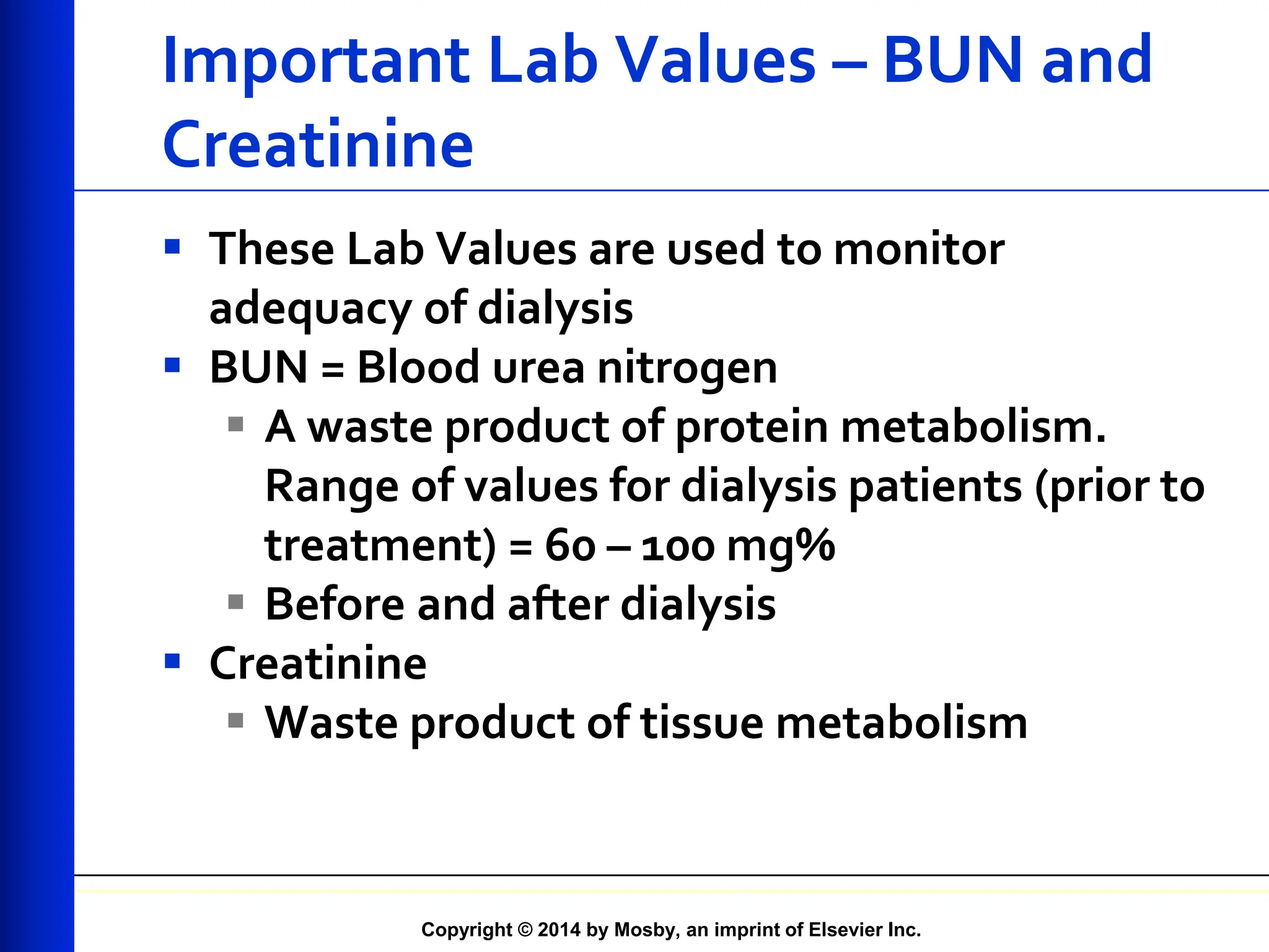
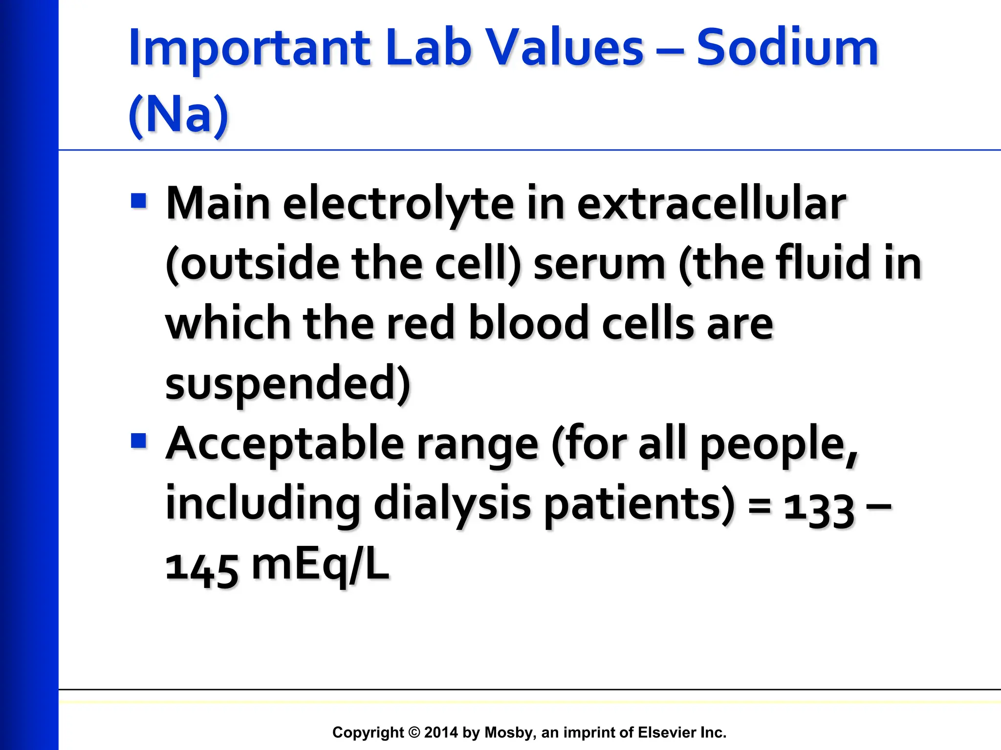
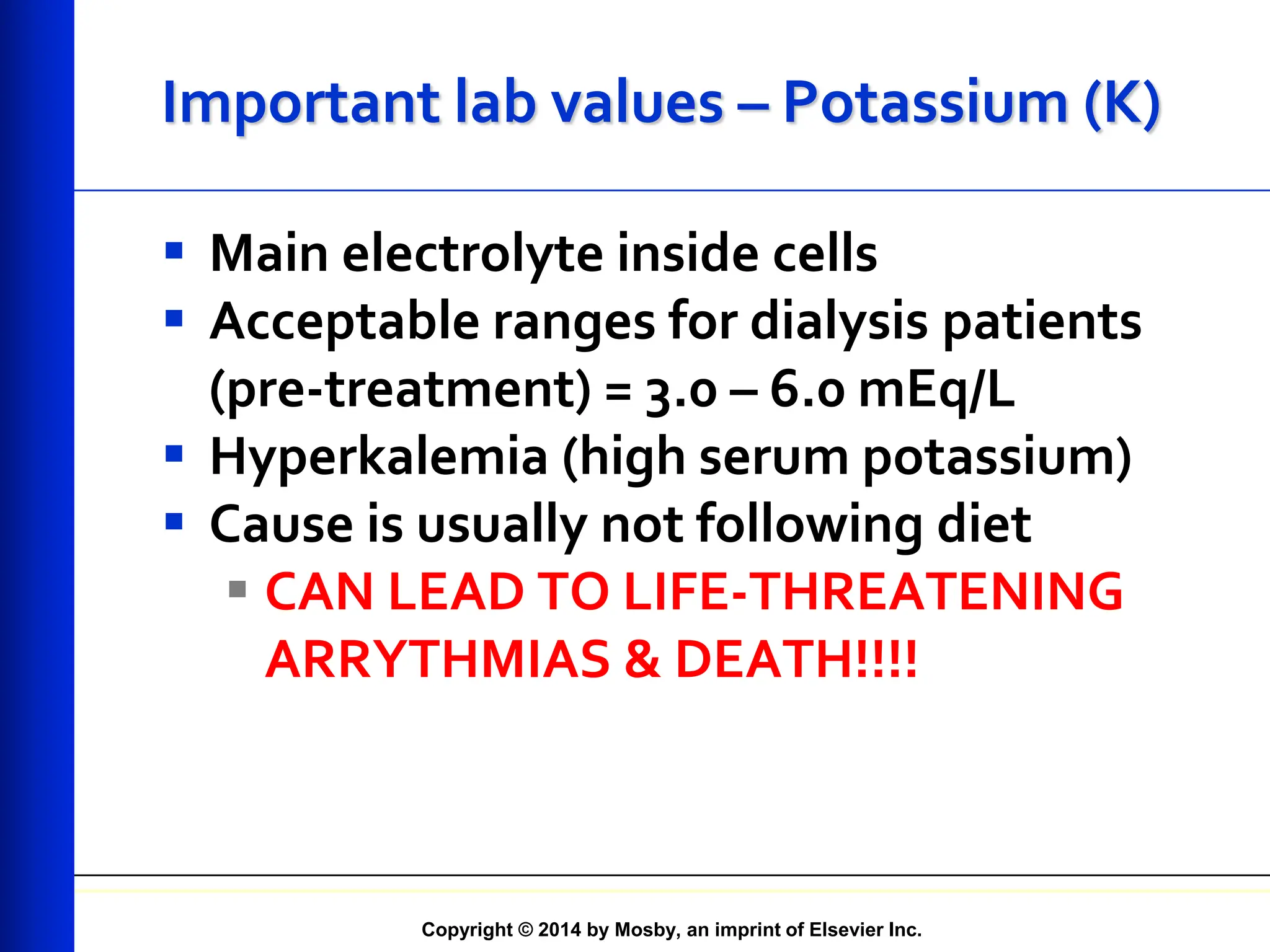
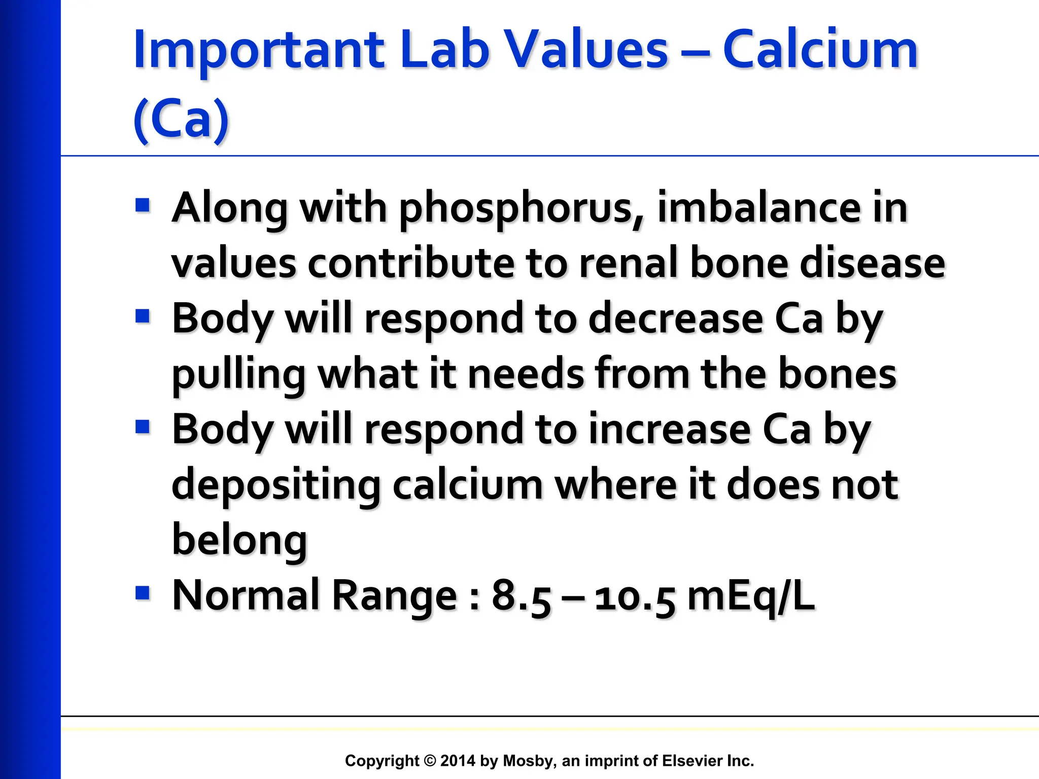
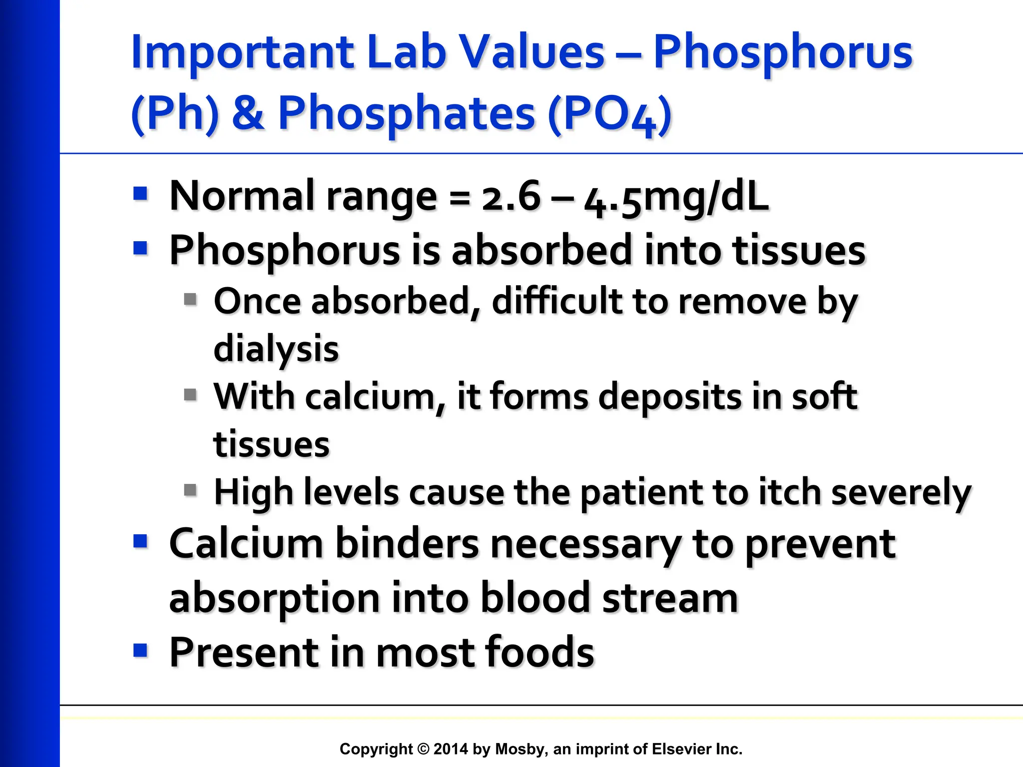
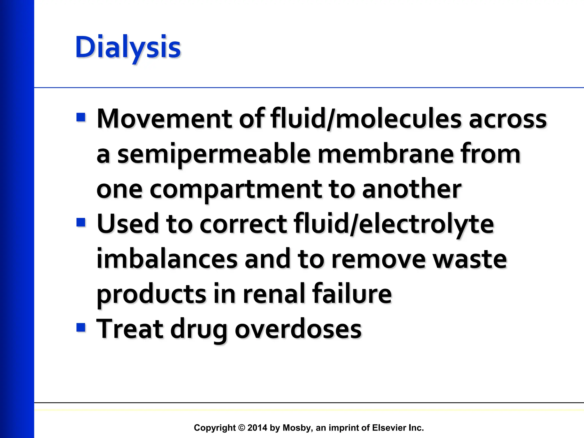
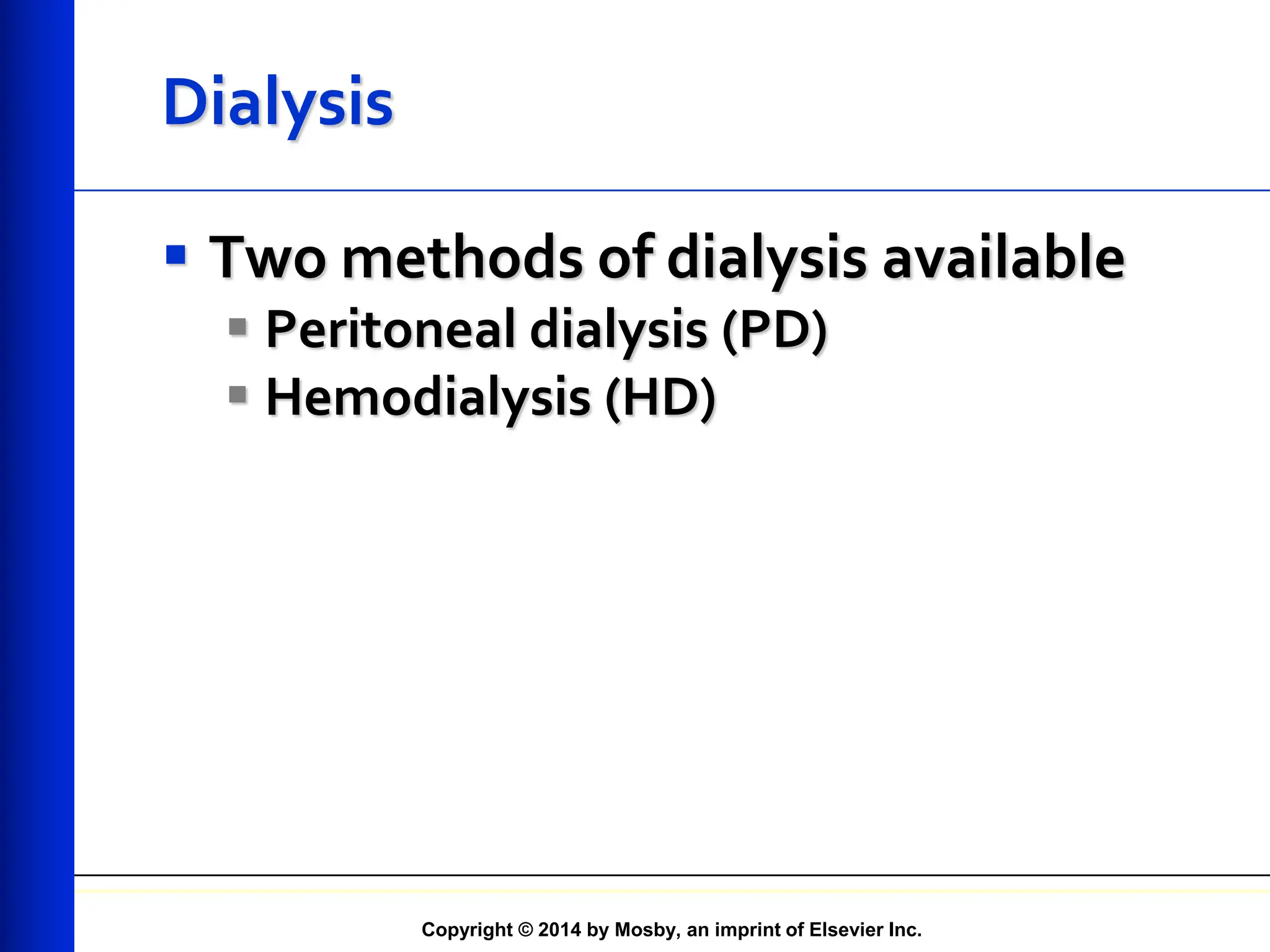
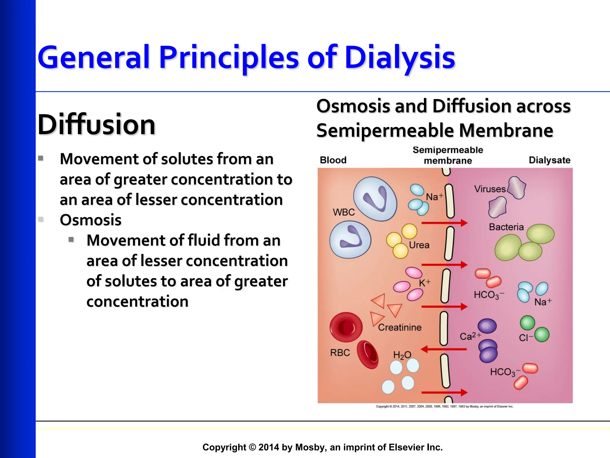
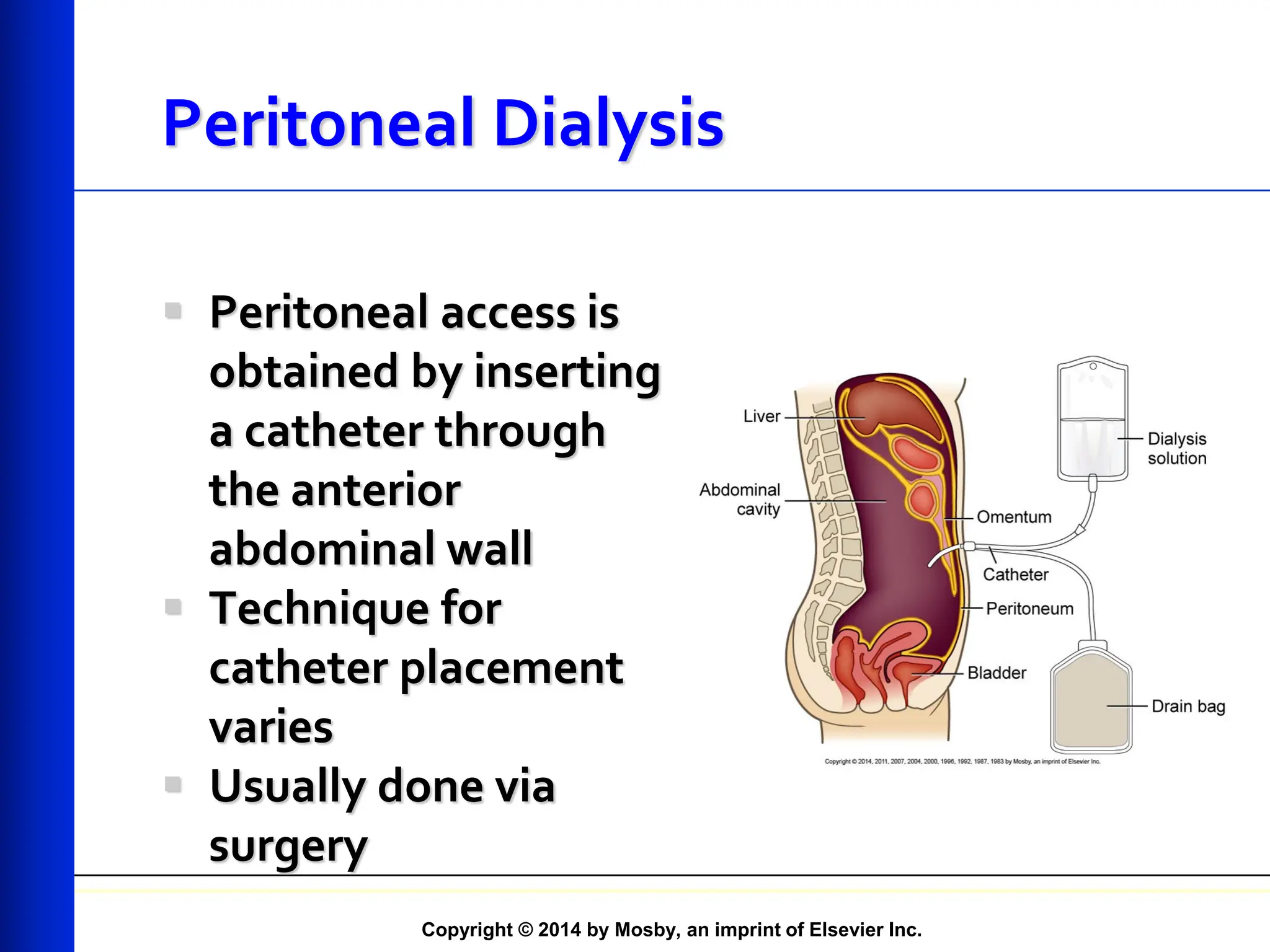
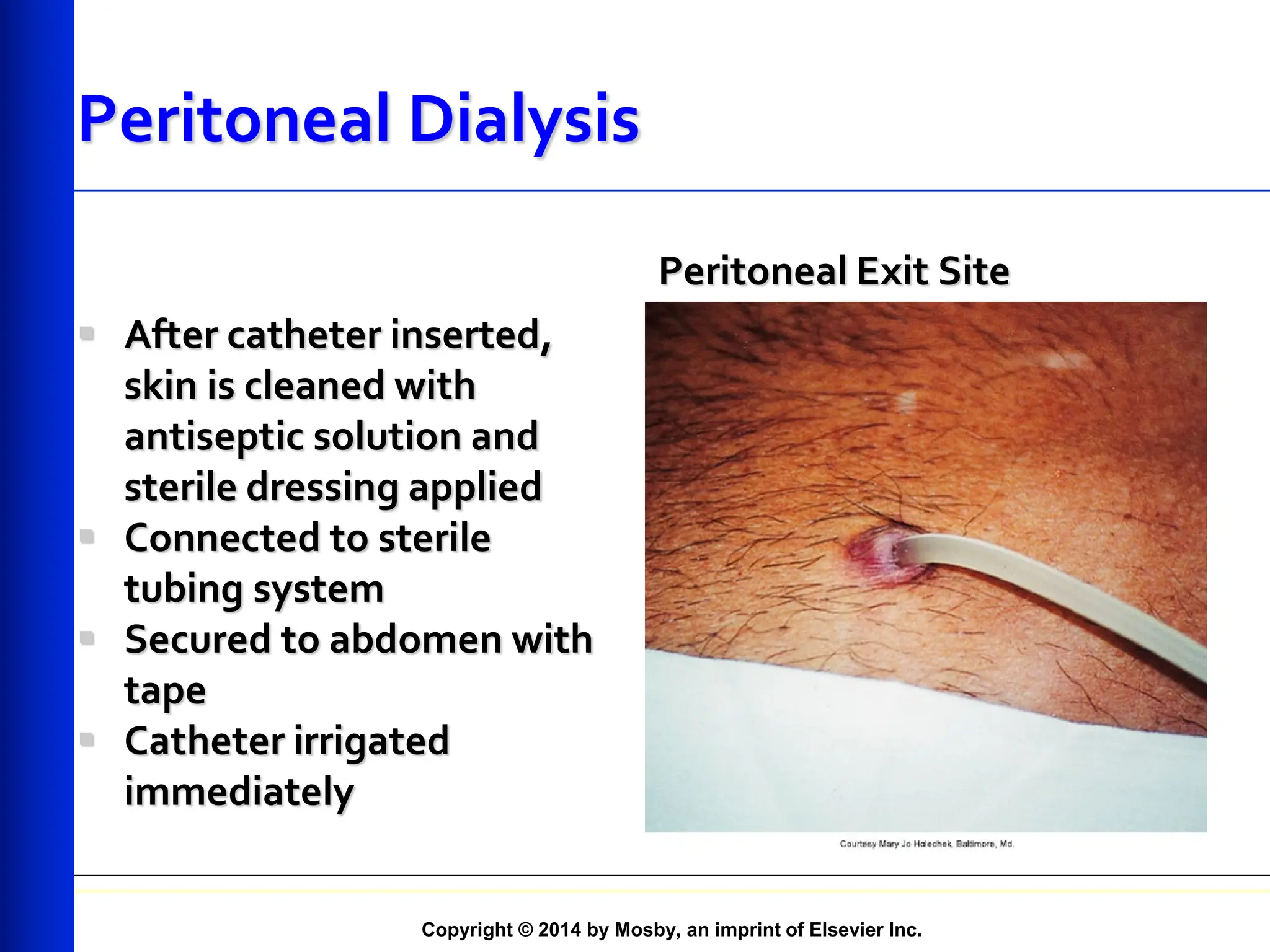
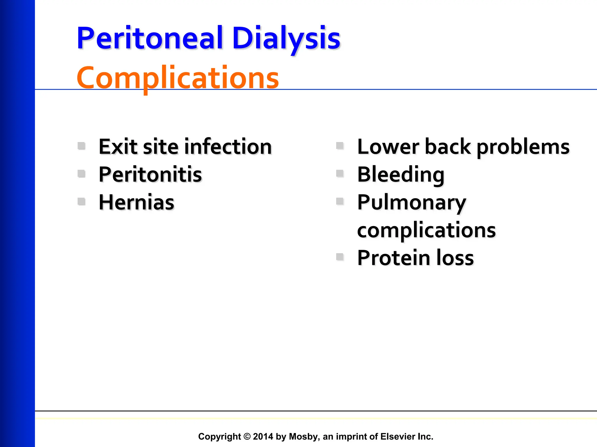
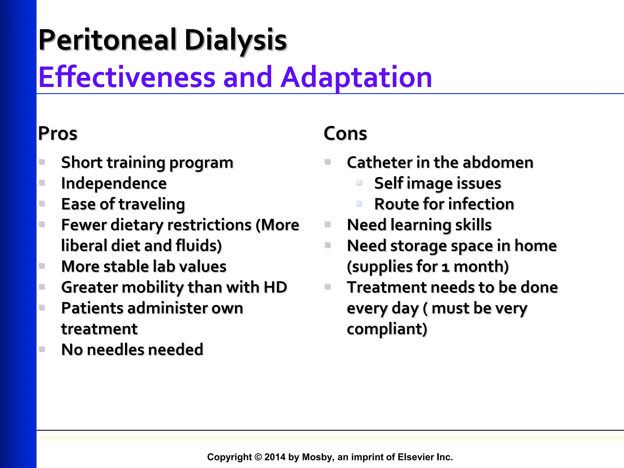
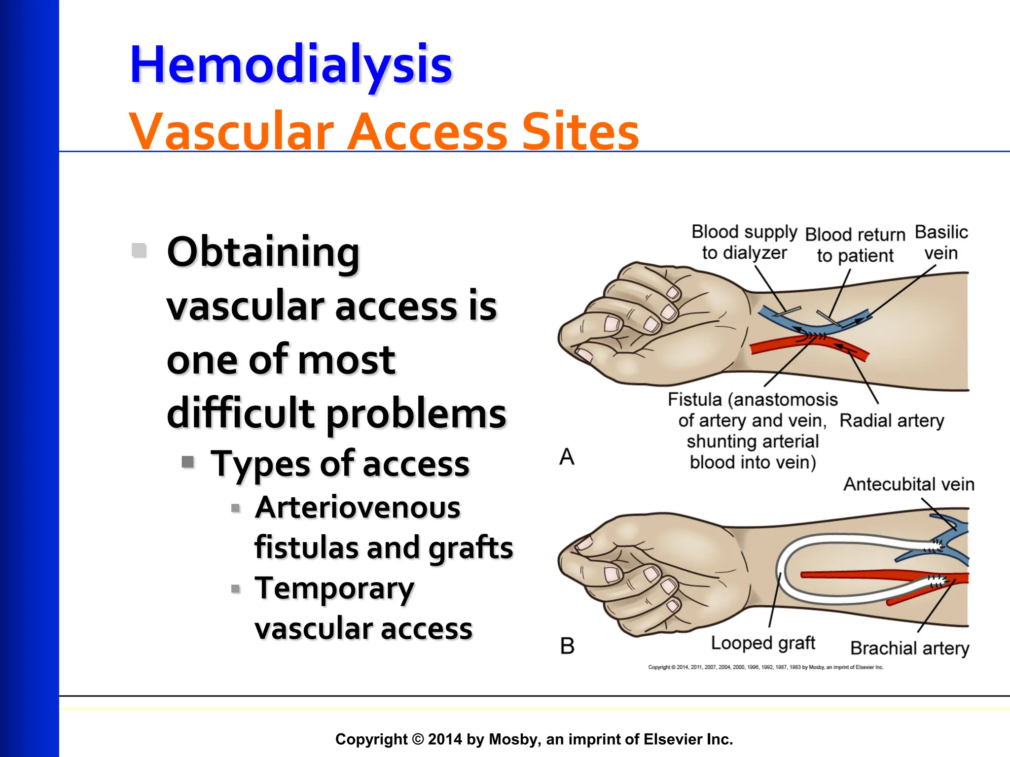
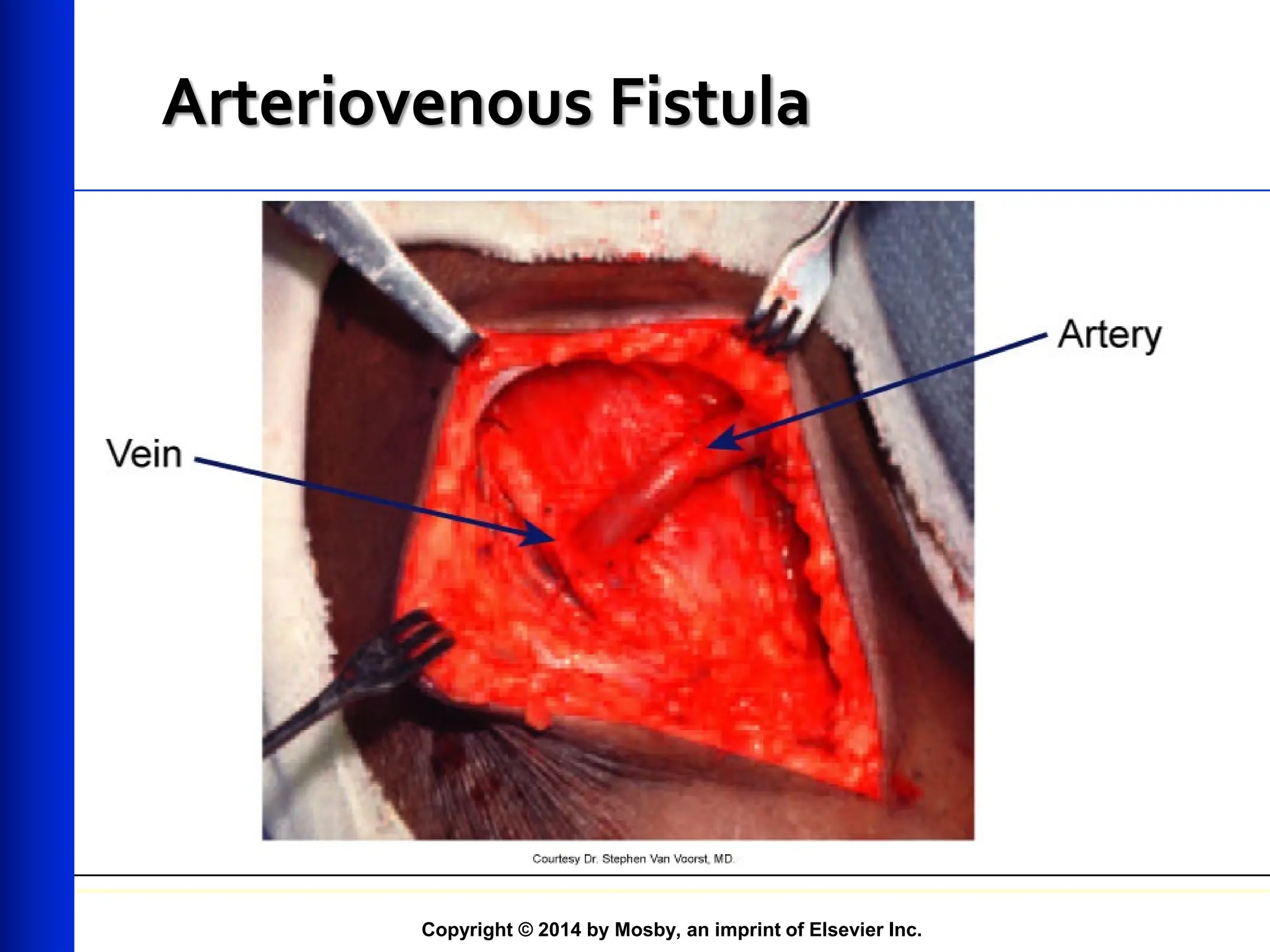
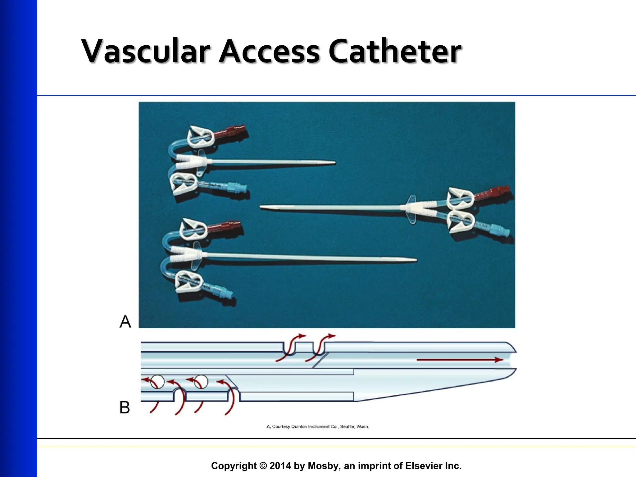
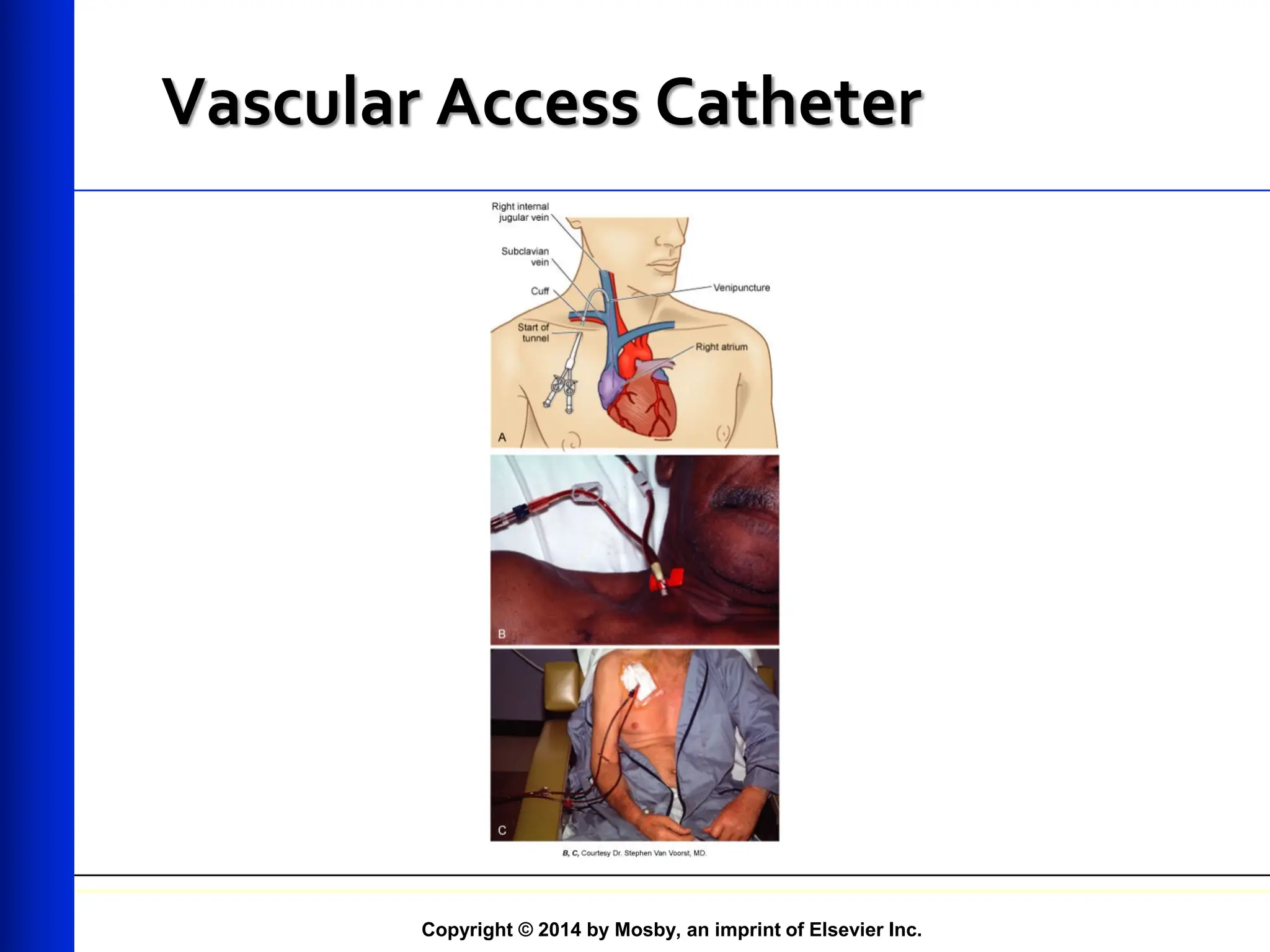
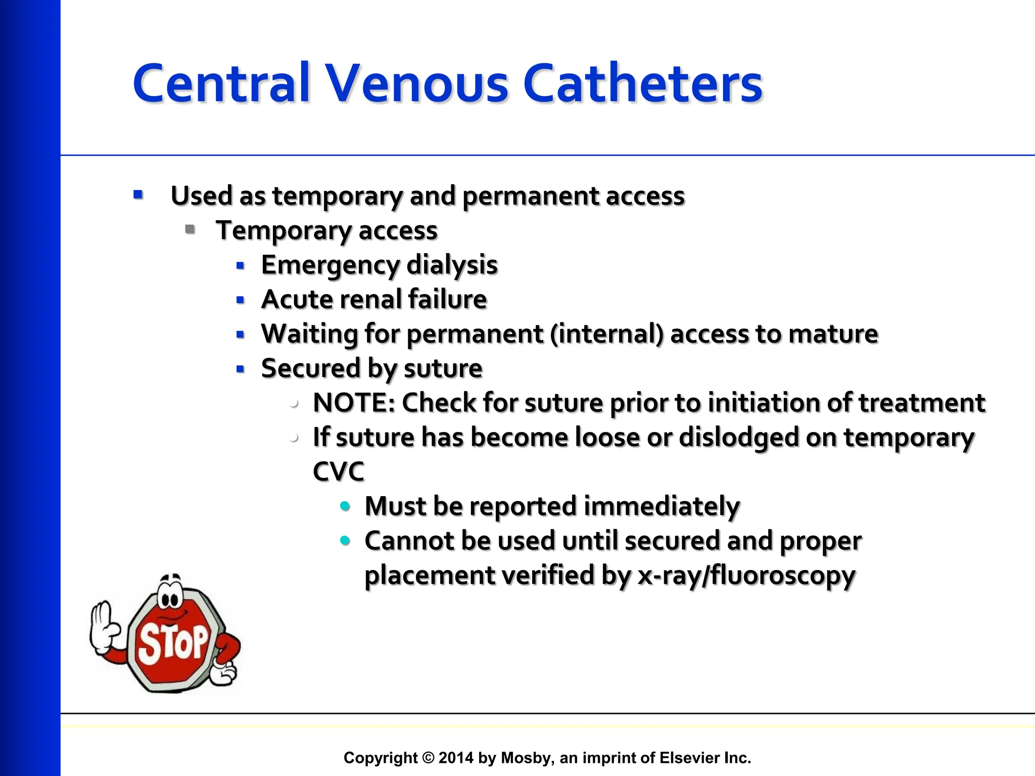
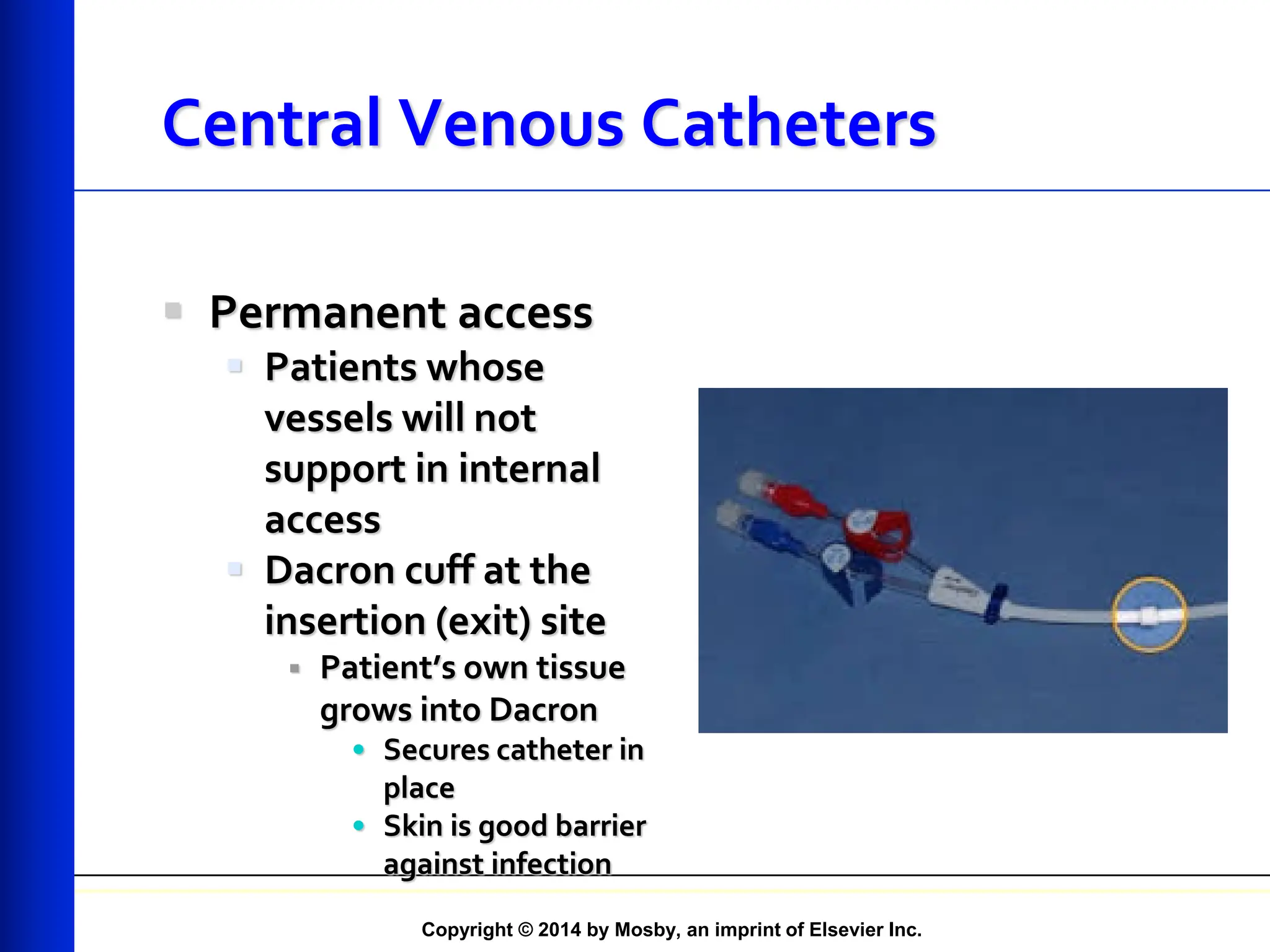
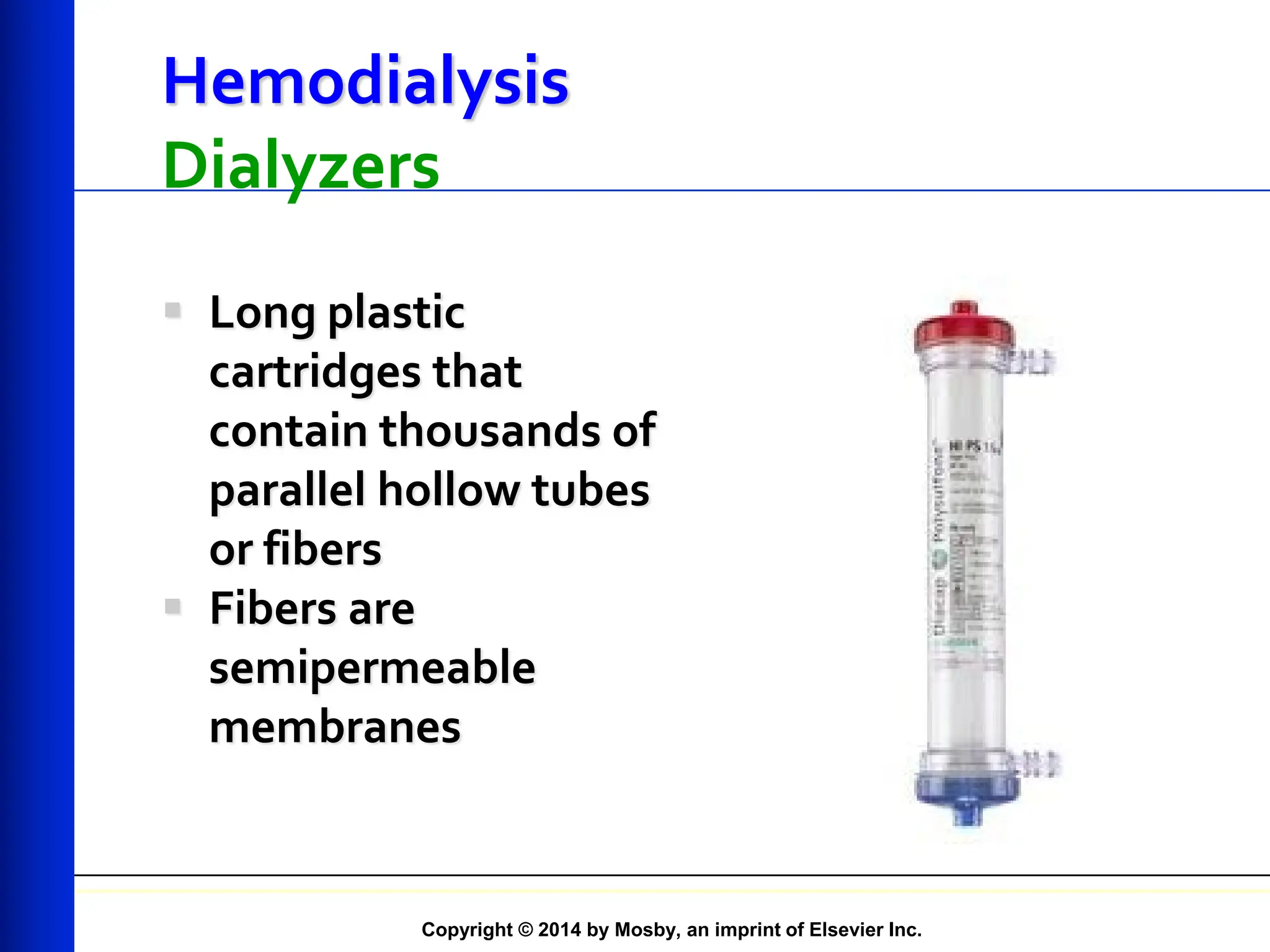
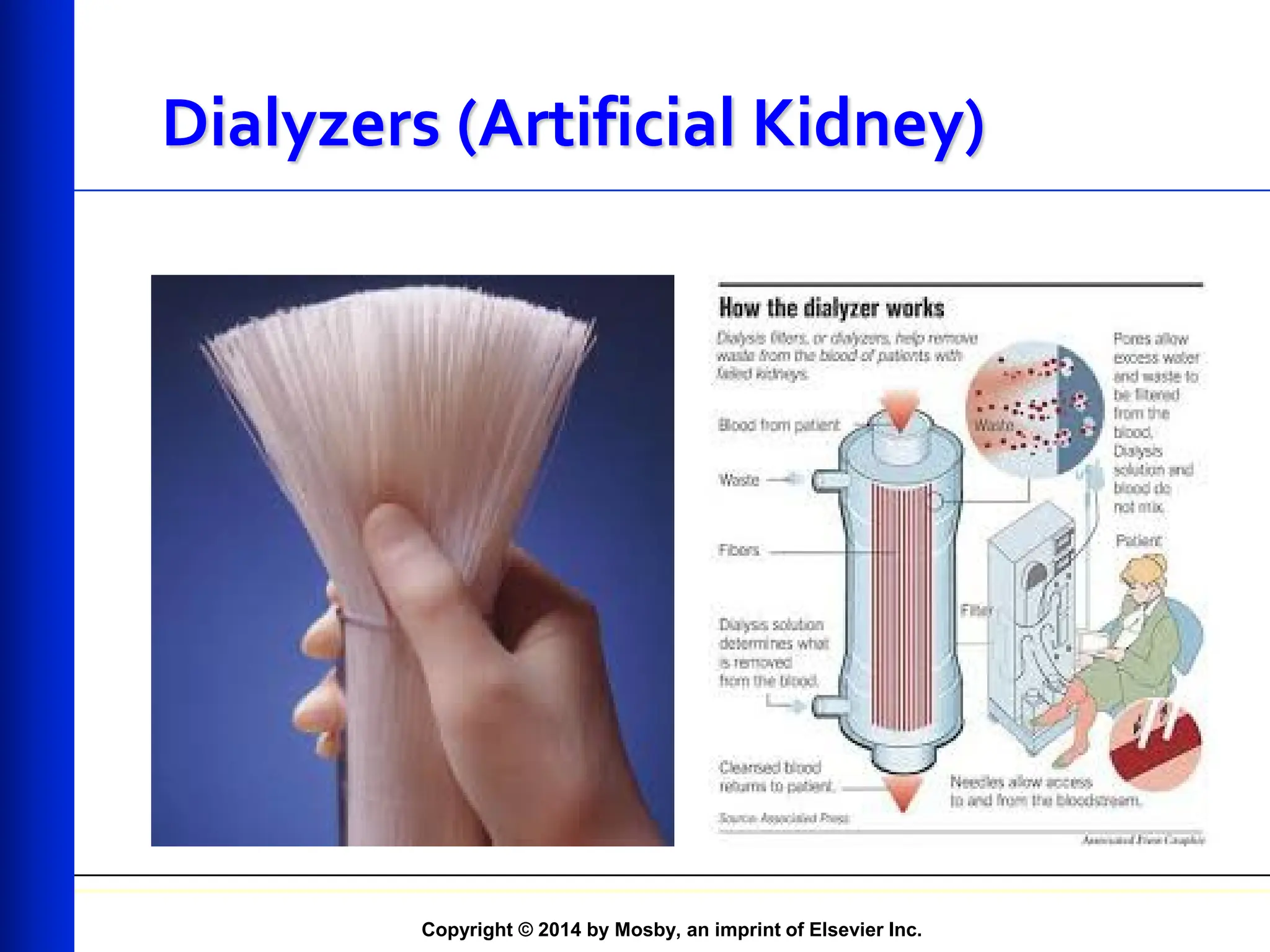
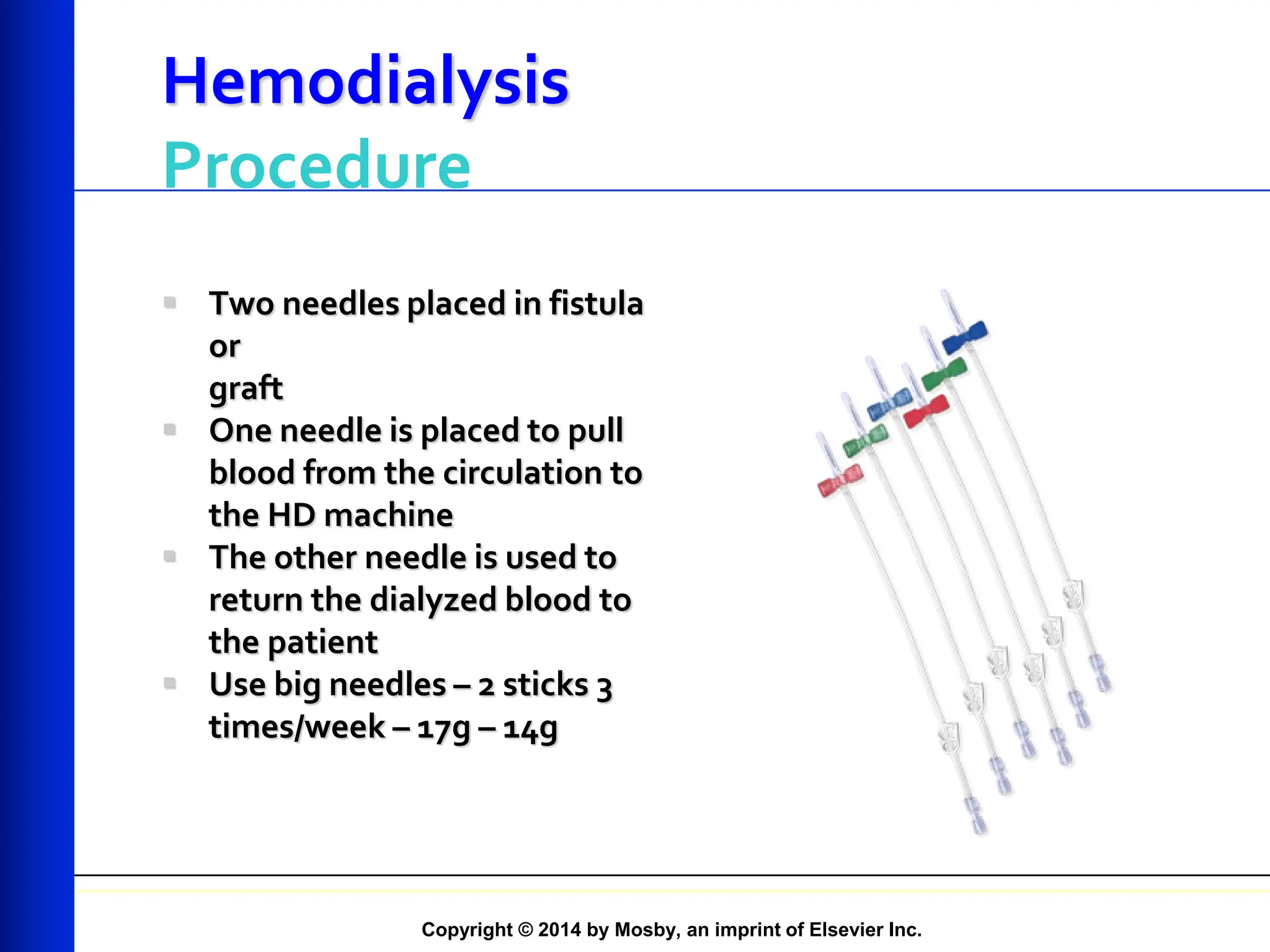
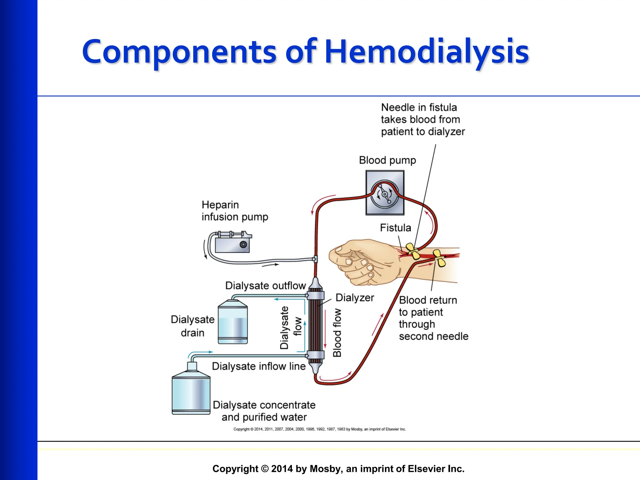
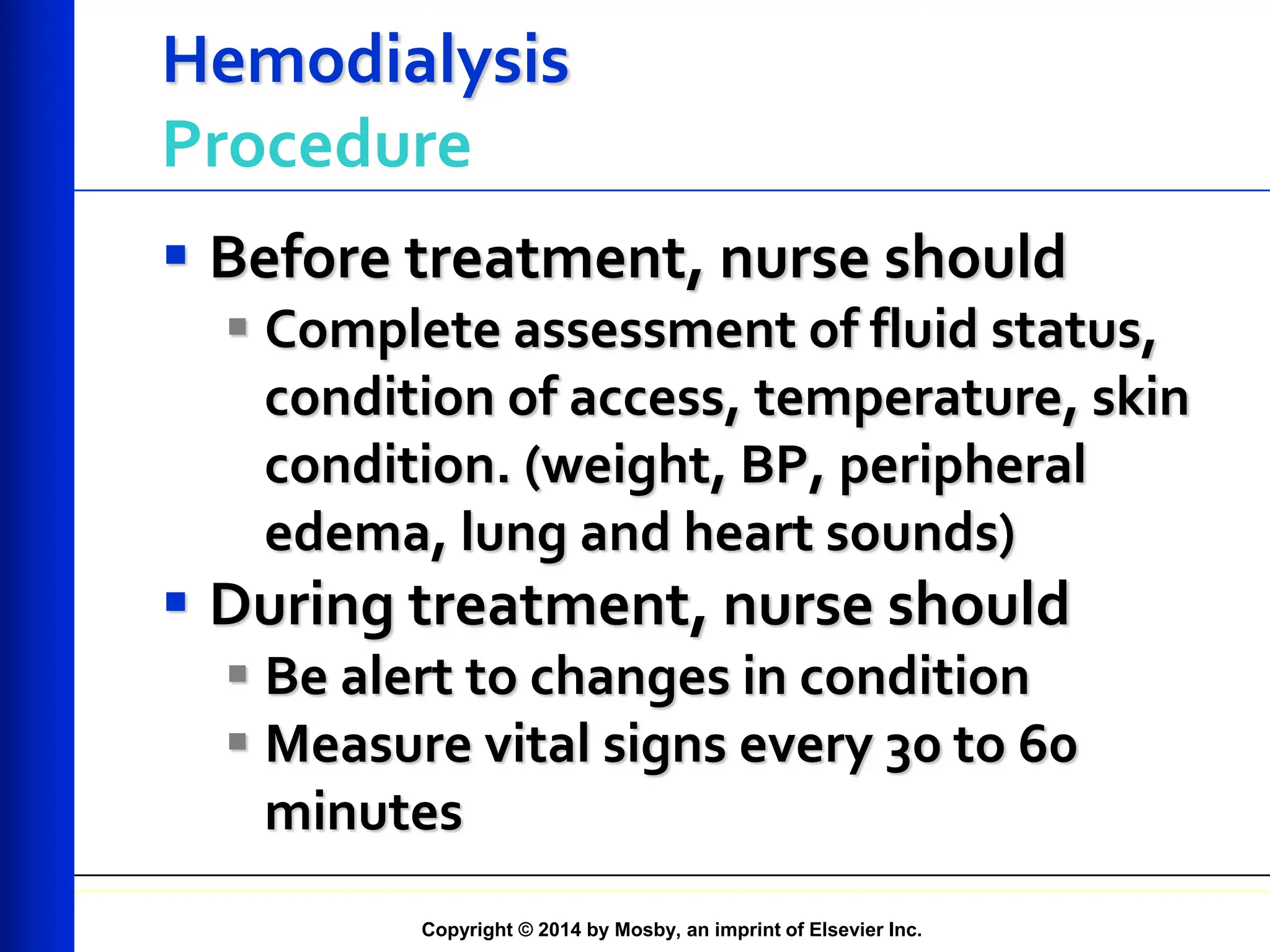
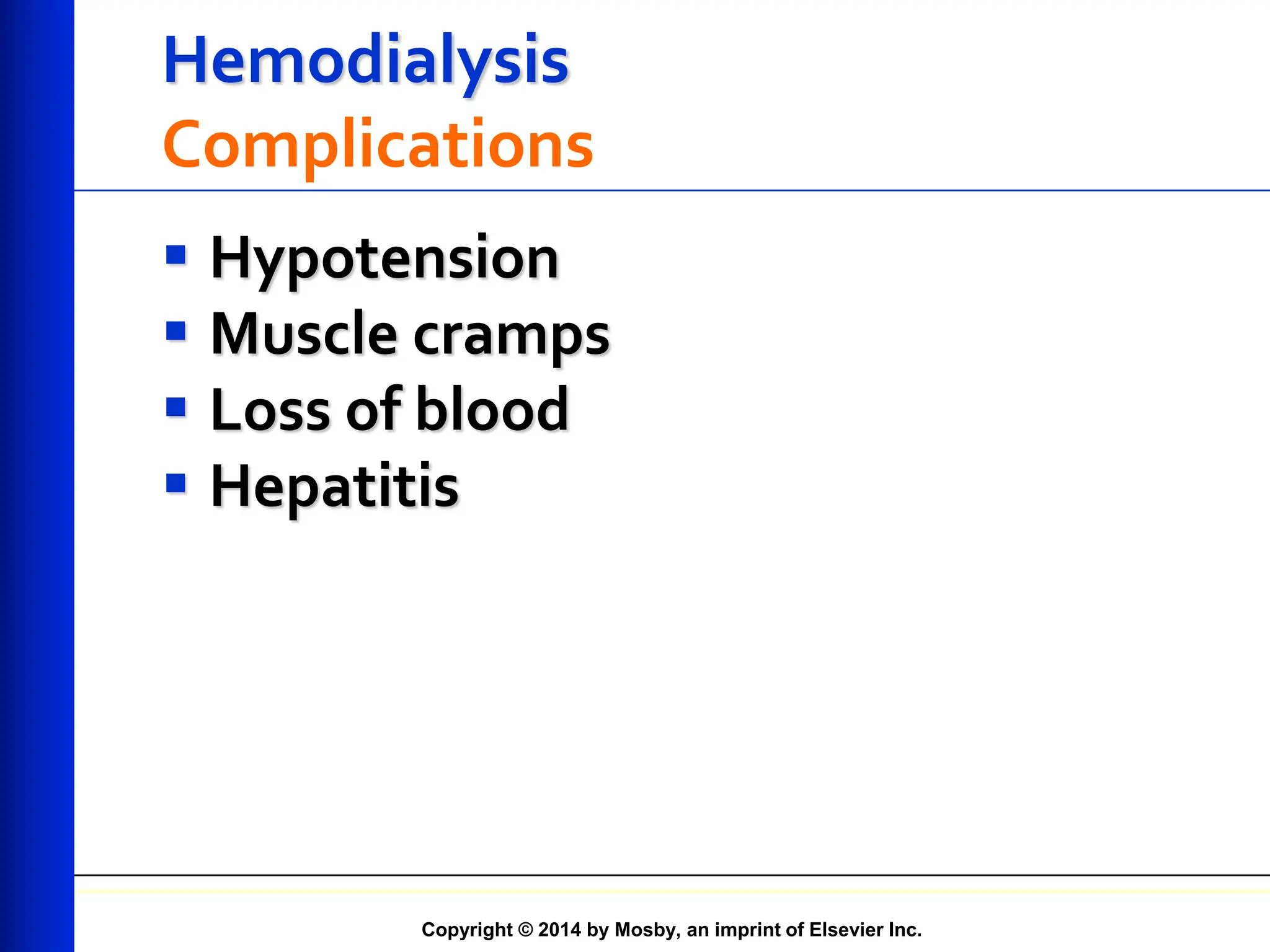
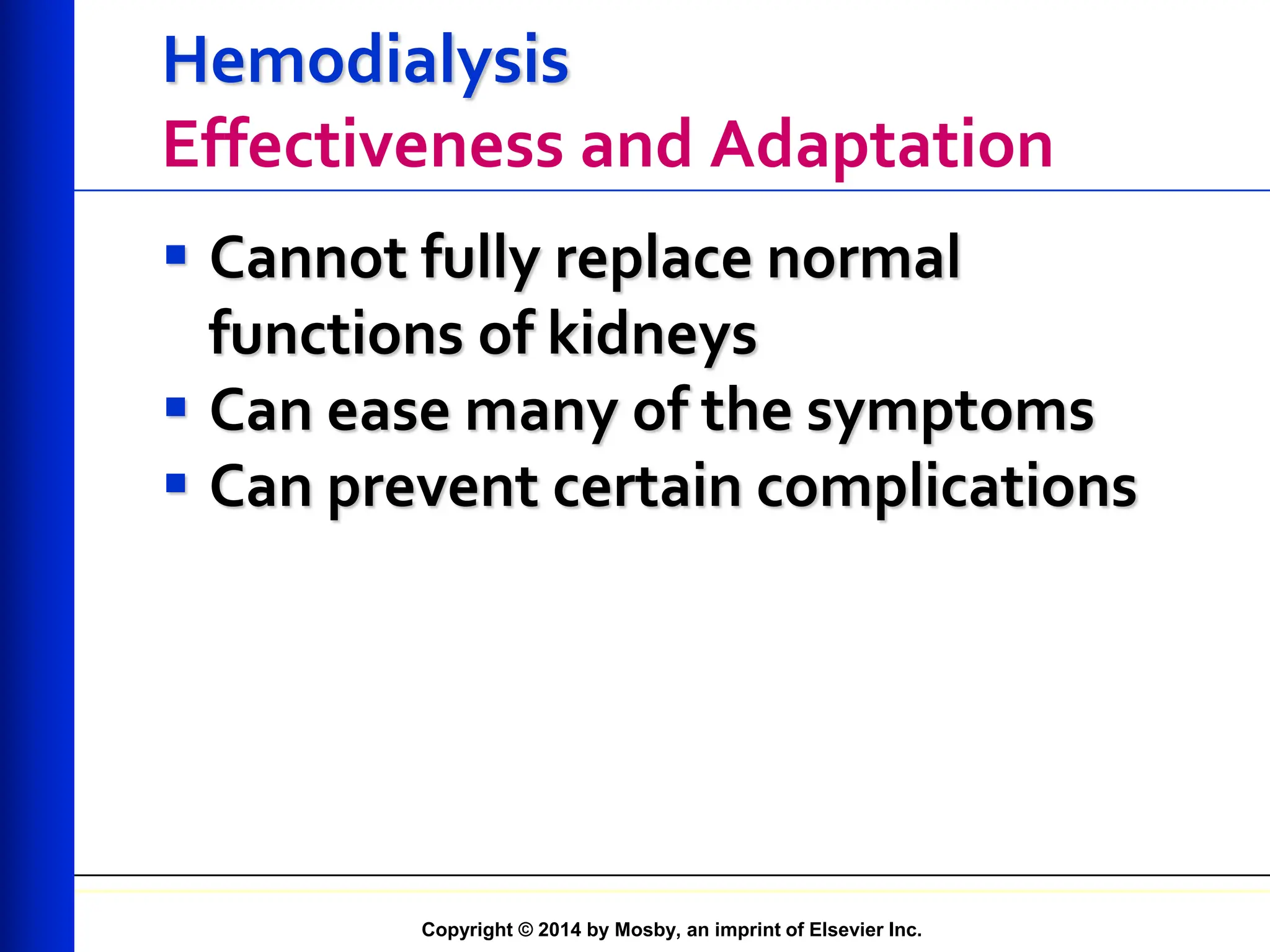
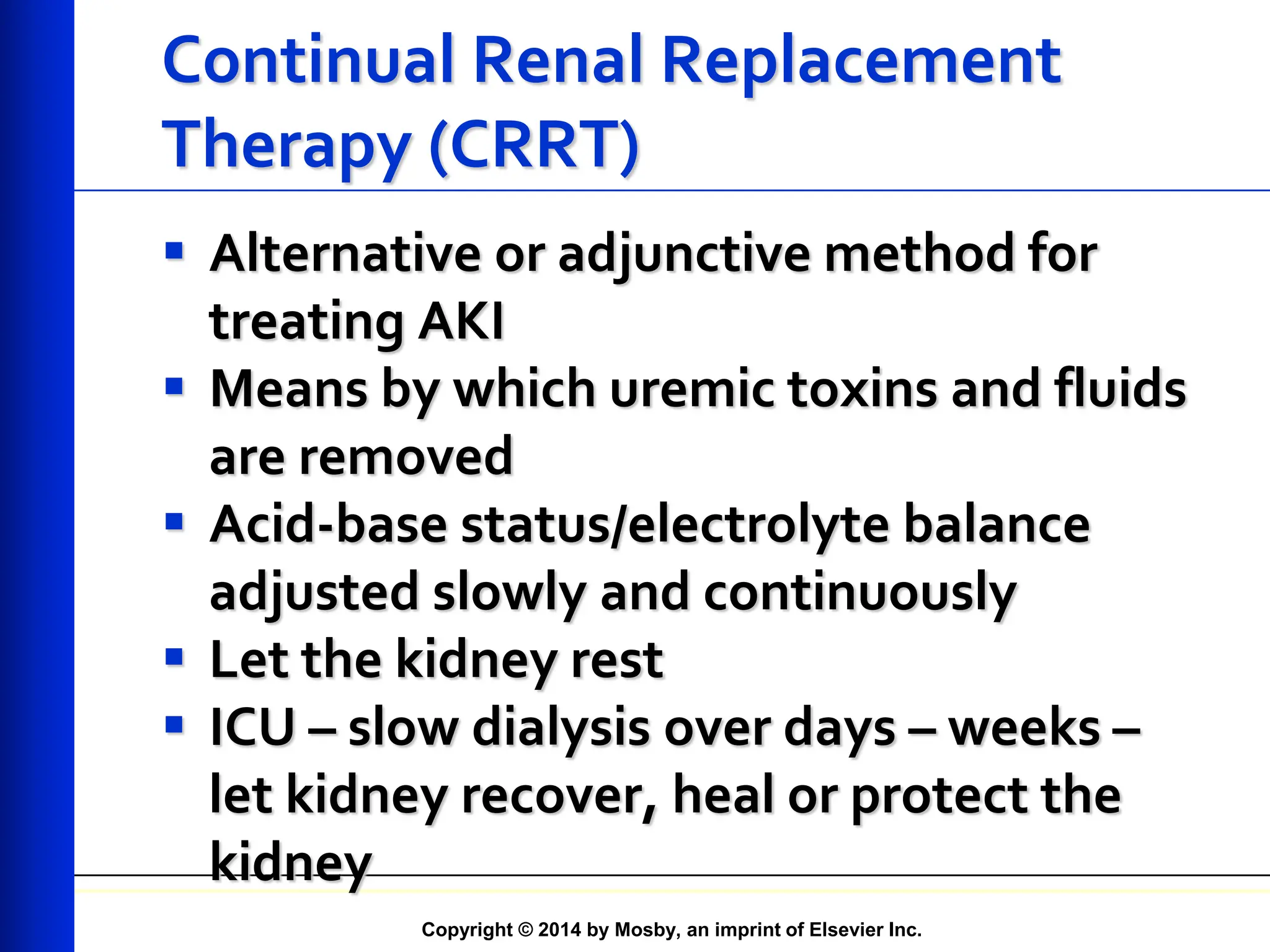
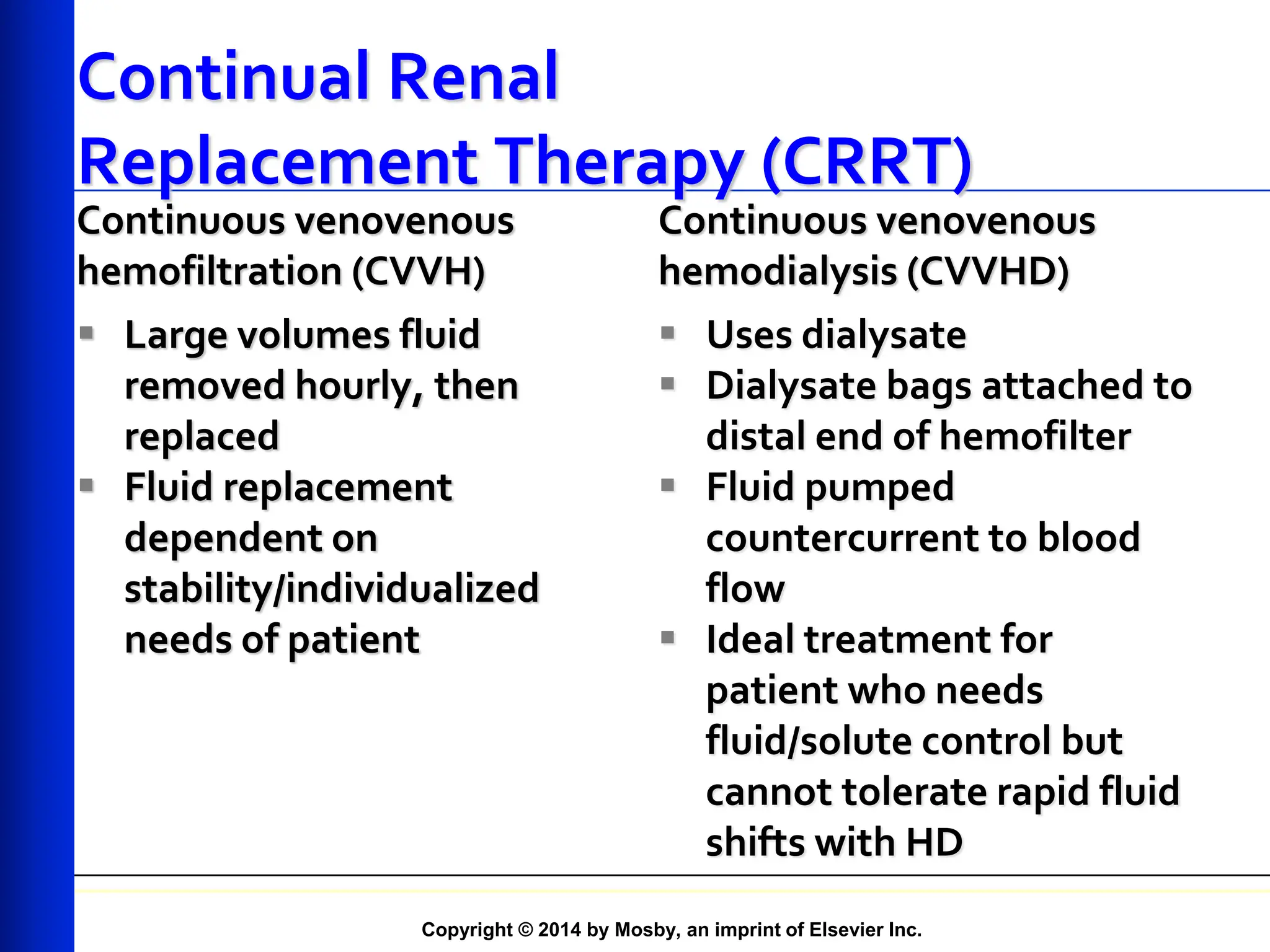
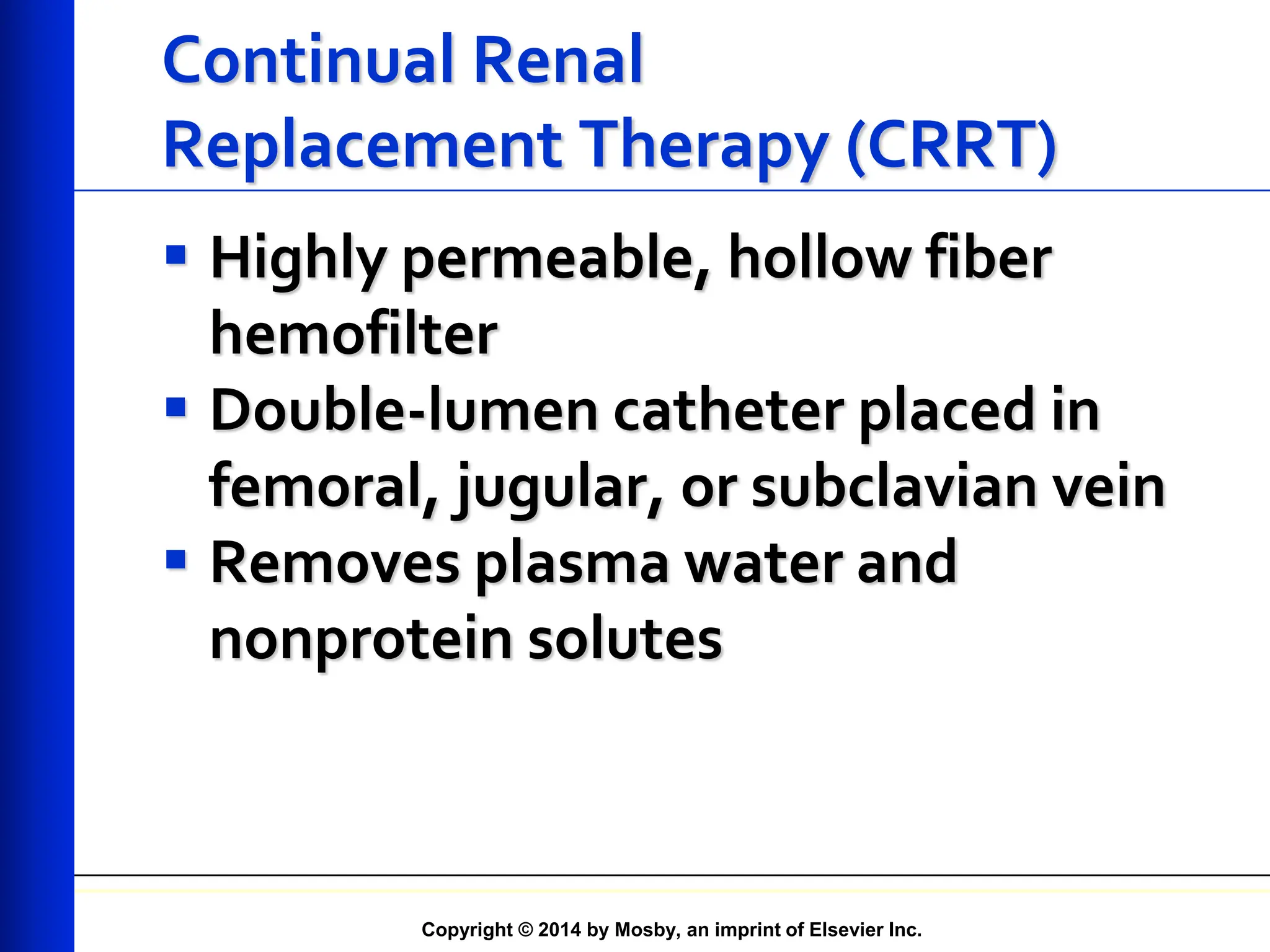
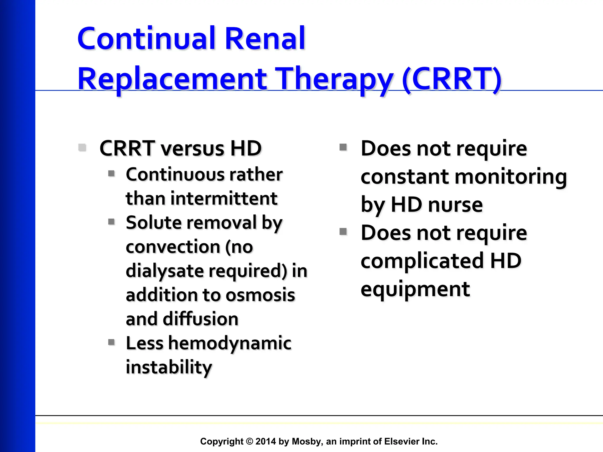
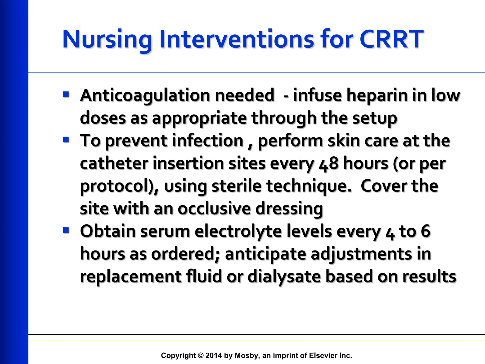
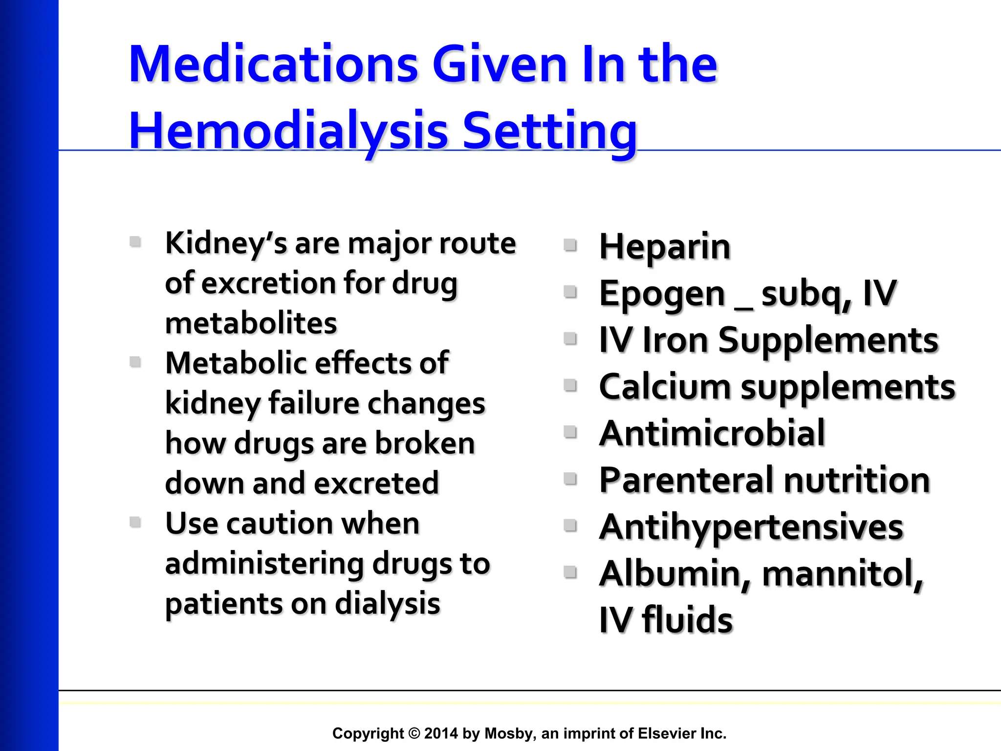
Copyright © 2014 by Mosby, an imprint of Elsevier Inc.
Dialysis
Chapter 47
Copyright © 2014 by Mosby, an imprint of Elsevier Inc.
Important Lab Values – BUN and
Creatinine
 These Lab Values are used to monitor
adequacy of dialysis
 BUN = Blood urea nitrogen
 A waste product of protein metabolism.
Range of values for dialysis patients (prior to
treatment) = 60 – 100 mg%
 Before and after dialysis
 Creatinine
 Waste product of tissue metabolism
Copyright © 2014 by Mosby, an imprint of Elsevier Inc.
Important Lab Values – Sodium
(Na)
 Main electrolyte in extracellular
(outside the cell) serum (the fluid in
which the red blood cells are
suspended)
 Acceptable range (for all people,
including dialysis patients) = 133 –
145 mEq/L
Copyright © 2014 by Mosby, an imprint of Elsevier Inc.
Important lab values – Potassium (K)
 Main electrolyte inside cells
 Acceptable ranges for dialysis patients
(pre-treatment) = 3.0 – 6.0 mEq/L
 Hyperkalemia (high serum potassium)
 Cause is usually not following diet
 CAN LEAD TO LIFE-THREATENING
ARRYTHMIAS & DEATH!!!!
Copyright © 2014 by Mosby, an imprint of Elsevier Inc.
Important Lab Values – Calcium
(Ca)
 Along with phosphorus, imbalance in
values contribute to renal bone disease
 Body will respond to decrease Ca by
pulling what it needs from the bones
 Body will respond to increase Ca by
depositing calcium where it does not
belong
 Normal Range : 8.5 – 10.5 mEq/L
Copyright © 2014 by Mosby, an imprint of Elsevier Inc.
Important Lab Values – Phosphorus
(Ph) & Phosphates (PO4)
 Normal range = 2.6 – 4.5mg/dL
 Phosphorus is absorbed into tissues
 Once absorbed, difficult to remove by
dialysis
 With calcium, it forms deposits in soft
tissues
 High levels cause the patient to itch severely
 Calcium binders necessary to prevent
absorption into blood stream
 Present in most foods
Copyright © 2014 by Mosby, an imprint of Elsevier Inc.
Dialysis
 Movement of fluid/molecules across
a semipermeable membrane from
one compartment to another
 Used to correct fluid/electrolyte
imbalances and to remove waste
products in renal failure
 Treat drug overdoses
Copyright © 2014 by Mosby, an imprint of Elsevier Inc.
Dialysis
 Two methods of dialysis available
 Peritoneal dialysis (PD)
 Hemodialysis (HD)
Copyright © 2014 by Mosby, an imprint of Elsevier Inc.
Dialysis
 Begun when patient’s uremia can
no longer be adequately managed
conservatively
 Initiated when GFR (or creatinine
clearance) is less than 15 mL/min
Copyright © 2014 by Mosby, an imprint of Elsevier Inc.
Dialysis
 ESKD treated with dialysis because
 There is a lack of donated organs
 Some patients are physically or
mentally unsuitable for
transplantation
 Some patients do not want transplants
Copyright © 2014 by Mosby, an imprint of Elsevier Inc.
General Principles of Dialysis
Diffusion
 Movement of solutes from an
area of greater concentration to
an area of lesser concentration
 Osmosis
 Movement of fluid from an
area of lesser concentration
of solutes to area of greater
concentration
Osmosis and Diffusion across
Semipermeable Membrane
Copyright © 2014 by Mosby, an imprint of Elsevier Inc.
General Principles of Dialysis
 Ultrafiltration
 Water and fluid removal
 Results when there is an osmotic
gradient across the membrane
Copyright © 2014 by Mosby, an imprint of Elsevier Inc.
Peritoneal Dialysis
 Peritoneal access is
obtained by inserting
a catheter through
the anterior
abdominal wall
 Technique for
catheter placement
varies
 Usually done via
surgery
Copyright © 2014 by Mosby, an imprint of Elsevier Inc.
Peritoneal Dialysis
 After catheter inserted,
skin is cleaned with
antiseptic solution and
sterile dressing applied
 Connected to sterile
tubing system
 Secured to abdomen with
tape
 Catheter irrigated
immediately
Peritoneal Exit Site
Copyright © 2014 by Mosby, an imprint of Elsevier Inc.
Peritoneal Dialysis
 Waiting period of 7 to 14 days
preferable
 Two to 4 weeks after implantation,
exit site should be clean, dry, and
free of redness/tenderness
 Once site healed, patient may
shower and pat dry
Copyright © 2014 by Mosby, an imprint of Elsevier Inc.
Peritoneal Dialysis
Dialysis Solutions and Cycles
 Available in 1- or 2-L plastic bags
with glucose concentrations of 1.5%,
2.5%, and 4.25%
 Electrolyte composition similar to
that of plasma
 Solution warmed to body
temperature
Copyright © 2014 by Mosby, an imprint of Elsevier Inc.
Peritoneal Dialysis
Dialysis Solutions and Cycles
 Three phases of PD cycle
 Inflow (fill)
 Dwell (equilibration)
 Drain
 Called an exchange
Copyright © 2014 by Mosby, an imprint of Elsevier Inc.
Peritoneal Dialysis
Dialysis Solutions and Cycles
Inflow (Fill)
 Prescribed amount of
solution infused
through established
catheter over about 10
minutes
 After solution infused,
inflow clamp closed to
prevent air from
entering tubing
Dwell
 Period of time when the
dialysate solution sits in the
peritoneum
 Allows toxin, excess fluid to
pass into the dialysate solution
 Also known as equilibration
 Diffusion and osmosis occur
between patient’s blood and
peritoneal cavity
 Duration of time varies,
depending on method
Copyright © 2014 by Mosby, an imprint of Elsevier Inc.
Peritoneal Dialysis
Dialysis Solutions and Cycles
 Drain
 Lasts 15 to 30 minutes
 May be facilitated by
gently
massaging abdomen or
changing position
 Turn the patient from side
to side and change
positions to allow pockets
of fluid to drain out if all
the dialysate fluid does
not return
 Exchange
 Word used to
describe the entire
fill-dwell-drain cycle
Copyright © 2014 by Mosby, an imprint of Elsevier Inc.
Peritoneal Dialysis
Systems
 Automated peritoneal
dialysis
(APD)
 Cycler delivers the
dialysate
 Times and controls fill,
dwell, and drain
 Continuous ambulatory
peritoneal dialysis
(CAPD)
 Manual exchange
Copyright © 2014 by Mosby, an imprint of Elsevier Inc.
Peritoneal Dialysis
Complications
 Exit site infection
 Peritonitis
 Hernias
 Lower back problems
 Bleeding
 Pulmonary
complications
 Protein loss
Copyright © 2014 by Mosby, an imprint of Elsevier Inc.
Peritoneal Dialysis
Effectiveness and Adaptation
Pros
 Short training program
 Independence
 Ease of traveling
 Fewer dietary restrictions (More
liberal diet and fluids)
 More stable lab values
 Greater mobility than with HD
 Patients administer own
treatment
 No needles needed
Cons
 Catheter in the abdomen
 Self image issues
 Route for infection
 Need learning skills
 Need storage space in home
(supplies for 1 month)
 Treatment needs to be done
every day ( must be very
compliant)
Copyright © 2014 by Mosby, an imprint of Elsevier Inc.
Hemodialysis
Vascular Access Sites
 Obtaining
vascular access is
one of most
difficult problems
 Types of access
 Arteriovenous
fistulas and grafts
 Temporary
vascular access
Copyright © 2014 by Mosby, an imprint of Elsevier Inc.
Arteriovenous Fistula
Copyright © 2014 by Mosby, an imprint of Elsevier Inc.
Assessment of the Internal Access
 Other Assessments
 Bleeding
 Swelling
 Bruising
 Redness
 Drainage
 Pain
 Change in thrill
Copyright © 2014 by Mosby, an imprint of Elsevier Inc.
Patient Education
 Lifeline – protect it
 Teach patient how to protect it and check daily to see if it is
working properly
 Feel over site and check “thrill”
 Contact the HCP or hemodialysis nurse immediately if there is a
change in the quality of the thrill
 Avoid any pressure or constriction on the access arm.
Don’t let anyone take a blood pressure or draw blood
 Don’t wear tight clothing (elastic sleeves, wear a
watch, carry a purse, or sleep on the the access arm
 Do not carry heavy objects with access arm
 Do not allow venipuncture to the done by anyone
other the an trained dialysis personnel
Copyright © 2014 by Mosby, an imprint of Elsevier Inc.
Patient Education (cont.)
 Adhere to diet – between treatments waste products
won’t build up in blood to quickly
 PD patient should have increase fiber in the diet to
offset constipation from decreased peristalsis. ( The
intestines are floating in the hypertonic dialysate
solution
 PD patients must strictly adhere to handwashing and
asepsis, and follow guidelines to access abdominal
catheter
 Epogen (erythropoietin) injections (subcutaneous or
IV) as prescribed increase energy and endurance
because increased RBCs means better oxygenation and
less fatigue
Copyright © 2014 by Mosby, an imprint of Elsevier Inc.
Vascular Access Catheter
Copyright © 2014 by Mosby, an imprint of Elsevier Inc.
Vascular Access Catheter
Copyright © 2014 by Mosby, an imprint of Elsevier Inc.
Central Venous Catheters
 Used as temporary and permanent access
 Temporary access
 Emergency dialysis
 Acute renal failure
 Waiting for permanent (internal) access to mature
 Secured by suture
• NOTE: Check for suture prior to initiation of treatment
• If suture has become loose or dislodged on temporary
CVC
• Must be reported immediately
• Cannot be used until secured and proper
placement verified by x-ray/fluoroscopy
Copyright © 2014 by Mosby, an imprint of Elsevier Inc.
Central Venous Catheters
 Permanent access
 Patients whose
vessels will not
support in internal
access
 Dacron cuff at the
insertion (exit) site
 Patient’s own tissue
grows into Dacron
• Secures catheter in
place
• Skin is good barrier
against infection
Copyright © 2014 by Mosby, an imprint of Elsevier Inc.
Hemodialysis
Dialyzers
 Long plastic
cartridges that
contain thousands of
parallel hollow tubes
or fibers
 Fibers are
semipermeable
membranes
Copyright © 2014 by Mosby, an imprint of Elsevier Inc.
Dialyzers (Artificial Kidney)
Copyright © 2014 by Mosby, an imprint of Elsevier Inc.
Hemodialysis
Procedure
 Two needles placed in fistula
or
graft
 One needle is placed to pull
blood from the circulation to
the HD machine
 The other needle is used to
return the dialyzed blood to
the patient
 Use big needles – 2 sticks 3
times/week – 17g – 14g
Copyright © 2014 by Mosby, an imprint of Elsevier Inc.
Components of Hemodialysis
Copyright © 2014 by Mosby, an imprint of Elsevier Inc.
Hemodialysis
Procedure
 Dialyzer/blood lines primed with
saline solution to eliminate air
 Terminated by flushing dialyzer with
saline to remove all blood
 Needles removed and firm pressure
applied
Copyright © 2014 by Mosby, an imprint of Elsevier Inc.
Hemodialysis
Procedure
 Before treatment, nurse should
 Complete assessment of fluid status,
condition of access, temperature, skin
condition. (weight, BP, peripheral
edema, lung and heart sounds)
 During treatment, nurse should
 Be alert to changes in condition
 Measure vital signs every 30 to 60
minutes
Copyright © 2014 by Mosby, an imprint of Elsevier Inc.
Hemodialysis
Complications
 Hypotension
 Muscle cramps
 Loss of blood
 Hepatitis
Copyright © 2014 by Mosby, an imprint of Elsevier Inc.
Hemodialysis
Effectiveness and Adaptation
 Cannot fully replace normal
functions of kidneys
 Can ease many of the symptoms
 Can prevent certain complications
Copyright © 2014 by Mosby, an imprint of Elsevier Inc.
Continual Renal Replacement
Therapy (CRRT)
 Alternative or adjunctive method for
treating AKI
 Means by which uremic toxins and fluids
are removed
 Acid-base status/electrolyte balance
adjusted slowly and continuously
 Let the kidney rest
 ICU – slow dialysis over days – weeks –
let kidney recover, heal or protect the
kidney
Copyright © 2014 by Mosby, an imprint of Elsevier Inc.
Continual Renal Replacement
Therapy (CRRT)
 Can be used in conjunction with HD
 Contraindication
 Presence of manifestations of uremia
(hyperkalemia, pericarditis) that necessitate
rapid resolution
 Continued for 30 to 40 days
 Hemofilter change every 24 to 48 hours
 Ultrafiltrate should be clear yellow
 Specimens may be obtained for
evaluation
Copyright © 2014 by Mosby, an imprint of Elsevier Inc.
Continual Renal
Replacement Therapy (CRRT)
 Two types of CRRT
 Venous access
 Arterial access
Copyright © 2014 by Mosby, an imprint of Elsevier Inc.
Continual Renal
Replacement Therapy (CRRT)
 Most common approaches:
venovenous
 Continuous venovenous
hemofiltration (CVVH)
 Continuous venovenous hemodialysis
(CVVHD)
 Refer to pg. 1123 Table 47-13
Copyright © 2014 by Mosby, an imprint of Elsevier Inc.
Continuous Venovenous
Therapies
Copyright © 2014 by Mosby, an imprint of Elsevier Inc.
Continual Renal
Replacement Therapy (CRRT)
 Large volumes fluid
removed hourly, then
replaced
 Fluid replacement
dependent on
stability/individualized
needs of patient
Continuous venovenous
hemodialysis (CVVHD)
 Uses dialysate
 Dialysate bags attached to
distal end of hemofilter
 Fluid pumped
countercurrent to blood
flow
 Ideal treatment for
patient who needs
fluid/solute control but
cannot tolerate rapid fluid
shifts with HD
Continuous venovenous
hemofiltration (CVVH)
Copyright © 2014 by Mosby, an imprint of Elsevier Inc.
Continual Renal
Replacement Therapy (CRRT)
 Highly permeable, hollow fiber
hemofilter
 Double-lumen catheter placed in
femoral, jugular, or subclavian vein
 Removes plasma water and
nonprotein solutes
Copyright © 2014 by Mosby, an imprint of Elsevier Inc.
Continual Renal
Replacement Therapy (CRRT)
 CRRT versus HD
 Continuous rather
than intermittent
 Solute removal by
convection (no
dialysate required) in
addition to osmosis
and diffusion
 Less hemodynamic
instability
 Does not require
constant monitoring
by HD nurse
 Does not require
complicated HD
equipment
Copyright © 2014 by Mosby, an imprint of Elsevier Inc.
Nursing Interventions for CRRT
 Anticoagulation needed - infuse heparin in low
doses as appropriate through the setup
 To prevent infection , perform skin care at the
catheter insertion sites every 48 hours (or per
protocol), using sterile technique. Cover the
site with an occlusive dressing
 Obtain serum electrolyte levels every 4 to 6
hours as ordered; anticipate adjustments in
replacement fluid or dialysate based on results
Copyright © 2014 by Mosby, an imprint of Elsevier Inc.
Medications Given In the
Hemodialysis Setting
 Kidney’s are major route
of excretion for drug
metabolites
 Metabolic effects of
kidney failure changes
how drugs are broken
down and excreted
 Use caution when
administering drugs to
patients on dialysis
 Heparin
 Epogen _ subq, IV
 IV Iron Supplements
 Calcium supplements
 Antimicrobial
 Parenteral nutrition
 Antihypertensives
 Albumin, mannitol,
IV fluids
Copyright © 2014 by Mosby, an imprint of Elsevier Inc.
Antihypertensives
 Points to remember:
 Some are not removed by dialysis
 May cause decrease in B/P during
dialysis
 Most MD’s recommend delaying dose
until after dialysis
 Instruct patient to take medication so
that peak action does not occur during
dialysis

Chapter 47 Dialysis with lab values and catheter info.pdf

  • 1.
    Copyright © 2014by Mosby, an imprint of Elsevier Inc. Dialysis Chapter 47
  • 2.
    Copyright © 2014by Mosby, an imprint of Elsevier Inc. Important Lab Values – BUN and Creatinine  These Lab Values are used to monitor adequacy of dialysis  BUN = Blood urea nitrogen  A waste product of protein metabolism. Range of values for dialysis patients (prior to treatment) = 60 – 100 mg%  Before and after dialysis  Creatinine  Waste product of tissue metabolism
  • 3.
    Copyright © 2014by Mosby, an imprint of Elsevier Inc. Important Lab Values – Sodium (Na)  Main electrolyte in extracellular (outside the cell) serum (the fluid in which the red blood cells are suspended)  Acceptable range (for all people, including dialysis patients) = 133 – 145 mEq/L
  • 4.
    Copyright © 2014by Mosby, an imprint of Elsevier Inc. Important lab values – Potassium (K)  Main electrolyte inside cells  Acceptable ranges for dialysis patients (pre-treatment) = 3.0 – 6.0 mEq/L  Hyperkalemia (high serum potassium)  Cause is usually not following diet  CAN LEAD TO LIFE-THREATENING ARRYTHMIAS & DEATH!!!!
  • 5.
    Copyright © 2014by Mosby, an imprint of Elsevier Inc. Important Lab Values – Calcium (Ca)  Along with phosphorus, imbalance in values contribute to renal bone disease  Body will respond to decrease Ca by pulling what it needs from the bones  Body will respond to increase Ca by depositing calcium where it does not belong  Normal Range : 8.5 – 10.5 mEq/L
  • 6.
    Copyright © 2014by Mosby, an imprint of Elsevier Inc. Important Lab Values – Phosphorus (Ph) & Phosphates (PO4)  Normal range = 2.6 – 4.5mg/dL  Phosphorus is absorbed into tissues  Once absorbed, difficult to remove by dialysis  With calcium, it forms deposits in soft tissues  High levels cause the patient to itch severely  Calcium binders necessary to prevent absorption into blood stream  Present in most foods
  • 7.
    Copyright © 2014by Mosby, an imprint of Elsevier Inc. Dialysis  Movement of fluid/molecules across a semipermeable membrane from one compartment to another  Used to correct fluid/electrolyte imbalances and to remove waste products in renal failure  Treat drug overdoses
  • 8.
    Copyright © 2014by Mosby, an imprint of Elsevier Inc. Dialysis  Two methods of dialysis available  Peritoneal dialysis (PD)  Hemodialysis (HD)
  • 9.
    Copyright © 2014by Mosby, an imprint of Elsevier Inc. Dialysis  Begun when patient’s uremia can no longer be adequately managed conservatively  Initiated when GFR (or creatinine clearance) is less than 15 mL/min
  • 10.
    Copyright © 2014by Mosby, an imprint of Elsevier Inc. Dialysis  ESKD treated with dialysis because  There is a lack of donated organs  Some patients are physically or mentally unsuitable for transplantation  Some patients do not want transplants
  • 11.
    Copyright © 2014by Mosby, an imprint of Elsevier Inc. General Principles of Dialysis Diffusion  Movement of solutes from an area of greater concentration to an area of lesser concentration  Osmosis  Movement of fluid from an area of lesser concentration of solutes to area of greater concentration Osmosis and Diffusion across Semipermeable Membrane
  • 12.
    Copyright © 2014by Mosby, an imprint of Elsevier Inc. General Principles of Dialysis  Ultrafiltration  Water and fluid removal  Results when there is an osmotic gradient across the membrane
  • 13.
    Copyright © 2014by Mosby, an imprint of Elsevier Inc. Peritoneal Dialysis  Peritoneal access is obtained by inserting a catheter through the anterior abdominal wall  Technique for catheter placement varies  Usually done via surgery
  • 14.
    Copyright © 2014by Mosby, an imprint of Elsevier Inc. Peritoneal Dialysis  After catheter inserted, skin is cleaned with antiseptic solution and sterile dressing applied  Connected to sterile tubing system  Secured to abdomen with tape  Catheter irrigated immediately Peritoneal Exit Site
  • 15.
    Copyright © 2014by Mosby, an imprint of Elsevier Inc. Peritoneal Dialysis  Waiting period of 7 to 14 days preferable  Two to 4 weeks after implantation, exit site should be clean, dry, and free of redness/tenderness  Once site healed, patient may shower and pat dry
  • 16.
    Copyright © 2014by Mosby, an imprint of Elsevier Inc. Peritoneal Dialysis Dialysis Solutions and Cycles  Available in 1- or 2-L plastic bags with glucose concentrations of 1.5%, 2.5%, and 4.25%  Electrolyte composition similar to that of plasma  Solution warmed to body temperature
  • 17.
    Copyright © 2014by Mosby, an imprint of Elsevier Inc. Peritoneal Dialysis Dialysis Solutions and Cycles  Three phases of PD cycle  Inflow (fill)  Dwell (equilibration)  Drain  Called an exchange
  • 18.
    Copyright © 2014by Mosby, an imprint of Elsevier Inc. Peritoneal Dialysis Dialysis Solutions and Cycles Inflow (Fill)  Prescribed amount of solution infused through established catheter over about 10 minutes  After solution infused, inflow clamp closed to prevent air from entering tubing Dwell  Period of time when the dialysate solution sits in the peritoneum  Allows toxin, excess fluid to pass into the dialysate solution  Also known as equilibration  Diffusion and osmosis occur between patient’s blood and peritoneal cavity  Duration of time varies, depending on method
  • 19.
    Copyright © 2014by Mosby, an imprint of Elsevier Inc. Peritoneal Dialysis Dialysis Solutions and Cycles  Drain  Lasts 15 to 30 minutes  May be facilitated by gently massaging abdomen or changing position  Turn the patient from side to side and change positions to allow pockets of fluid to drain out if all the dialysate fluid does not return  Exchange  Word used to describe the entire fill-dwell-drain cycle
  • 20.
    Copyright © 2014by Mosby, an imprint of Elsevier Inc. Peritoneal Dialysis Systems  Automated peritoneal dialysis (APD)  Cycler delivers the dialysate  Times and controls fill, dwell, and drain  Continuous ambulatory peritoneal dialysis (CAPD)  Manual exchange
  • 21.
    Copyright © 2014by Mosby, an imprint of Elsevier Inc. Peritoneal Dialysis Complications  Exit site infection  Peritonitis  Hernias  Lower back problems  Bleeding  Pulmonary complications  Protein loss
  • 22.
    Copyright © 2014by Mosby, an imprint of Elsevier Inc. Peritoneal Dialysis Effectiveness and Adaptation Pros  Short training program  Independence  Ease of traveling  Fewer dietary restrictions (More liberal diet and fluids)  More stable lab values  Greater mobility than with HD  Patients administer own treatment  No needles needed Cons  Catheter in the abdomen  Self image issues  Route for infection  Need learning skills  Need storage space in home (supplies for 1 month)  Treatment needs to be done every day ( must be very compliant)
  • 23.
    Copyright © 2014by Mosby, an imprint of Elsevier Inc. Hemodialysis Vascular Access Sites  Obtaining vascular access is one of most difficult problems  Types of access  Arteriovenous fistulas and grafts  Temporary vascular access
  • 24.
    Copyright © 2014by Mosby, an imprint of Elsevier Inc. Arteriovenous Fistula
  • 25.
    Copyright © 2014by Mosby, an imprint of Elsevier Inc. Assessment of the Internal Access  Other Assessments  Bleeding  Swelling  Bruising  Redness  Drainage  Pain  Change in thrill
  • 26.
    Copyright © 2014by Mosby, an imprint of Elsevier Inc. Patient Education  Lifeline – protect it  Teach patient how to protect it and check daily to see if it is working properly  Feel over site and check “thrill”  Contact the HCP or hemodialysis nurse immediately if there is a change in the quality of the thrill  Avoid any pressure or constriction on the access arm. Don’t let anyone take a blood pressure or draw blood  Don’t wear tight clothing (elastic sleeves, wear a watch, carry a purse, or sleep on the the access arm  Do not carry heavy objects with access arm  Do not allow venipuncture to the done by anyone other the an trained dialysis personnel
  • 27.
    Copyright © 2014by Mosby, an imprint of Elsevier Inc. Patient Education (cont.)  Adhere to diet – between treatments waste products won’t build up in blood to quickly  PD patient should have increase fiber in the diet to offset constipation from decreased peristalsis. ( The intestines are floating in the hypertonic dialysate solution  PD patients must strictly adhere to handwashing and asepsis, and follow guidelines to access abdominal catheter  Epogen (erythropoietin) injections (subcutaneous or IV) as prescribed increase energy and endurance because increased RBCs means better oxygenation and less fatigue
  • 28.
    Copyright © 2014by Mosby, an imprint of Elsevier Inc. Vascular Access Catheter
  • 29.
    Copyright © 2014by Mosby, an imprint of Elsevier Inc. Vascular Access Catheter
  • 30.
    Copyright © 2014by Mosby, an imprint of Elsevier Inc. Central Venous Catheters  Used as temporary and permanent access  Temporary access  Emergency dialysis  Acute renal failure  Waiting for permanent (internal) access to mature  Secured by suture • NOTE: Check for suture prior to initiation of treatment • If suture has become loose or dislodged on temporary CVC • Must be reported immediately • Cannot be used until secured and proper placement verified by x-ray/fluoroscopy
  • 31.
    Copyright © 2014by Mosby, an imprint of Elsevier Inc. Central Venous Catheters  Permanent access  Patients whose vessels will not support in internal access  Dacron cuff at the insertion (exit) site  Patient’s own tissue grows into Dacron • Secures catheter in place • Skin is good barrier against infection
  • 32.
    Copyright © 2014by Mosby, an imprint of Elsevier Inc. Hemodialysis Dialyzers  Long plastic cartridges that contain thousands of parallel hollow tubes or fibers  Fibers are semipermeable membranes
  • 33.
    Copyright © 2014by Mosby, an imprint of Elsevier Inc. Dialyzers (Artificial Kidney)
  • 34.
    Copyright © 2014by Mosby, an imprint of Elsevier Inc. Hemodialysis Procedure  Two needles placed in fistula or graft  One needle is placed to pull blood from the circulation to the HD machine  The other needle is used to return the dialyzed blood to the patient  Use big needles – 2 sticks 3 times/week – 17g – 14g
  • 35.
    Copyright © 2014by Mosby, an imprint of Elsevier Inc. Components of Hemodialysis
  • 36.
    Copyright © 2014by Mosby, an imprint of Elsevier Inc. Hemodialysis Procedure  Dialyzer/blood lines primed with saline solution to eliminate air  Terminated by flushing dialyzer with saline to remove all blood  Needles removed and firm pressure applied
  • 37.
    Copyright © 2014by Mosby, an imprint of Elsevier Inc. Hemodialysis Procedure  Before treatment, nurse should  Complete assessment of fluid status, condition of access, temperature, skin condition. (weight, BP, peripheral edema, lung and heart sounds)  During treatment, nurse should  Be alert to changes in condition  Measure vital signs every 30 to 60 minutes
  • 38.
    Copyright © 2014by Mosby, an imprint of Elsevier Inc. Hemodialysis Complications  Hypotension  Muscle cramps  Loss of blood  Hepatitis
  • 39.
    Copyright © 2014by Mosby, an imprint of Elsevier Inc. Hemodialysis Effectiveness and Adaptation  Cannot fully replace normal functions of kidneys  Can ease many of the symptoms  Can prevent certain complications
  • 40.
    Copyright © 2014by Mosby, an imprint of Elsevier Inc. Continual Renal Replacement Therapy (CRRT)  Alternative or adjunctive method for treating AKI  Means by which uremic toxins and fluids are removed  Acid-base status/electrolyte balance adjusted slowly and continuously  Let the kidney rest  ICU – slow dialysis over days – weeks – let kidney recover, heal or protect the kidney
  • 41.
    Copyright © 2014by Mosby, an imprint of Elsevier Inc. Continual Renal Replacement Therapy (CRRT)  Can be used in conjunction with HD  Contraindication  Presence of manifestations of uremia (hyperkalemia, pericarditis) that necessitate rapid resolution  Continued for 30 to 40 days  Hemofilter change every 24 to 48 hours  Ultrafiltrate should be clear yellow  Specimens may be obtained for evaluation
  • 42.
    Copyright © 2014by Mosby, an imprint of Elsevier Inc. Continual Renal Replacement Therapy (CRRT)  Two types of CRRT  Venous access  Arterial access
  • 43.
    Copyright © 2014by Mosby, an imprint of Elsevier Inc. Continual Renal Replacement Therapy (CRRT)  Most common approaches: venovenous  Continuous venovenous hemofiltration (CVVH)  Continuous venovenous hemodialysis (CVVHD)  Refer to pg. 1123 Table 47-13
  • 44.
    Copyright © 2014by Mosby, an imprint of Elsevier Inc. Continuous Venovenous Therapies
  • 45.
    Copyright © 2014by Mosby, an imprint of Elsevier Inc. Continual Renal Replacement Therapy (CRRT)  Large volumes fluid removed hourly, then replaced  Fluid replacement dependent on stability/individualized needs of patient Continuous venovenous hemodialysis (CVVHD)  Uses dialysate  Dialysate bags attached to distal end of hemofilter  Fluid pumped countercurrent to blood flow  Ideal treatment for patient who needs fluid/solute control but cannot tolerate rapid fluid shifts with HD Continuous venovenous hemofiltration (CVVH)
  • 46.
    Copyright © 2014by Mosby, an imprint of Elsevier Inc. Continual Renal Replacement Therapy (CRRT)  Highly permeable, hollow fiber hemofilter  Double-lumen catheter placed in femoral, jugular, or subclavian vein  Removes plasma water and nonprotein solutes
  • 47.
    Copyright © 2014by Mosby, an imprint of Elsevier Inc. Continual Renal Replacement Therapy (CRRT)  CRRT versus HD  Continuous rather than intermittent  Solute removal by convection (no dialysate required) in addition to osmosis and diffusion  Less hemodynamic instability  Does not require constant monitoring by HD nurse  Does not require complicated HD equipment
  • 48.
    Copyright © 2014by Mosby, an imprint of Elsevier Inc. Nursing Interventions for CRRT  Anticoagulation needed - infuse heparin in low doses as appropriate through the setup  To prevent infection , perform skin care at the catheter insertion sites every 48 hours (or per protocol), using sterile technique. Cover the site with an occlusive dressing  Obtain serum electrolyte levels every 4 to 6 hours as ordered; anticipate adjustments in replacement fluid or dialysate based on results
  • 49.
    Copyright © 2014by Mosby, an imprint of Elsevier Inc. Medications Given In the Hemodialysis Setting  Kidney’s are major route of excretion for drug metabolites  Metabolic effects of kidney failure changes how drugs are broken down and excreted  Use caution when administering drugs to patients on dialysis  Heparin  Epogen _ subq, IV  IV Iron Supplements  Calcium supplements  Antimicrobial  Parenteral nutrition  Antihypertensives  Albumin, mannitol, IV fluids
  • 50.
    Copyright © 2014by Mosby, an imprint of Elsevier Inc. Antihypertensives  Points to remember:  Some are not removed by dialysis  May cause decrease in B/P during dialysis  Most MD’s recommend delaying dose until after dialysis  Instruct patient to take medication so that peak action does not occur during dialysis