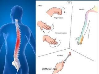
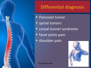
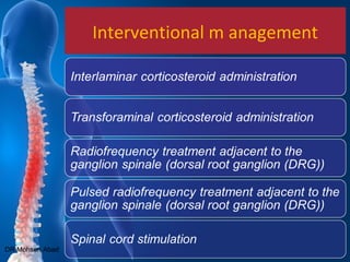
cervical radicular pain is
defined as pain perceived as arising in the upper limb caused by ectopic activation of nociceptive afferent fibers in a spinal nerve or its roots or other neuropathic mechanisms
Cervical disc disorders include cervical spondylosis, radiculopathy, and myelopathy. Cervical spondylosis is a general term referring to degenerative changes in the cervical spine. It commonly causes neck pain but can also cause radiculopathy or myelopathy. Cervical radiculopathy involves compression of a cervical nerve root, causing pain and weakness along the nerve distribution. Cervical myelopathy refers to compression of the spinal cord, which can cause gait abnormalities, leg weakness, and hand/arm symptoms. Management involves conservative treatments like physical therapy initially, with surgery considered for worsening or persistent symptoms.
Meralgia Paresthetica (MP) is a condition caused by impingement of the lateral femoral cutaneous nerve, causing numbness and pain along the front of the thigh. It is often caused by entrapment of the nerve under the inguinal ligament. Diagnosis involves history, physical exam including the pelvic compression test, and may include imaging or nerve blocks. Treatment options include removing any underlying causes, medications, physical therapy, injections, or surgery.
This document discusses failed back syndrome, specifically defining it as any condition where there is failure to improve satisfactorily following back surgery. It then classifies failures into those with no immediate improvement and temporary relief but recurrence of pain within weeks, months, or years. Causes of failures include wrong diagnosis, technical errors during surgery, infection, arachnoiditis, and recurrent stenosis. The document also discusses juxtafacet cysts near facet joints.
The document discusses spinal canal stenosis, including:
1. It describes spinal canal stenosis as the narrowing of the spinal canal and compression of the spinal cord and nerve roots, most commonly occurring in the lumbar vertebrae.
2. Symptoms include back pain radiating into the legs, numbness, and weakness that is relieved by bending forward and made worse by standing upright or walking.
3. Treatment options range from non-surgical approaches like medication, physical therapy, and epidural injections for mild-to-moderate cases to surgical decompression like laminectomy or the X-STOP implant for more severe cases.
This document provides information about Perthes' disease, including:
- It is characterized by avascular necrosis of the femoral head in children.
- Risk factors include being male and between ages 5-10 years old.
- Imaging studies like x-rays are used to diagnose and monitor the stages of avascular necrosis, fragmentation, ossification, and remodeling.
- Differential diagnosis depends on whether the condition is unilateral or bilateral.
- Treatment aims to prevent deformity through nonsurgical or surgical methods depending on the severity.
Cervical radiculopathy is a peripheral nerve syndrome caused by mechanical injury or chemical irritation of the cervical nerve roots. It typically affects the C5-T1 nerve roots, accounting for 5-30% of radiculopathy cases. Common causes include spondylosis, cervical disc disease, disc herniation, and biochemically-induced inflammation. Symptoms include neck pain radiating into the arms and hands with weakness, numbness, or tingling. Diagnosis involves electrodiagnostic tests, imaging like MRI, and the Spurling maneuver. Initial treatment focuses on conservative options like medication and physical therapy, with surgery reserved for cases that fail non-operative management.
This document discusses coxa vara, which is a hip deformity characterized by an abnormal decrease in the femoral neck-shaft angle. It classifies coxa vara as congenital, developmental, or acquired. Developmental coxa vara is the most common type and is caused by a primary cartilage defect in the femoral neck. Clinical features include limping and pain. Treatment involves corrective valgus osteotomies to restore the neck-shaft angle and relieve stress on the femoral physis. The document describes several techniques for valgus osteotomy including Pauwel's, Borden's, and subtrochanteric osteotomy. The goal of surgery is to stimulate healing of the femoral neck defect and restore normal
Cervical disc prolapse occurs when a cervical disc herniates and compresses the nerve root. The cervical spine has 7 vertebrae and 6 intervertebral discs that act as shock absorbers and allow motion. A disc is composed of an inner nucleus pulposus surrounded by the outer annulus fibrosus. Common sites of prolapse are C5-C6 and C6-C7. Clinical features include neck pain radiating to the arm. Imaging like MRI or CT is used to confirm prolapse. Treatment involves rest, medications, traction and surgery like anterior cervical discectomy if non-operative measures fail.
SPONDYLOLISTHESIS: DIAGNOSIS, CLASSIFICATION, EVALUATION AND MANAGEMENTBenthungo Tungoe
Spondylolisthesis is the forward slippage of one vertebra on another. There are several classifications including developmental, isthmic, degenerative, traumatic, and postsurgical types. Developmental spondylolisthesis is usually asymptomatic and rarely progresses after adulthood. Isthmic spondylolisthesis has a risk of progression over 25% slippage or in symptomatic cases. Degenerative spondylolisthesis results from sagittal facet orientation or disc degeneration and increases in older females. The natural history depends on factors like age, gender, slip severity and progression.
This document discusses different types of shoulder dislocations including acute, recurrent, anterior, posterior, and inferior dislocations. It covers the anatomy of the shoulder joint, mechanisms of injury, clinical presentation, treatment including closed and open reduction techniques, and complications. Recurrent dislocations are more common in younger patients. Posterior dislocations are rare but the diagnosis is often missed on initial x-ray. Inferior dislocations involve severe abduction forces and risk neurovascular injury.
This document provides an overview of congenital muscular torticollis (CMT). CMT is caused by shortening of the sternocleidomastoid muscle at birth and results in an inclined neck. It affects 0.3-2.0% of live births. Diagnosis is based on history and examination. Treatment involves gentle stretching exercises before age 12 months. Surgery to lengthen the muscle may be considered for older children if conservative treatment fails. Surgical techniques aim to release the tight sternocleidomastoid muscle while avoiding injury to nearby nerves and vessels. Post-operative immobilization and exercises are used to prevent recontracture. Early treatment generally results in over 90% success rate.
Any buldge around disc causing compression of nerve root.
Herniation of disc is of 4 types:-
Contact:- No rupture in outer layer of NP within their limit, discogenic pain & deep dull pain.
Complete rupture /protruded disc :- Outer most layer is intact & inner layer of AF is ruptured.
Herniated Sequestered disc:- Outer
most layer is also ruptured & nerve root
compression (NRC) is there.
4 No buldge:- Nuclear Matrix comes out
but no rupture of AF, No NRC but
sequestration NRC is there.
Osteochondritis Dessicans is a pathological condition characterized by separation of articular cartilage and subchondral bone from the joint surface. It most commonly affects the knee, especially the medial femoral condyle. The exact etiology is unknown but repetitive trauma is a major risk factor. Diagnosis is made through imaging like MRI and arthroscopy. Treatment depends on the age, location, and stability of the lesion. Conservative treatment is usually attempted first for juvenile OCD while unstable or large lesions in adults typically require surgical intervention such as drilling, fixation, or restorative techniques like microfracture or osteochondral grafting.
This document discusses spondylolisthesis, including its anatomy, classification, natural history, and management. Some key points include:
- Spondylolisthesis is the forward translation of one vertebra on another, often caused by a defect in the pars interarticularis. It is classified by its cause and severity.
- Symptoms range from low back pain to neurological deficits depending on grade. Imaging helps assess severity and complications.
- Conservative care focuses on symptom relief but surgery may be needed for progressive slippage, deformity, or neurological problems.
- Surgical options include decompression with or without fusion to improve stability. Fusion techniques include posterolateral, anterior, or circumferential
The document summarizes evidence from studies on the indications and timing of surgery for lumbar disc herniation. It finds that while early surgery (within 6-12 weeks of symptoms) provides faster relief of sciatic leg pain compared to conservative treatment, there is no difference in long-term outcomes between early surgery and conservative treatment. The optimal timing for surgery is not clearly defined by the evidence presented.
Recurrent shoulder dislocation and managementAnshul Sethi
This document provides an overview of recurrent shoulder dislocations. It discusses the anatomy of the shoulder joint and its stabilizers. The glenohumeral ligaments, labrum, rotator cuff muscles, and negative intra-articular pressure provide static stability, while dynamic stability comes from the rotator cuff and scapulo-thoracic motion. Younger age, returning to collision sports, and bone defects increase risk of recurrence. Evaluation involves assessing range of motion, translation, and special tests like the anterior drawer and sulcus sign. History and physical exam help determine treatment which may include rehabilitation or surgery to address labral tears or bone loss.
Frozen shoulder, also known as adhesive capsulitis, is a condition characterized by pain and stiffness in the shoulder joint that limits range of motion. It involves thickening and scarring of the shoulder joint capsule. Treatment involves conservative measures like oral anti-inflammatory drugs, corticosteroid injections into the joint, physical therapy including heat therapy and gentle range of motion exercises, and manipulation under anesthesia for refractory cases. Physical therapy aims to reduce pain and inflammation in the early stage and increase mobility in the stiffening stage through heat, passive range of motion, and home exercises.
1. Melorheostosis is a rare sclerosing bone disorder of unknown cause that produces thickened bone along the surface and inner lining of bones.
2. It most commonly affects the lower extremities in individuals between 5-20 years old and presents with bone overgrowth patterns resembling candle wax or myositis ossificans on imaging.
3. Affected individuals may experience pain, joint stiffness, deformities, and leg length discrepancies. Skin thickening and vascular abnormalities can also occur.
This document provides information on meniscal tears of the knee. It begins with an introduction stating that meniscal tears are common injuries responsible for many arthroscopies annually. It then covers anatomy of the medial and lateral menisci, blood supply, classification of tears, mechanisms of injury, clinical features, investigations like MRI and arthroscopy, and treatment options including non-operative care, meniscectomy, and meniscal repair. The focus is on providing detailed information on meniscal anatomy, tears, and surgical and non-surgical management.
Mallet finger, or drop finger, is a deformity of the finger caused by damage to the extensor tendon below the DIP joint, preventing straightening of the fingertip. It most commonly occurs in the long, ring, or small finger of the dominant hand in young males after the fingertip is forcibly bent backwards. Treatment depends on the severity of the injury but generally involves splinting the finger to keep the DIP joint straight as the tendon heals, usually for 6-8 weeks. Surgery may be needed for open injuries, large bone fragments, or if non-surgical treatment is unsuccessful. Complications can include an extensor lag deformity or swan neck deformity if not properly
Dr. Mahak Jain presented on spondylolisthesis. Key points include:
1) Spondylolisthesis is the forward translation of one vertebra on another, commonly caused by defects in the pars interarticularis known as spondylolysis.
2) It is classified based on etiology, with dysplastic, isthmic, degenerative, traumatic, and pathological types.
3) Treatment depends on factors like grade, symptoms, and etiology, ranging from conservative care to surgical options like decompression, fusion, and instrumentation.
4) Studies show surgery with fusion has better outcomes for pain and function than nonsurgical treatment or decompression alone for degenerative
The document discusses the anatomy and biomechanics of the shoulder joint and various types of shoulder dislocations. It describes the glenohumeral joint as a ball and socket joint between the humeral head and glenoid cavity. It then covers the different ligaments and muscles that support the shoulder joint. The rest of the document discusses the mechanisms, clinical presentations, investigations, and treatments for the main types of shoulder dislocations including anterior, posterior, inferior dislocations. It also notes potential complications of shoulder dislocations.
Cervical myelopathy is caused by compression of the cervical spinal cord, most commonly from cervical spondylosis. Cervical spondylosis involves degenerative changes to the spine that decrease space for the spinal cord. This can lead to static or dynamic compression of the cord, impairing circulation. Patients experience symptoms like neck pain, weakness, spasticity and sensory changes. Diagnosis involves assessing severity using scales and investigating spinal changes through imaging like CT which shows stenosis and compression more clearly than x-rays.
1) This document provides guidance on evaluating and differentiating the causes of low back pain through patient history, physical examination, and imaging.
2) The differential diagnosis depends on characteristics of the pain such as duration, location, radiation, and aggravating/relieving factors. Common etiologies include inflammatory, mechanical, and radicular causes.
3) The physical examination focuses on the spine, hips, and tests to reproduce pain including range of motion and provocative maneuvers. Red flags are identified.
4) Imaging like MRI can identify abnormalities in the discs, vertebrae, nerves and surrounding tissues that provide diagnostic clues. Both regular sequences and enhanced images after contrast are useful.
Coccydynia is pain arising from the coccyx or tailbone that is commonly caused by trauma, infection, or idiopathic factors. It presents as pain localized to the coccyx that is exacerbated by sitting, standing from sitting, intercourse, defecation, and menstruation. Diagnosis involves physical exam, x-rays, CT, or MRI. Conservative treatments like anti-inflammatories, cushions, and physical therapy resolve most cases, but injections or coccygectomy may be used if conservative options fail.
Evaluation of Lumbar Spine Disease starts with understanding the clinical back grounds. It starts with good history and physical examination. This is a teaching lecture given twice by Prof. Dr. Mohamed Mohi Eldin, professor of neurosurgery, in the Basic Spine Course, Egyptian Medical Syndicate, Cairo, March 2009 and in 2010.
This document provides information about spondylolisthesis, including:
- It is a condition where one vertebra slips out of position, usually involving L5 slipping forward on S1.
- It can be caused by a defect in the pars interarticularis that allows slippage.
- Treatment options include non-operative measures like bracing or injections, or surgical options like decompression or fusion to correct the slippage and stabilize the spine.
- Surgical treatment is generally recommended for severe slips over 50% or when non-operative treatment fails to relieve pain. Different procedures are used depending on the severity and characteristics of the spondylolisthesis.
Cancer pain is caused by tumors invading tissues and pressing on nerves. There are three types of pain: nociceptive, inflammatory, and neuropathic. Pain signals travel along nerve pathways from tissues to the spinal cord and brain. Cancer pain management involves detailed assessment, analgesic drugs like opioids, and non-pharmacological treatments. Radiation, chemotherapy, surgery, nerve blocks, and cement injections can help reduce tumor size and pressure causing pain. The goal is comprehensive treatment of physical and psychological distress from cancer.
Shoulder Impingement Evidence Based Case Study Rumy Petkov
Used evidence based literature to compare laser therapy treatment versus corticosteroid injections, ultrasound, rehab exercises, and Kinesio taping to treat shoulder impingement.
Spinal cord stimulation- principles and clinical applications5g5b6xsphd
This document discusses spinal cord stimulation (SCS) therapy for treating chronic pain. It provides information on:
- SCS is an FDA-approved third-tier therapy used to treat chronic trunk and limb pain such as failed back surgery syndrome and complex regional pain syndrome.
- SCS works via the gate control theory and stimulating descending pain pathways in the spinal cord to reduce pain transmission.
- For SCS treatment, candidates undergo a trial period with temporary electrodes to determine effectiveness before receiving permanent implantation. Clinical studies show SCS provides effective pain relief and improved quality of life for various chronic pain conditions.
Ppt for cims con 2017 chronic pain algorythm drdipakdesai
This document discusses various interventional pain management techniques for chronic pain, including injection therapies, neuroaugmentation, and intrathecal drug delivery. It provides details on procedures like trigger point injections, epidurals, medial branch nerve blocks, sympathetic blocks, spinal cord stimulation, peripheral nerve stimulation, and implantable intrathecal pumps. The document emphasizes that a multi-disciplinary approach utilizing all available resources works best for effectively treating chronic pain.
This document provides information on lateral epicondylitis (tennis elbow), including its anatomy, causes, symptoms, diagnosis, and treatment options. It describes how lateral epicondylitis is an overuse injury caused by repetitive microtrauma to the common extensor tendon at the lateral epicondyle. The diagnosis is typically made based on physical examination findings of tenderness over the lateral epicondyle with resisted wrist and finger extension. Both non-operative treatments like physiotherapy, bracing, and steroid injections and surgical options are discussed for managing lateral epicondylitis.
This document provides information on cervical epidural anesthesia. It discusses the history and uses of cervical epidural, including for bilateral upper limb surgery, mastectomy, thyroid surgery, and chronic pain management. Risks like spinal cord injury and neurological complications are addressed. Techniques to increase safety are covered, such as using fluoroscopy, avoiding levels above C6-7, and low injection volumes. Drugs commonly used include ropivacaine, lidocaine, and bupivacaine. Overall, the document outlines the applications and techniques of cervical epidural anesthesia while also discussing risks and safety considerations.
This document provides information on neck pain, including causes, symptoms, classifications, clinical examination, investigations, and treatment. It discusses how neck pain can result from disorders of structures in the neck like bones, joints, nerves and muscles. Examination involves inspection, palpation, and assessment of movement and neurological function. Investigations may include x-rays, MRI, and blood tests. Treatment depends on the underlying cause but typically involves rest, medications, physiotherapy including exercises and manual therapy, and surgery if conservative treatments fail. Prevention emphasizes proper posture, exercise and ergonomics to avoid strain on the neck structures.
Lumbar spinal stenosis, laminectomy, prolapsed intervertebral discYangtze university
Lumbar spinal stenosis is a narrowing of the spinal canal in the lower back that puts pressure on the spinal cord and nerves. It commonly occurs in people over 50 due to age-related wear and tear causing bone spurs or thickened ligaments. The best test for diagnosis is an MRI of the lumbar spine, which will show if there is compression of the spinal cord or nerves. Conservative treatment includes medications like NSAIDs, muscle relaxants, and epidural steroid injections, as well as physical therapy. Surgery such as laminectomy or discectomy may be considered if conservative measures fail to provide relief from pain and symptoms.
This document provides information on patient selection criteria, candidate types, and contraindications for spinal cord stimulation (SCS). Good candidates tend to have neuropathic or complex regional pain syndrome. Psychological screening is important, and those with untreated disorders like depression are poor candidates. The document also summarizes SCS techniques, types of implant systems, lead types, and trial lead placement procedures.
Brachial plexus injuries can damage the nerves that control the shoulder, arm, and hand. They can be classified as pre-ganglionic or post-ganglionic based on whether the injury occurs before or after the dorsal root ganglion. Investigation may include imaging like CT myelography or MRI as well as electrodiagnostic testing. Treatment depends on the type and severity of injury but may include conservative care, nerve grafting, nerve transfer, tendon transfer, or arthrodesis. The prognosis depends on factors like the specific nerves injured and whether recovery is possible.
Diagnostic & Therapeutic Updates of Discogenic Low Back PainReza Aminnejad
Neurogenic inflammation is the basis of discogenic low back pain. New advances in treating degenerative disk disease focus on biological reconstructive measures.
Neck pain is an aching, burning, stabbing, shooting, or cramping pain. PCI is well known Neck Pain Treatment Clinic in Mumbai. Visit http://goo.gl/pqRBvJ & Get Relief from Neck Pain
This document provides descriptions of various orthopedic tests of the cervical spine. It describes tests such as cervical compression, flexion, distraction, and resisted muscle tests. For each test, it provides the indications for use, procedures, mechanisms, and interpretations. The tests are used to evaluate patients with neck and arm pain, particularly to identify conditions like radiculopathy or nerve root irritation.
The Indian Dental Academy is the Leader in continuing dental education , training dentists in all aspects of dentistry and
offering a wide range of dental certified courses in different formats.for more details please visit
www.indiandentalacademy.com
This document provides an overview of regional anesthesia techniques, focusing on spinal anesthesia. It describes the anatomy of the spinal canal and meninges. It identifies landmarks for spinal needle placement and discusses factors that influence the level and duration of spinal blocks. It explains the pharmacology of local anesthetics used in spinal blocks and covers cardiovascular and respiratory effects. The document outlines proper technique for midline and paramedian spinal needle approaches. Potential complications like hypotension and spinal headache are discussed along with appropriate treatments.
This document discusses wrist drop, which refers to an inability to actively extend the hand at the wrist due to weakness of the wrist extensor muscles innervated by the radial nerve. Risk factors include occupations involving repetitive motion, injuries like fractures or dislocations. Signs include numbness from the triceps down and an inability to lift the wrist. Diagnosis involves physical exams, EMGs, and nerve conduction studies. Treatment may include medication, physical therapy, splinting, and in some cases surgery, with recovery times ranging from weeks to months depending on nerve damage severity. Complications can include loss of feeling or movement in the hand or deformities.
A study of core decompression & free fibular strut grafting in the management...Vltech Knr
Core decompression and free fibular strut grafting were studied as a treatment for osteonecrosis of the femoral head. In the study of 28 hips with Ficat-Arlet grade 1-3 osteonecrosis, 67.86% of patients experienced pain relief after the procedure. At the 6-month follow up, 82.61% of patients were considered surgical successes based on Harris Hip Scores and radiographic evidence. However, 8 hips showed further advancement of osteonecrosis despite the procedure. The study concluded that core decompression with fibular grafting can effectively treat early stage osteonecrosis, but patient factors like age, hip flexibility, and adherence to post-op care affected outcomes.
This document discusses various types of cervical (neck) pain including strains, sprains, radiculopathy, joint pain, internal disk disruption, and cervicogenic headaches. It describes symptoms, potential causes, diagnostic tests like MRI and nerve blocks, and treatment options for each type of pain such as medications, physical therapy, injections, and in rare cases surgery. The focus is on assessing patients, making an accurate diagnosis, and applying multimodal conservative treatments before more invasive options are considered.
Adhd Medication Shortage Uk - trinexpharmacy.comreignlana06
The UK is currently facing a Adhd Medication Shortage Uk, which has left many patients and their families grappling with uncertainty and frustration. ADHD, or Attention Deficit Hyperactivity Disorder, is a chronic condition that requires consistent medication to manage effectively. This shortage has highlighted the critical role these medications play in the daily lives of those affected by ADHD. Contact : +1 (747) 209 – 3649 E-mail : sales@trinexpharmacy.com
Recomendações da OMS sobre cuidados maternos e neonatais para uma experiência pós-natal positiva.
Em consonância com os ODS – Objetivos do Desenvolvimento Sustentável e a Estratégia Global para a Saúde das Mulheres, Crianças e Adolescentes, e aplicando uma abordagem baseada nos direitos humanos, os esforços de cuidados pós-natais devem expandir-se para além da cobertura e da simples sobrevivência, de modo a incluir cuidados de qualidade.
Estas diretrizes visam melhorar a qualidade dos cuidados pós-natais essenciais e de rotina prestados às mulheres e aos recém-nascidos, com o objetivo final de melhorar a saúde e o bem-estar materno e neonatal.
Uma “experiência pós-natal positiva” é um resultado importante para todas as mulheres que dão à luz e para os seus recém-nascidos, estabelecendo as bases para a melhoria da saúde e do bem-estar a curto e longo prazo. Uma experiência pós-natal positiva é definida como aquela em que as mulheres, pessoas que gestam, os recém-nascidos, os casais, os pais, os cuidadores e as famílias recebem informação consistente, garantia e apoio de profissionais de saúde motivados; e onde um sistema de saúde flexível e com recursos reconheça as necessidades das mulheres e dos bebês e respeite o seu contexto cultural.
Estas diretrizes consolidadas apresentam algumas recomendações novas e já bem fundamentadas sobre cuidados pós-natais de rotina para mulheres e neonatos que recebem cuidados no pós-parto em unidades de saúde ou na comunidade, independentemente dos recursos disponíveis.
É fornecido um conjunto abrangente de recomendações para cuidados durante o período puerperal, com ênfase nos cuidados essenciais que todas as mulheres e recém-nascidos devem receber, e com a devida atenção à qualidade dos cuidados; isto é, a entrega e a experiência do cuidado recebido. Estas diretrizes atualizam e ampliam as recomendações da OMS de 2014 sobre cuidados pós-natais da mãe e do recém-nascido e complementam as atuais diretrizes da OMS sobre a gestão de complicações pós-natais.
O estabelecimento da amamentação e o manejo das principais intercorrências é contemplada.
Recomendamos muito.
Vamos discutir essas recomendações no nosso curso de pós-graduação em Aleitamento no Instituto Ciclos.
Esta publicação só está disponível em inglês até o momento.
Prof. Marcus Renato de Carvalho
www.agostodourado.com
Histololgy of Female Reproductive System.pptxAyeshaZaid1
Dive into an in-depth exploration of the histological structure of female reproductive system with this comprehensive lecture. Presented by Dr. Ayesha Irfan, Assistant Professor of Anatomy, this presentation covers the Gross anatomy and functional histology of the female reproductive organs. Ideal for students, educators, and anyone interested in medical science, this lecture provides clear explanations, detailed diagrams, and valuable insights into female reproductive system. Enhance your knowledge and understanding of this essential aspect of human biology.
Cell Therapy Expansion and Challenges in Autoimmune DiseaseHealth Advances
There is increasing confidence that cell therapies will soon play a role in the treatment of autoimmune disorders, but the extent of this impact remains to be seen. Early readouts on autologous CAR-Ts in lupus are encouraging, but manufacturing and cost limitations are likely to restrict access to highly refractory patients. Allogeneic CAR-Ts have the potential to broaden access to earlier lines of treatment due to their inherent cost benefits, however they will need to demonstrate comparable or improved efficacy to established modalities.
In addition to infrastructure and capacity constraints, CAR-Ts face a very different risk-benefit dynamic in autoimmune compared to oncology, highlighting the need for tolerable therapies with low adverse event risk. CAR-NK and Treg-based therapies are also being developed in certain autoimmune disorders and may demonstrate favorable safety profiles. Several novel non-cell therapies such as bispecific antibodies, nanobodies, and RNAi drugs, may also offer future alternative competitive solutions with variable value propositions.
Widespread adoption of cell therapies will not only require strong efficacy and safety data, but also adapted pricing and access strategies. At oncology-based price points, CAR-Ts are unlikely to achieve broad market access in autoimmune disorders, with eligible patient populations that are potentially orders of magnitude greater than the number of currently addressable cancer patients. Developers have made strides towards reducing cell therapy COGS while improving manufacturing efficiency, but payors will inevitably restrict access until more sustainable pricing is achieved.
Despite these headwinds, industry leaders and investors remain confident that cell therapies are poised to address significant unmet need in patients suffering from autoimmune disorders. However, the extent of this impact on the treatment landscape remains to be seen, as the industry rapidly approaches an inflection point.
Rasamanikya is a excellent preparation in the field of Rasashastra, it is used in various Kushtha Roga, Shwasa, Vicharchika, Bhagandara, Vatarakta, and Phiranga Roga. In this article Preparation& Comparative analytical profile for both Formulationon i.e Rasamanikya prepared by Kushmanda swarasa & Churnodhaka Shodita Haratala. The study aims to provide insights into the comparative efficacy and analytical aspects of these formulations for enhanced therapeutic outcomes.
Promoting Wellbeing - Applied Social Psychology - Psychology SuperNotesPsychoTech Services
A proprietary approach developed by bringing together the best of learning theories from Psychology, design principles from the world of visualization, and pedagogical methods from over a decade of training experience, that enables you to: Learn better, faster!
2. Introduction
pain perceived in the upper
limb, shooting or electric in
quality, caused by irritation
and or injury of a cervical
spinal nerve
DR Mohsen Abad
3. International
Association for the Study of Pain
cervical radicular pain is
defined as pain perceived as
arising in the upper limb
caused by ectopic activation
of nociceptive afferent fibers
in a spinal nerve or its roots
or other neuropathic
mechanisms
DR Mohsen Abad
4. Definition
• Cervical radicular pain must be
distinguished from cervical
radiculopathy.
• In the latter disorder there is
an objective loss of sensory
and/or motor function
• Radicular pain and
radiculopathy are not
synonymous
DR Mohsen Abad
5. Definition
• Radicular pain is a symptom
that is caused by ectopic
impulse formation, while
radiculopathy also includes
neurologic signs such as
sensory or motor changes
• These two disorders may occur
simultaneously.
DR Mohsen Abad
6. Diagnosis
• characterized by pain in the neck that
radiates over the posterior shoulder
into the arm and sometimes into the
hand
• The radiation follows a segment –
specific pattern.
• Pain from various dermatomes can
overlap and there is no specific region
of the arm
DR Mohsen Abad
7. Diagnosis
• Pain originating from C4 is confined to
the neck and suprascapular region
• C5 radiates up to the upper arm
• C6 and C7 radiates from the neck to
the shoulder, the forearm, and the
hand
DR Mohsen Abad
8. Diagnosis
• Radicular pain is not limited to a
particular dermatome and can be
perceived in all structures that are
innervated by the affected nerve
roots such as muscles, joints,
ligaments, and the skin
DR Mohsen Abad
9. Physical examination
• there is no gold standard for the
diagnosis of cervical radicular pain
• Diagnosis is made based on a
combination of history, clinical
examination, and additional test
• Sensation, strength, and tendon reflexes.
DR Mohsen Abad
10. Specific clinical tests
Spurling test:
Neck extended with head rotated to
affected shoulder while axially loaded.
Reproduction of the patient’s shoulder or
arm pain indicates possible cervical spinal
nerve root compression
DR Mohsen Abad
12. Specific clinical tests
Shoulder abduction test
•The patient lifts a hand above his or her
head. A positive result is the decrease or
disappearance of the radicular symptom.
•Once the patient is in the testing position,
the position should be held for 5-10
seconds to allow symptoms to dissipate or
resolve
DR Mohsen Abad
14. Specific clinical tests
Axial manual traction test
In supine position an axial traction force
corresponding to 10 to 15 kg is applied.
A positive finding is the decrease or
disappearance of the radicular symptom
DR Mohsen Abad
16. Specific clinical tests
• All of these tests have a high specifi city
(81% to 100%) but a low sensitivity.
• The Spurling test was similarly evaluated
using electromyography as the reference
test
• These three examinations are
considered valuable aids in the clinical
diagnosis of a patient with neck and arm
pain.
DR Mohsen Abad
17. Measurement of the
sagittal diameter of the spinal
canal is accomplished by
calculating the distance
between the posterior surface
of the vertebral body
and the spinolaminar line
(between the arrows). At the
C4-7 levels, spinal cord
compression is unlikely if the
diameter of the canal is
13 mm or more
DR Mohsen Abad
18. Sagittal multiple planar
gradient recalled (MPGR)
magnetic resonance image
reveals
stenosis of the lower cervical
spine related to the presence
of
osteophytes arising from the
posterior surface of the
vertebral
bodies
DR Mohsen Abad
19. Disc
level
Root Pain
distribution
Muscle
weakness
Sensory
loss
Reflex loss
C4 to C5 C5 Margo medialis
scapulae, lateral
upper arm to
elbow
M. deltoideus,
m.
supraspinatus,
m.
infraspinatus
Lateral
upper arm
Supinator
reflex
C5 to C6 C6 Lateral forearm,
thumb and index
finger
M. biceps
brachii, m.
brachioradialis,
wrist extensors
Thumb and
index finger
Biceps reflex
C6 to C7 C7 Medial scapula,
posterior arm,
dorsum of
forearm, third
finger
M. triceps
brachii, wrist
flexors, finger
extensors
Posterior
forearm,
third finger
Triceps
reflex
C7 to T1 C8 Shoulder, ulnar
side of forearm,
fifth finger
Thumb fl
exors,
abductors,
intrinsic hand
muscles
Fifth finger –
DR Mohsen Abad
24. Medical imaging
• used to exclude primary pathologies, the
so - called “ red flags ”
• tumor, infection, and fractures
• CT scans are able to reproduce the
changes in bone structure more
sensitively than MRI.
• MRI is currently regarded as the most
suitable medical imaging technique for
patients with cervical radicular pain
DR Mohsen Abad
25. Medical imaging
• no data available regarding the
sensitivity and specificity of the various
imaging techniques
• A direct link between the pain syndrome
and the results of medical imaging does
not exist
DR Mohsen Abad
26. Electrophysiologic tests
• can be requested when nerve damage is
suspected but will not provide any
information about the pain
• Quantitative Sensory Testing (QST) has
been recommended in the literature as
an electrophysiologic test that can
provide more specific information about
pain
DR Mohsen Abad
27. Diagnostic selective nerve root
blocks
• The diagnostic blocks are applied in
separate sessions per level
• Under radiological visualization using a
contrast dye (fluoroscopy), a small
amount of local anesthetic is injected
(0.5 mL)
• During a period of 30 to 60 minutes after
injection, the pain score is evaluated at
regular time intervals. When there is at
least a 50% decrease in pain,DR Mohsen Abad
29. Treatment options
Conservative management
•NSAIDS medications are primarily
recommended for short - term treatment
•Anticonvulsants: such as carbamazepine,
oxcarbamazepine , gabapentin, and
pregabalin
•Cochrane review: patient education for
neck pain: does not show effectiveness
•Cochrane review: mechanical traction for
neck pain: no evidence to supportDR Mohsen Abad
32. Epidural corticosteroid administration
• Rely on the anti - inflammatory
response induced by inhibition
of the phospholipase A2 -
initiated arachidonic acid
cascade
• There are no direct
comparisons available between
interlaminar and
transforaminal administration
at a cervical level.DR Mohsen Abad
33. Epidural corticosteroid administration
• randomized study comparing
interlaminar and intramuscular
corticosteroid administration
found that 68% significant pain
relief lasting at least 1 year
compared to 12% in the group
treated intramuscularly.
DR Mohsen Abad
34. Interlaminar administration:
complications
• Minor complications that
spontaneously disappeared,
often within 24 hours :
increasing axial neck pain,
• posture - independent
headache,
• facial flushing,
• vasovagal episodes
DR Mohsen Abad
36. Practical recommendations
• The positive RCT for interlaminar
administration
• There are no studies which have
investigated the effectiveness of
the various depot corticosteroids
• The particle size of the depot
corticosteroid is possibly related to
the reported neurologic
complications
• Currently there is no evidence that
a higher dose of corticosteroids will
result in a better clinical effect.
DR Mohsen Abad
38. efficacy
• Currently preference is given to PRF where
the tip temperature of the electrode does
not exceed the critical threshold of 42 ° C
and consequently there is minimal neuro -
destruction.
• In an RCT, PRF appeared to be more
effective than placebo 3 months post -
treatment. Also 6 months post - treatment
there was a positive trend in the PRF
treatment
DR Mohsen Abad
39. Surgical treatment
• is indicated in cervical radiculopathy with
spinal cord compression (myelomalacia)
because of the risk for possibly irreversible
neurologic deficiency.
• In a randomized study where surgical
treatment was compared with conservative
treatment a significant improvement in pain
relief was noted 3 months after the
intervention
• A year post - treatment however there was
no difference between the two groups
DR Mohsen Abad
40. Spinal cord stimulation
• Up until now there is no literature on the
outcome of SCS in the treatment of cervical
radicular pain.
• SCS can be considered in clinical practice for
chronic cervical radicular pain in well -
selected patients when other types of
treatment have failed, given that the efficacy
has been demonstrated in other comparable
neuropathic pain syndromes
DR Mohsen Abad