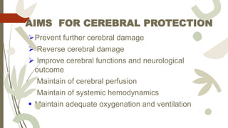
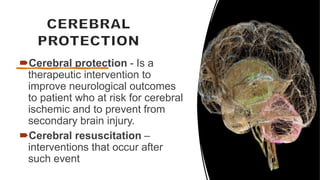
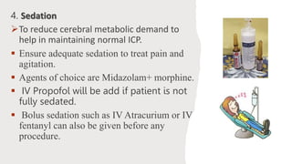
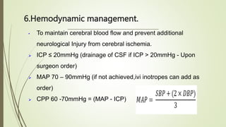
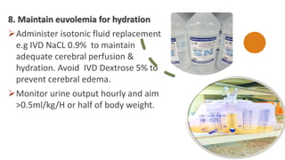
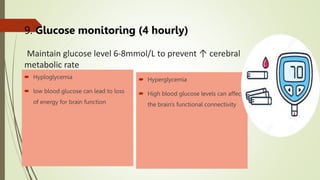
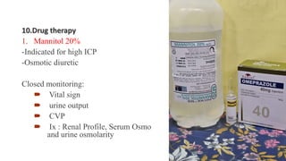
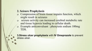
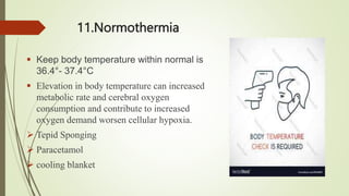
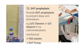
CEREBRAL
PROTECTION
OBJECTIVE
Define cerebral protection
Expose nurses with basic knowledge in care of
patient with cerebral protection
Able to identify the abnormalities
AIMS FOR CEREBRAL PROTECTION
Prevent further cerebral damage
 Reverse cerebral damage
 Improve cerebral functions and neurological
outcome
 Maintain of cerebral perfusion
 Maintain of systemic hemodynamics
 Maintain adequate oxygenation and ventilation
Cerebral protection - Is a
therapeutic intervention to
improve neurological outcomes
to patient who at risk for cerebral
ischemic and to prevent from
secondary brain injury.
Cerebral resuscitation –
interventions that occur after
such event
INDICATION CEREBRAL
PROTECTION
1. Head injury
2. Post cranial surgery
3. Cerebral edema
4. Seizures
5. Cerebral hypoxia
6. Space occupying lesion
7. Brain infection
8. Post cardio respiratory
arrest
1. Mechanical Ventilation
ABG
Pao2 80-100mmHg
- If <60mmHg -cause vasodilation lead to
hypoxemia causing cerebral edema
pCO² 30-35mmHg
- If <35mmHg can lead to vasoconstriction-
cerebral ischemic.
- If >45mmHg lead to vasodilation-cerebral
edema.
ETT tube
2.Suctioning
 To remove secretion to get adequate oxygenation and reduce the work of breathing.
 Secretion can stimulate a cough reflex, causing temporary hypoxia & hypercapnia with increase venous
pressure → can lead to increased ICP
• Performed suction PRN fast & gentle
• < 10 sec with pressure 80-120mmHg,
• Pre oxygenation 100% before suction,
• used correct size of catheter to maintain oxygenation.
• Maintain spo2 >95%
3.POSITION
 Nurse patient in neutral position with HOB 30º to facilitate
venous return to reduce intracranial pressure.
 This maximizes venous return from the head while
preserving CPP and CBF
REMINDER!!!
 Avoid extreme neck flexion, extension or rotation can
increase ICP.
 Cervical collars and ties on ETT (Endo Tracheal Tube) or
tracheostomy should be checked frequently to prevent
excessive pressure.
 Hip flexion to be avoided to provide venous drainage.
4. Sedation
To reduce cerebral metabolic demand to
help in maintaining normal ICP.
 Ensure adequate sedation to treat pain and
agitation.
 Agents of choice are Midazolam+ morphine.
 IV Propofol will be add if patient is not
fully sedated.
 Bolus sedation such as IV Atracurium or IV
fentanyl can also be given before any
procedure.
5. Avoid noxious stimuli
Administer IV Atracurium or IV
fentanyl bolus before any
procedure to reduce stimuli that
lead in increasing ICP.
Do suctioning PRN.
6.Hemodynamic management.
• To maintain cerebral blood flow and prevent additional
neurological Injury from cerebral ischemia.
 ICP ≤ 20mmHg (drainage of CSF if ICP > 20mmHg - Upon
surgeon order)
 MAP 70 – 90mmHg (if not achieved,ivi inotropes can add as
order)
 CPP 60 -70mmHg = (MAP - ICP)
7. If patient with EVD and for ICP
monitoring:
Monitor wave form
Maintain CPP 60-70mmHg, ICP <
20mmHg.
Level the transducer ordered.
Monitor patency of monitoring system.
Perform calibration and zeroing
SPIEGELBERG MONITORING DEVICE
8. Maintain euvolemia for hydration
Administer isotonic fluid replacement
e.g IVD NaCL 0.9% to maintain
adequate cerebral perfusion &
hydration. Avoid IVD Dextrose 5% to
prevent cerebral edema.
Monitor urine output hourly and aim
>0.5ml/kg/H or half of body weight.
9. Glucose monitoring (4 hourly)
Maintain glucose level 6-8mmol/L to prevent ↑ cerebral
metabolic rate
 Hyploglycemia
 low blood glucose can lead to loss
of energy for brain function
 Hyperglycemia
 High blood glucose levels can affect
the brain's functional connectivity
10.Drug therapy
1. Mannitol 20%
-Indicated for high ICP
-Osmotic diuretic
Closed monitoring:
 Vital sign
 urine output
 CVP
 Ix : Renal Profile, Serum Osmo
and urine osmolarity
2. Seizure Prophylaxis
 Compression of brain tissue impairs function, which
might result in seizures
 seizure activity can increased cerebral metabolic rate
and tissue hypoxia leading to cellular death.
 Example anticonvulsant : phenytoin sodium 100mg
TDS.
3.Stress ulcer prophylaxis with IV Omeprazole to prevent
stress ulcer.
11.Normothermia
 Keep body temperature within normal is
36.4°- 37.4°C
 Elevation in body temperature can increased
metabolic rate and cerebral oxygen
consumption and contribute to increased
oxygen demand worsen cellular hypoxia.
 Tepid Sponging
 Paracetamol
 cooling blanket
12. DVT prophylaxis
Provide DVT prophylaxis
to prevent deep vein
thrombosis
e.g S/C Clexane or S/C
Heparin if no
contraindications
mechanical
TED stockin
DVT Pump.
 60% of patients will suffer from electrolyte imbalances post TBI.
 Most commonly Na+.
 Hyponatremia reduce plasma osmolality and may cause brain
swelling.
 Syndrome of Inappropriate Antidiuretic Hormone (SIADH)
 Cerebral salt waste
13.Maintain electrolyte balance
14. Nutrition
Early feeding via
nasogastric tube
Early feeding to
promote recovery.
THANK YOU

cerebral protection.pptx

  • 1.
  • 2.
    OBJECTIVE Define cerebral protection Exposenurses with basic knowledge in care of patient with cerebral protection Able to identify the abnormalities
  • 3.
    AIMS FOR CEREBRALPROTECTION Prevent further cerebral damage  Reverse cerebral damage  Improve cerebral functions and neurological outcome  Maintain of cerebral perfusion  Maintain of systemic hemodynamics  Maintain adequate oxygenation and ventilation
  • 6.
    Cerebral protection -Is a therapeutic intervention to improve neurological outcomes to patient who at risk for cerebral ischemic and to prevent from secondary brain injury. Cerebral resuscitation – interventions that occur after such event
  • 7.
    INDICATION CEREBRAL PROTECTION 1. Headinjury 2. Post cranial surgery 3. Cerebral edema 4. Seizures 5. Cerebral hypoxia 6. Space occupying lesion 7. Brain infection 8. Post cardio respiratory arrest
  • 9.
    1. Mechanical Ventilation ABG Pao280-100mmHg - If <60mmHg -cause vasodilation lead to hypoxemia causing cerebral edema pCO² 30-35mmHg - If <35mmHg can lead to vasoconstriction- cerebral ischemic. - If >45mmHg lead to vasodilation-cerebral edema. ETT tube
  • 10.
    2.Suctioning  To removesecretion to get adequate oxygenation and reduce the work of breathing.  Secretion can stimulate a cough reflex, causing temporary hypoxia & hypercapnia with increase venous pressure → can lead to increased ICP • Performed suction PRN fast & gentle • < 10 sec with pressure 80-120mmHg, • Pre oxygenation 100% before suction, • used correct size of catheter to maintain oxygenation. • Maintain spo2 >95%
  • 11.
    3.POSITION  Nurse patientin neutral position with HOB 30º to facilitate venous return to reduce intracranial pressure.  This maximizes venous return from the head while preserving CPP and CBF
  • 12.
    REMINDER!!!  Avoid extremeneck flexion, extension or rotation can increase ICP.  Cervical collars and ties on ETT (Endo Tracheal Tube) or tracheostomy should be checked frequently to prevent excessive pressure.  Hip flexion to be avoided to provide venous drainage.
  • 13.
    4. Sedation To reducecerebral metabolic demand to help in maintaining normal ICP.  Ensure adequate sedation to treat pain and agitation.  Agents of choice are Midazolam+ morphine.  IV Propofol will be add if patient is not fully sedated.  Bolus sedation such as IV Atracurium or IV fentanyl can also be given before any procedure.
  • 14.
    5. Avoid noxiousstimuli Administer IV Atracurium or IV fentanyl bolus before any procedure to reduce stimuli that lead in increasing ICP. Do suctioning PRN.
  • 15.
    6.Hemodynamic management. • Tomaintain cerebral blood flow and prevent additional neurological Injury from cerebral ischemia.  ICP ≤ 20mmHg (drainage of CSF if ICP > 20mmHg - Upon surgeon order)  MAP 70 – 90mmHg (if not achieved,ivi inotropes can add as order)  CPP 60 -70mmHg = (MAP - ICP)
  • 16.
    7. If patientwith EVD and for ICP monitoring: Monitor wave form Maintain CPP 60-70mmHg, ICP < 20mmHg.
  • 17.
    Level the transducerordered. Monitor patency of monitoring system. Perform calibration and zeroing
  • 18.
  • 19.
    8. Maintain euvolemiafor hydration Administer isotonic fluid replacement e.g IVD NaCL 0.9% to maintain adequate cerebral perfusion & hydration. Avoid IVD Dextrose 5% to prevent cerebral edema. Monitor urine output hourly and aim >0.5ml/kg/H or half of body weight.
  • 20.
    9. Glucose monitoring(4 hourly) Maintain glucose level 6-8mmol/L to prevent ↑ cerebral metabolic rate  Hyploglycemia  low blood glucose can lead to loss of energy for brain function  Hyperglycemia  High blood glucose levels can affect the brain's functional connectivity
  • 21.
    10.Drug therapy 1. Mannitol20% -Indicated for high ICP -Osmotic diuretic Closed monitoring:  Vital sign  urine output  CVP  Ix : Renal Profile, Serum Osmo and urine osmolarity
  • 22.
    2. Seizure Prophylaxis Compression of brain tissue impairs function, which might result in seizures  seizure activity can increased cerebral metabolic rate and tissue hypoxia leading to cellular death.  Example anticonvulsant : phenytoin sodium 100mg TDS. 3.Stress ulcer prophylaxis with IV Omeprazole to prevent stress ulcer.
  • 23.
    11.Normothermia  Keep bodytemperature within normal is 36.4°- 37.4°C  Elevation in body temperature can increased metabolic rate and cerebral oxygen consumption and contribute to increased oxygen demand worsen cellular hypoxia.  Tepid Sponging  Paracetamol  cooling blanket
  • 24.
    12. DVT prophylaxis ProvideDVT prophylaxis to prevent deep vein thrombosis e.g S/C Clexane or S/C Heparin if no contraindications mechanical TED stockin DVT Pump.
  • 25.
     60% ofpatients will suffer from electrolyte imbalances post TBI.  Most commonly Na+.  Hyponatremia reduce plasma osmolality and may cause brain swelling.  Syndrome of Inappropriate Antidiuretic Hormone (SIADH)  Cerebral salt waste 13.Maintain electrolyte balance
  • 26.
    14. Nutrition Early feedingvia nasogastric tube Early feeding to promote recovery.
  • 28.