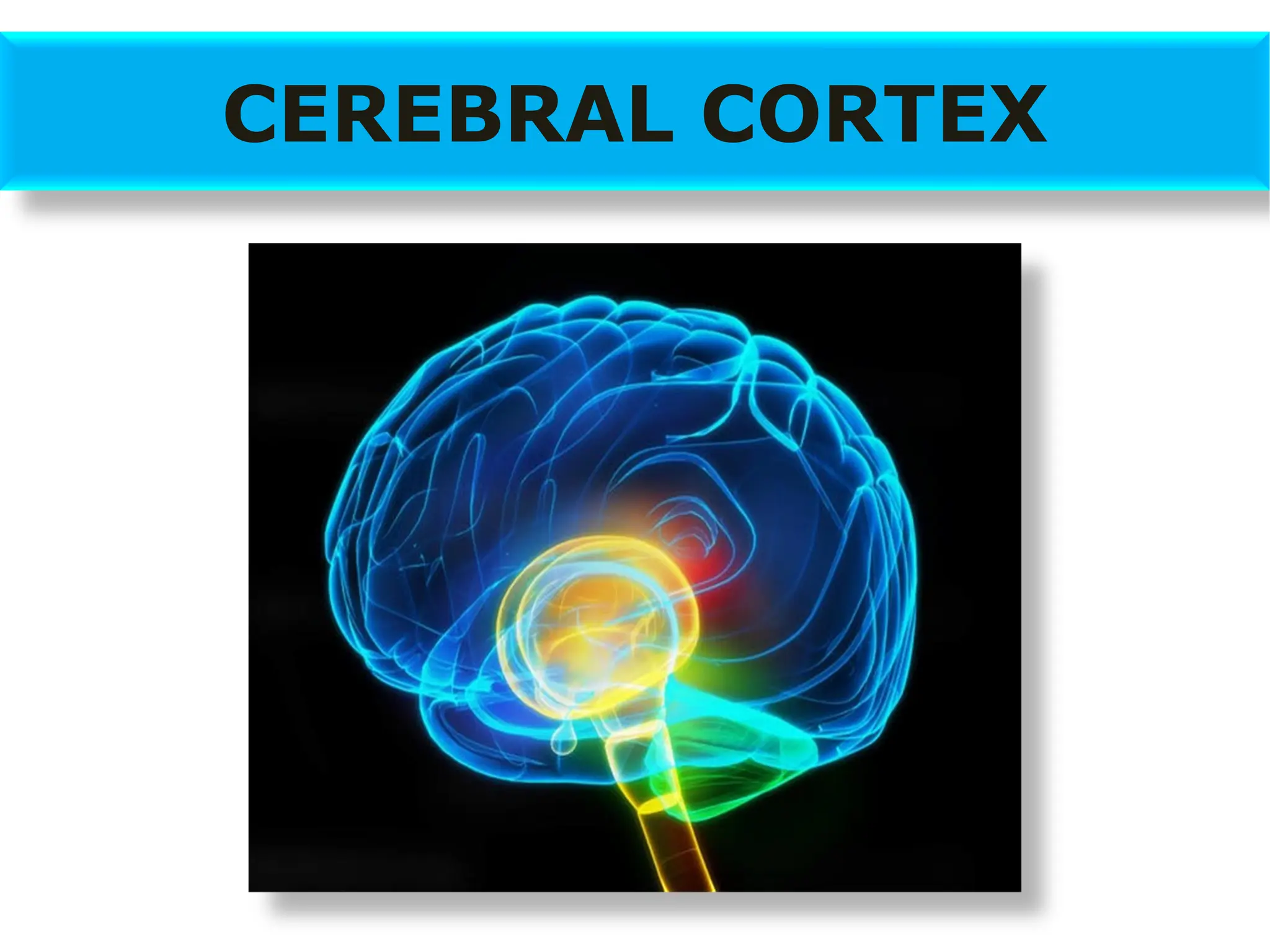
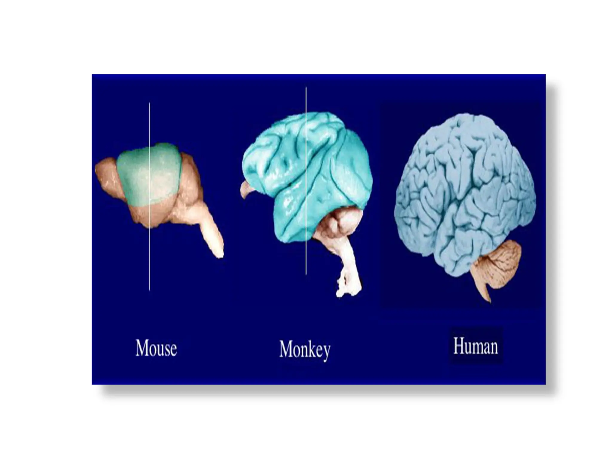
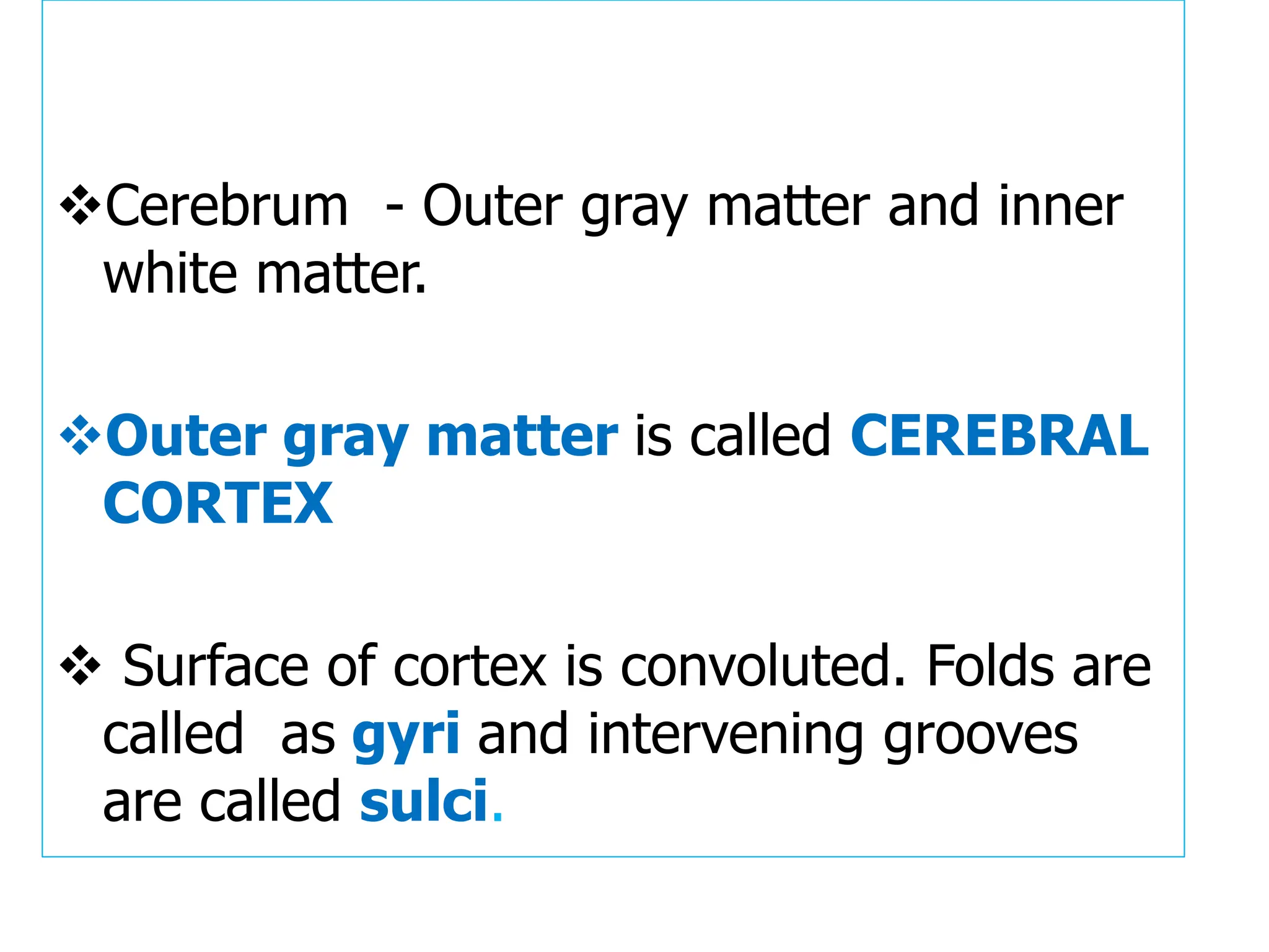
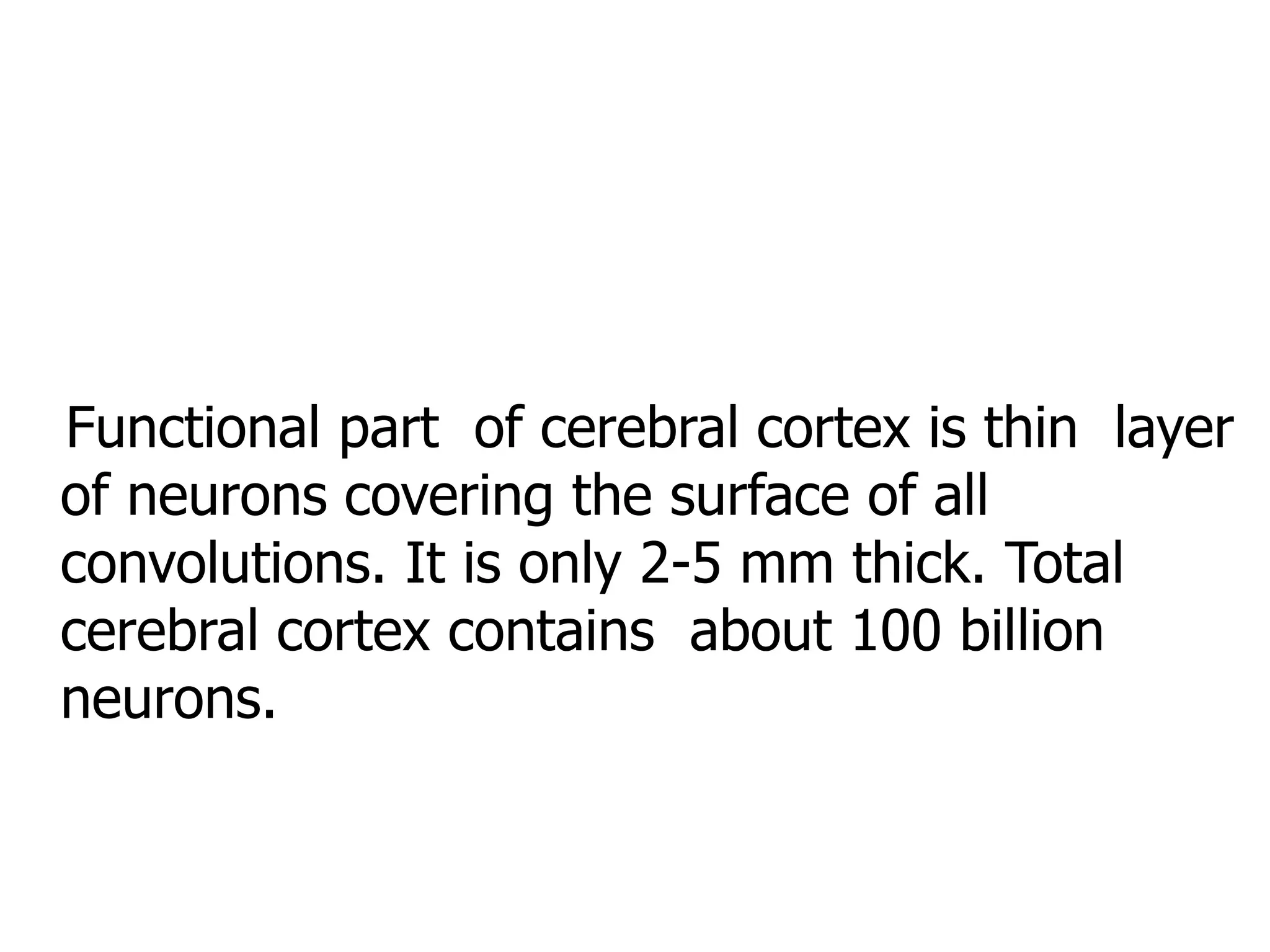
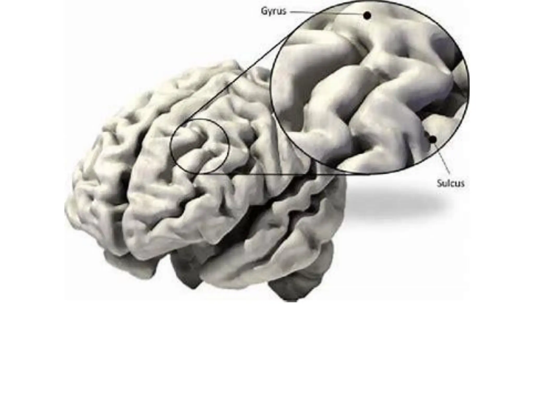
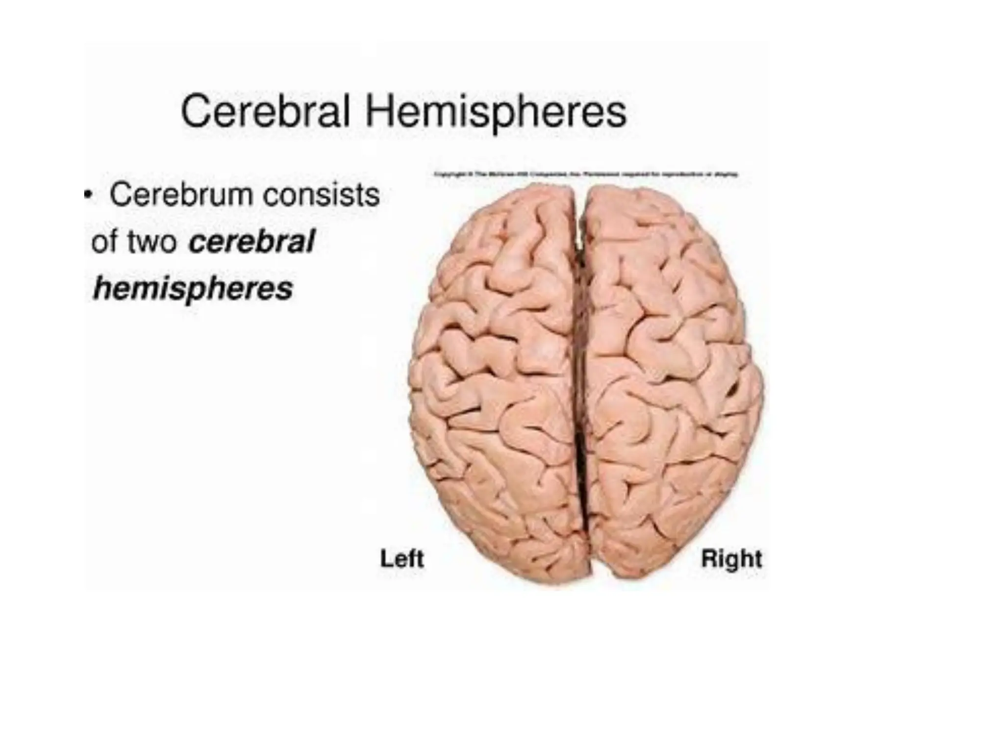
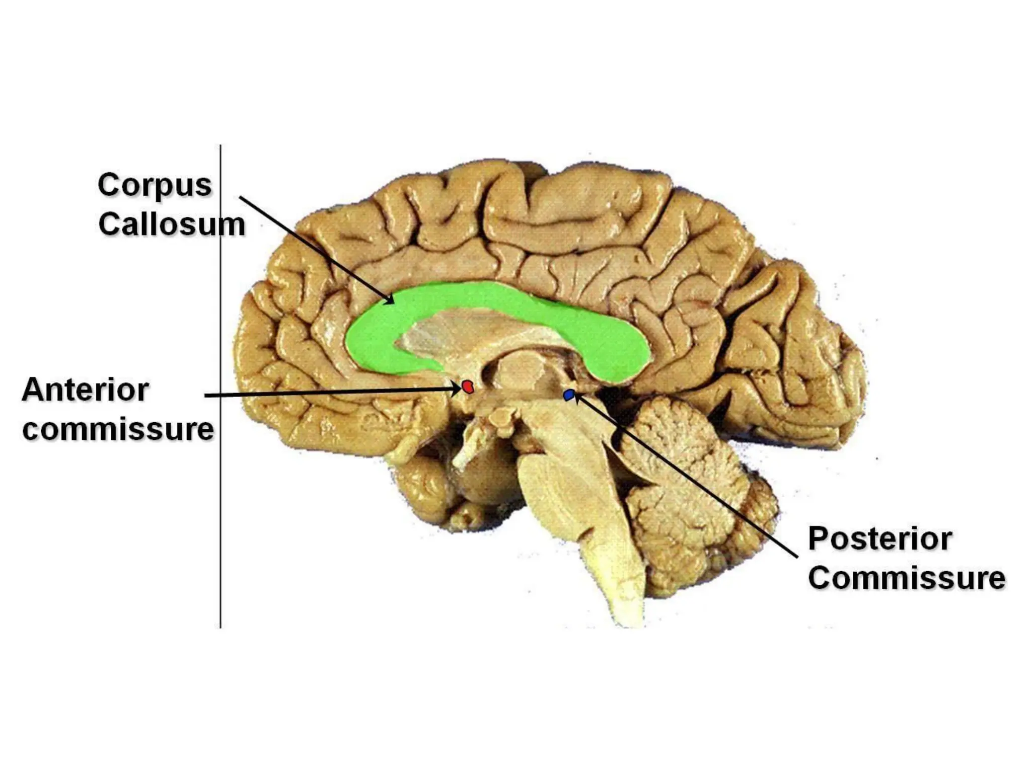
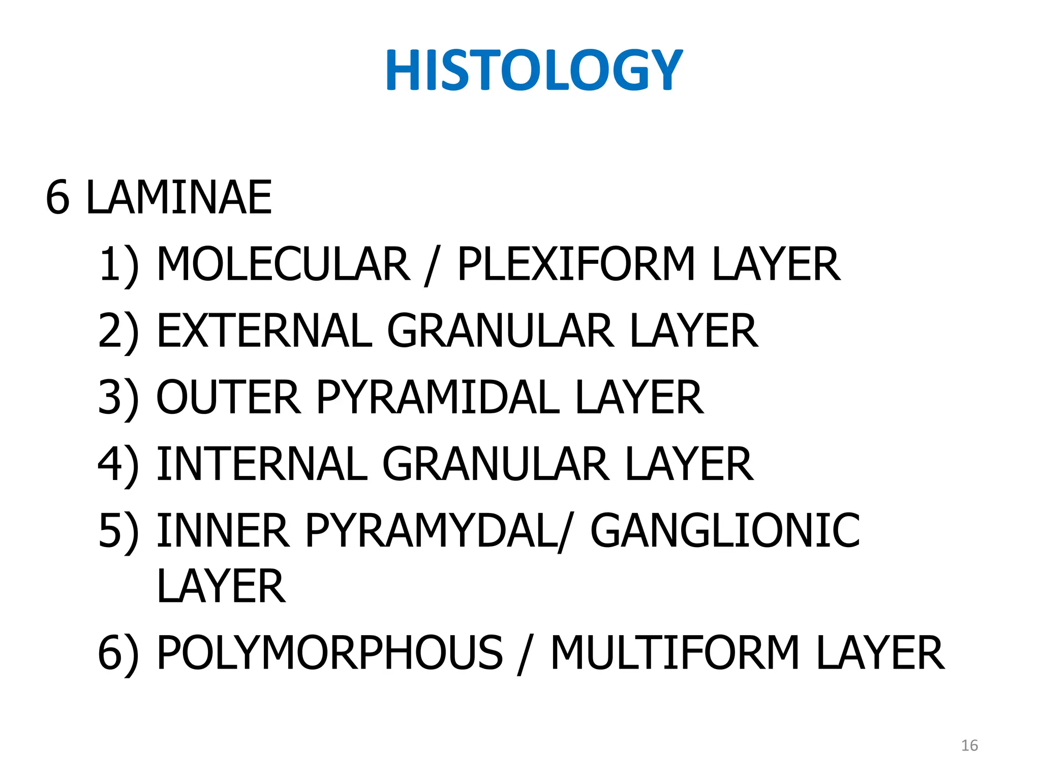
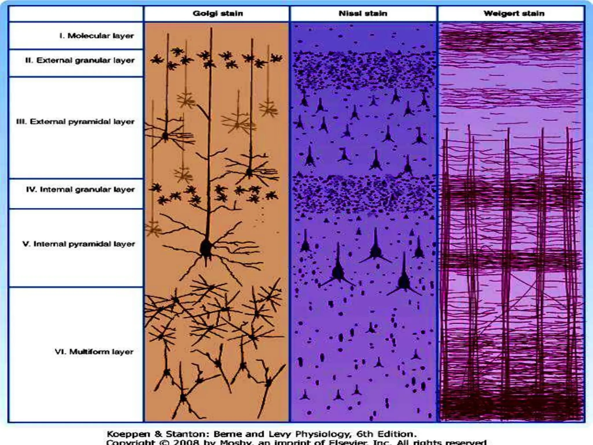
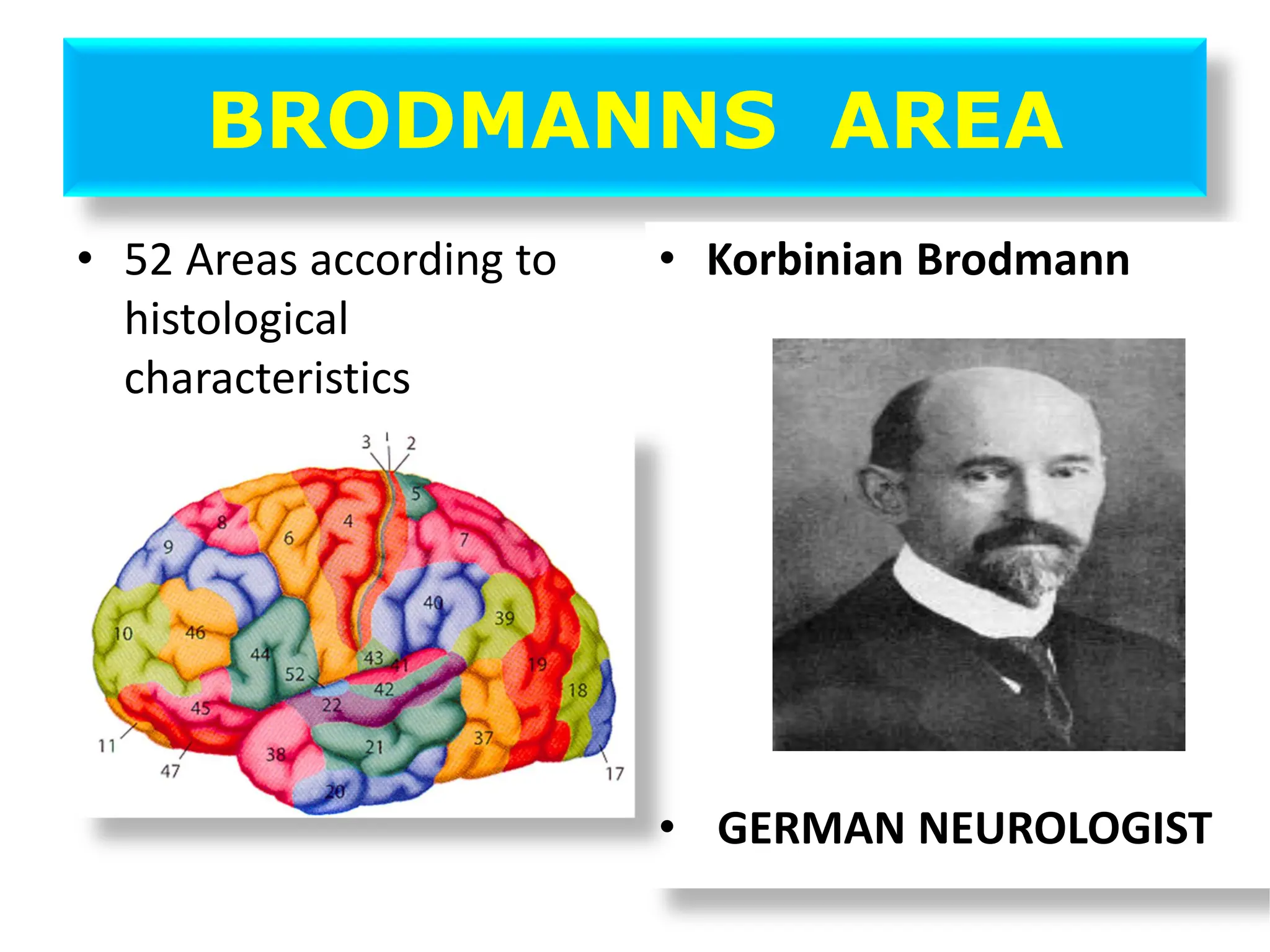
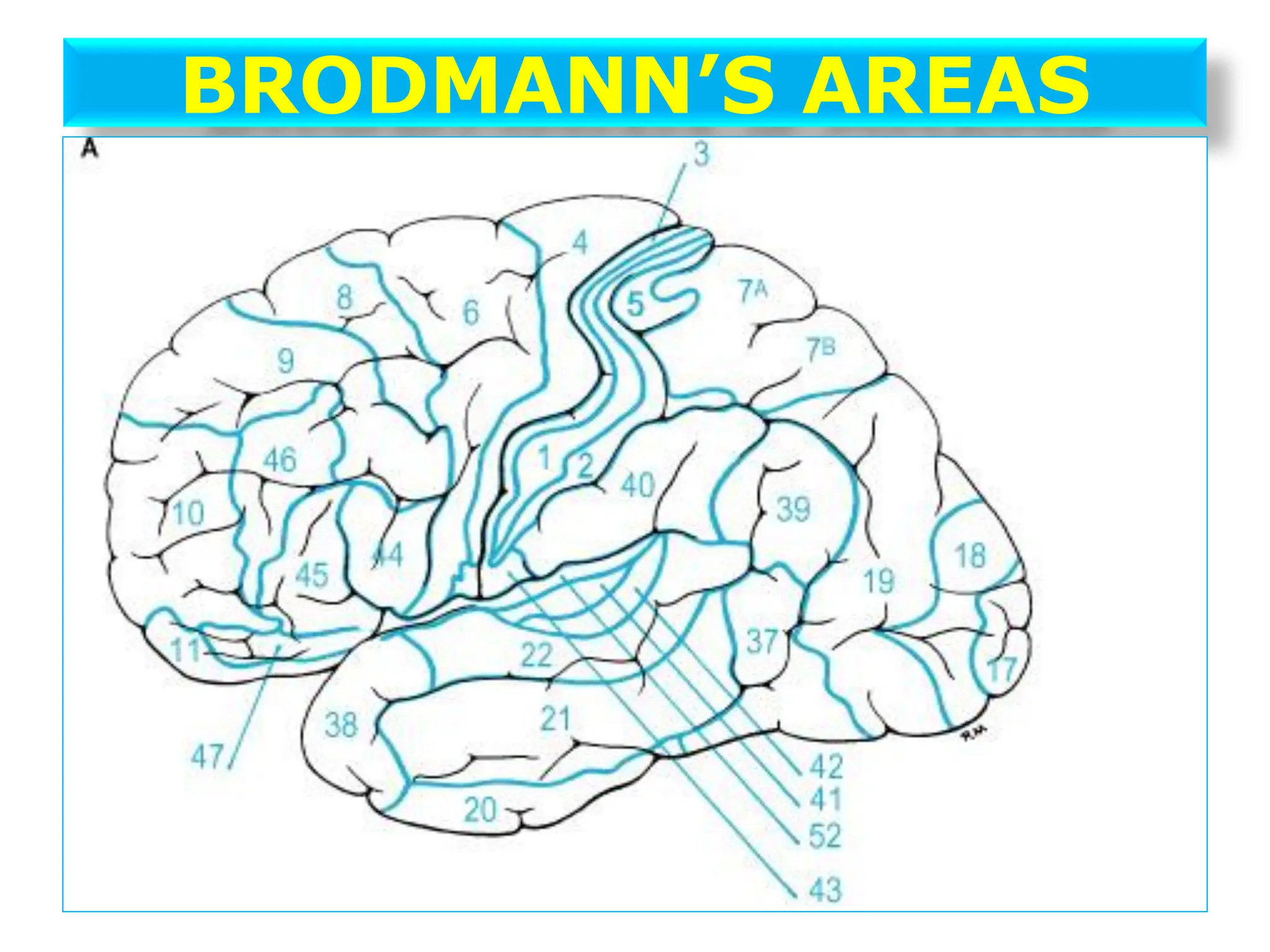
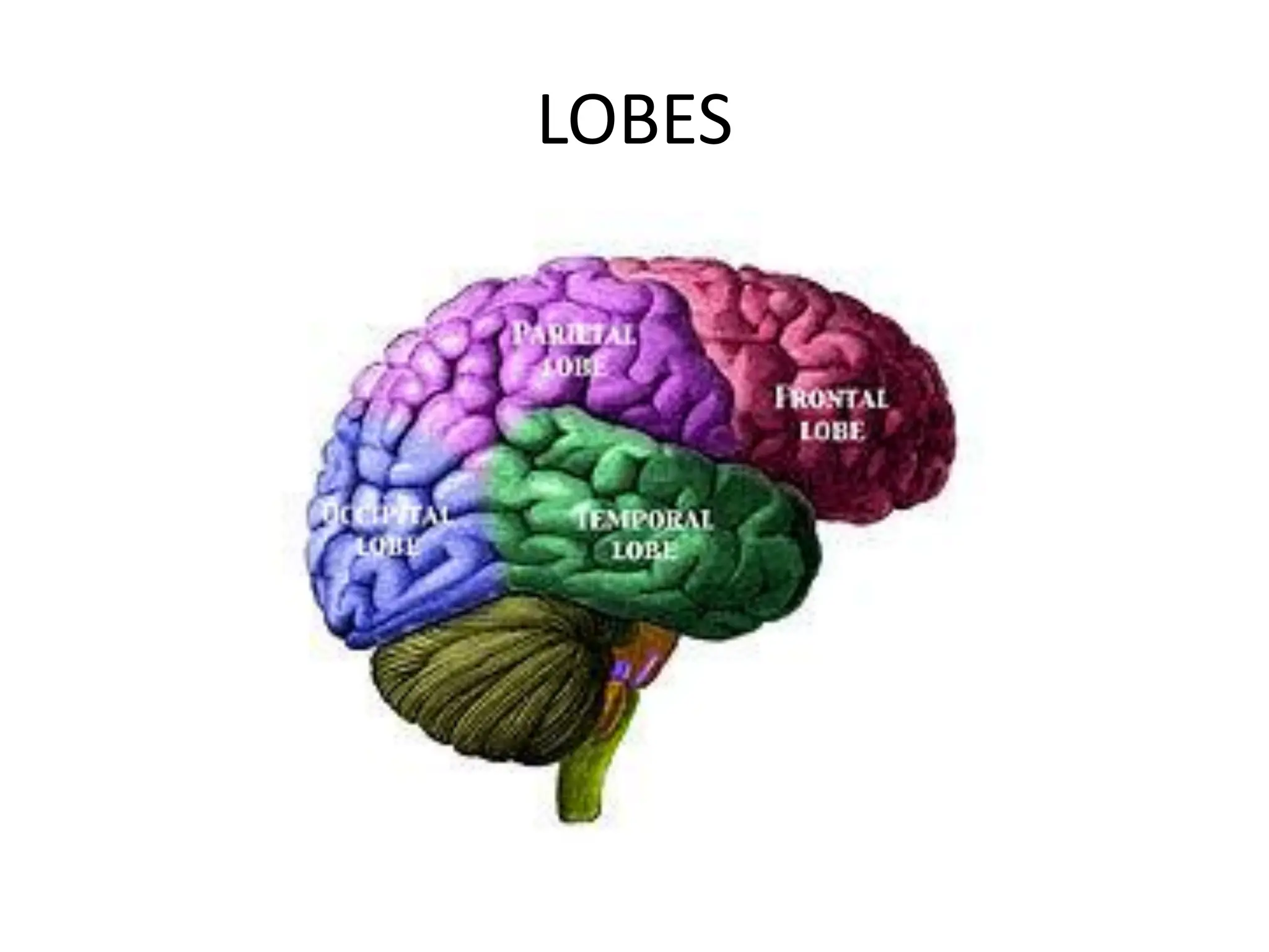
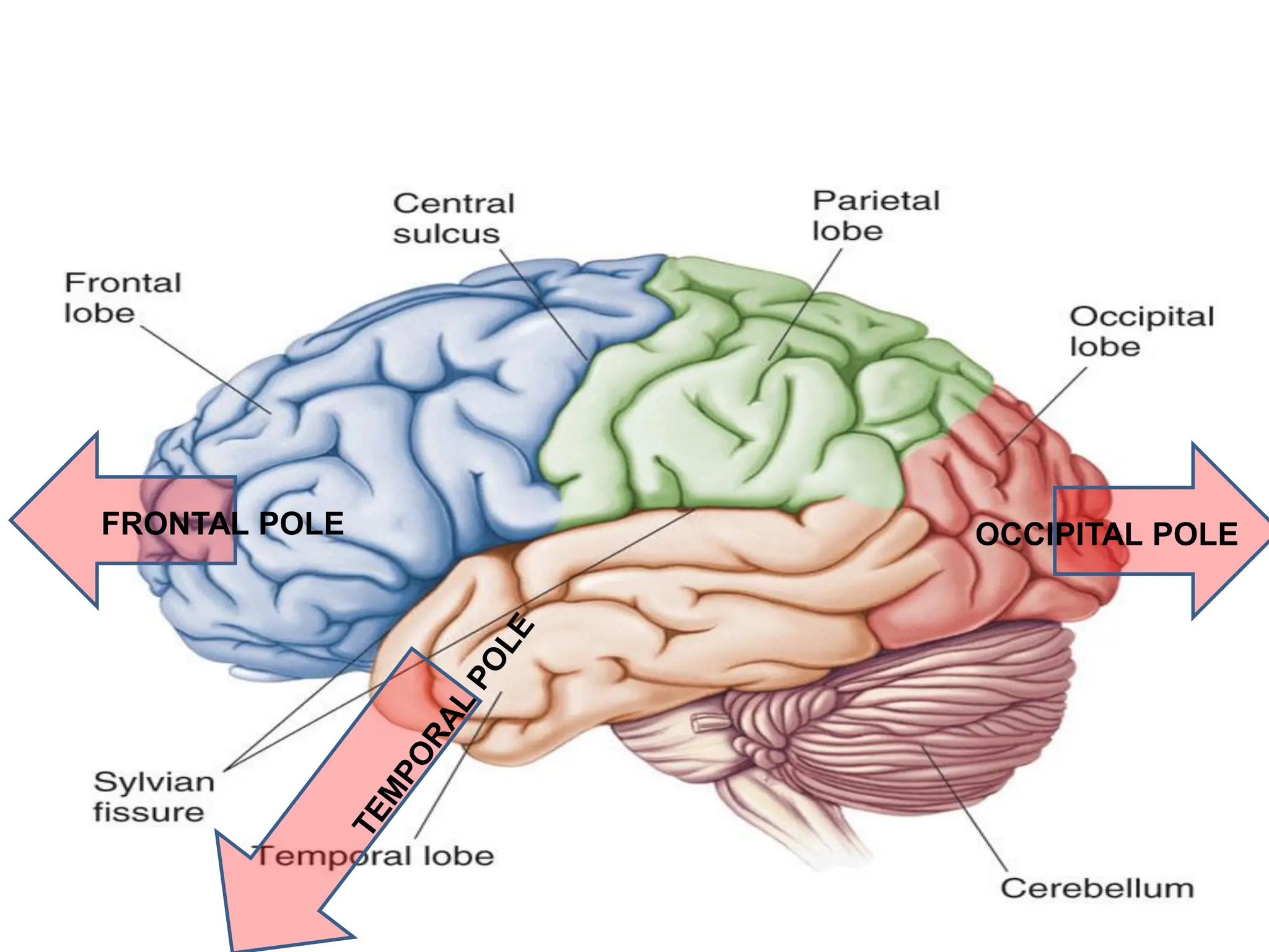
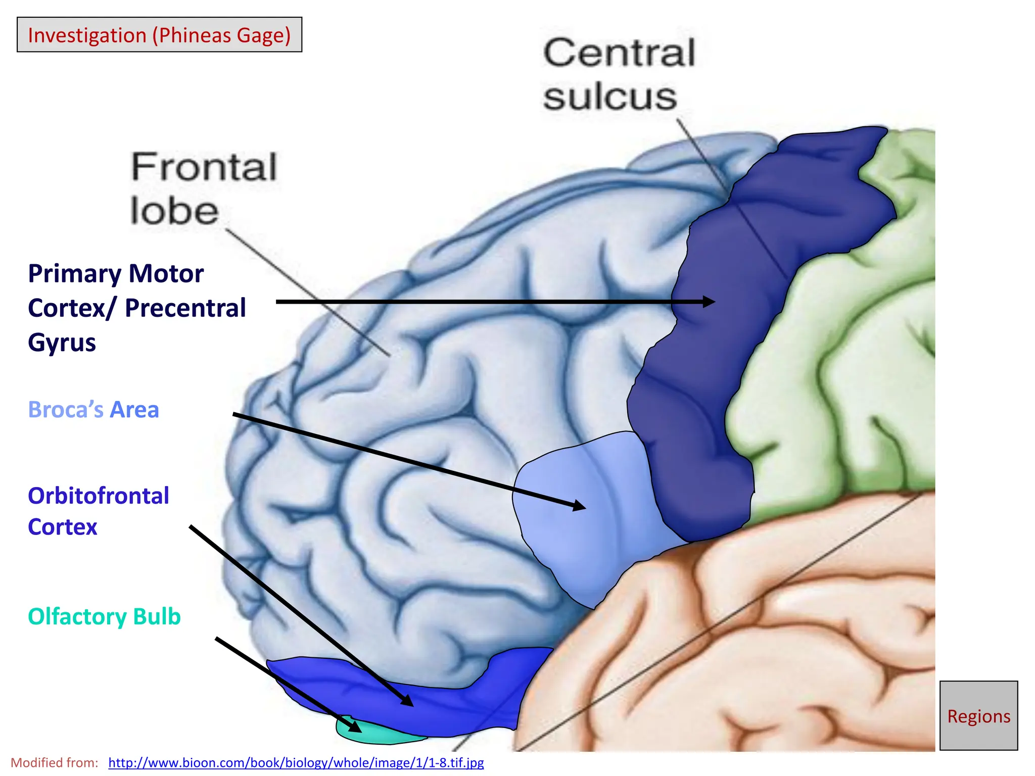
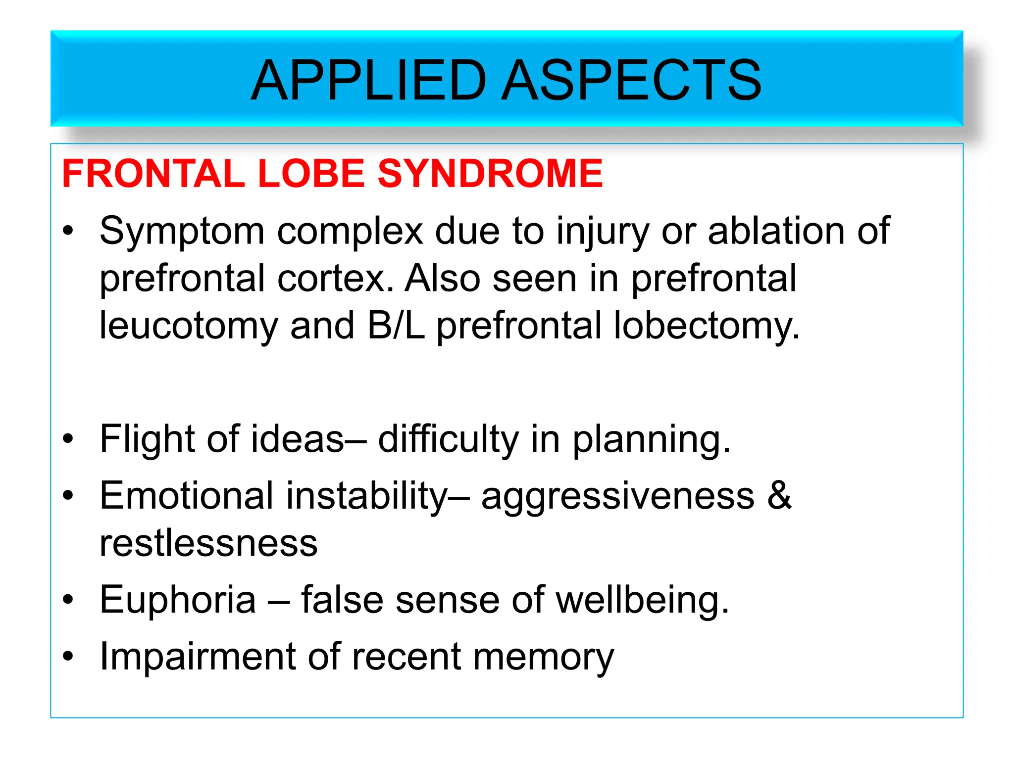
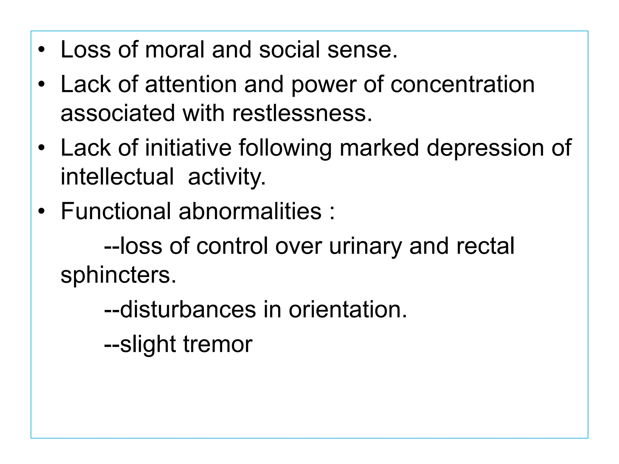
The document provides an overview of the cerebral cortex, detailing its anatomy, functional divisions, and specific lobes including the frontal, parietal, temporal, and occipital lobes. It discusses the roles of different areas, such as the primary motor area, Broca's area, and sensory regions, along with the consequences of frontal lobe damage. Additionally, it mentions Brodmann's areas and how brain functions are mapped across these regions.
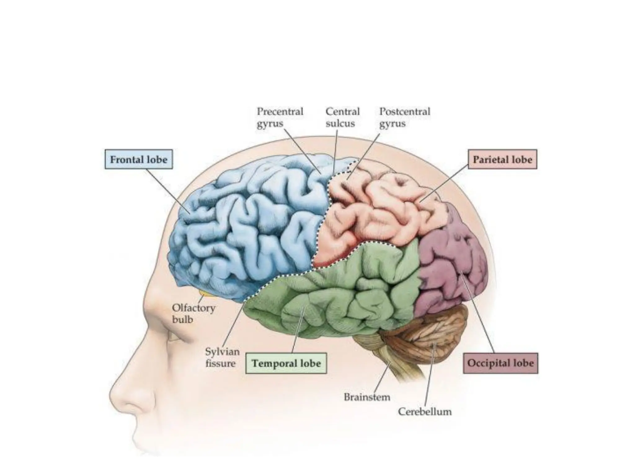
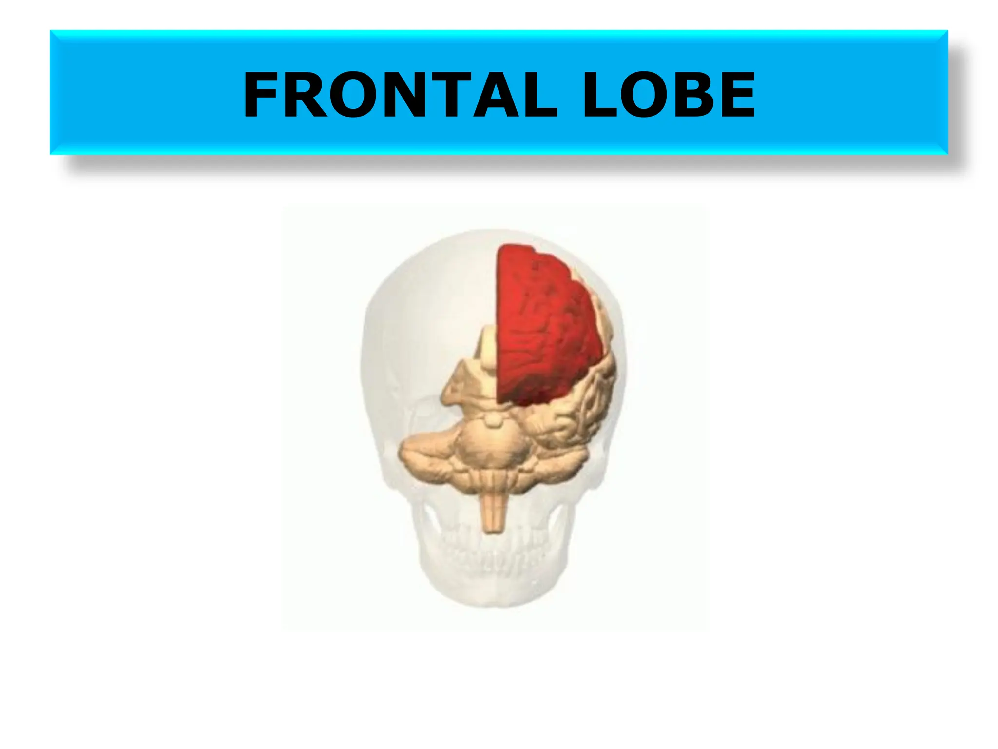
![FRONTAL LOBE
Subdivided into 2 according to function
• Precentral cortex
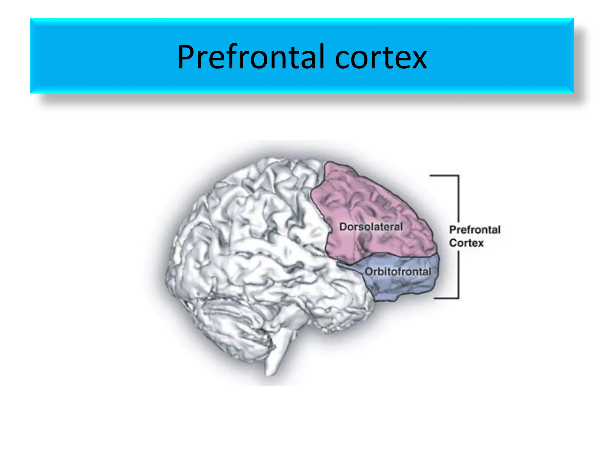
• Prefrontal cortex
Areas present in precentral cortex are
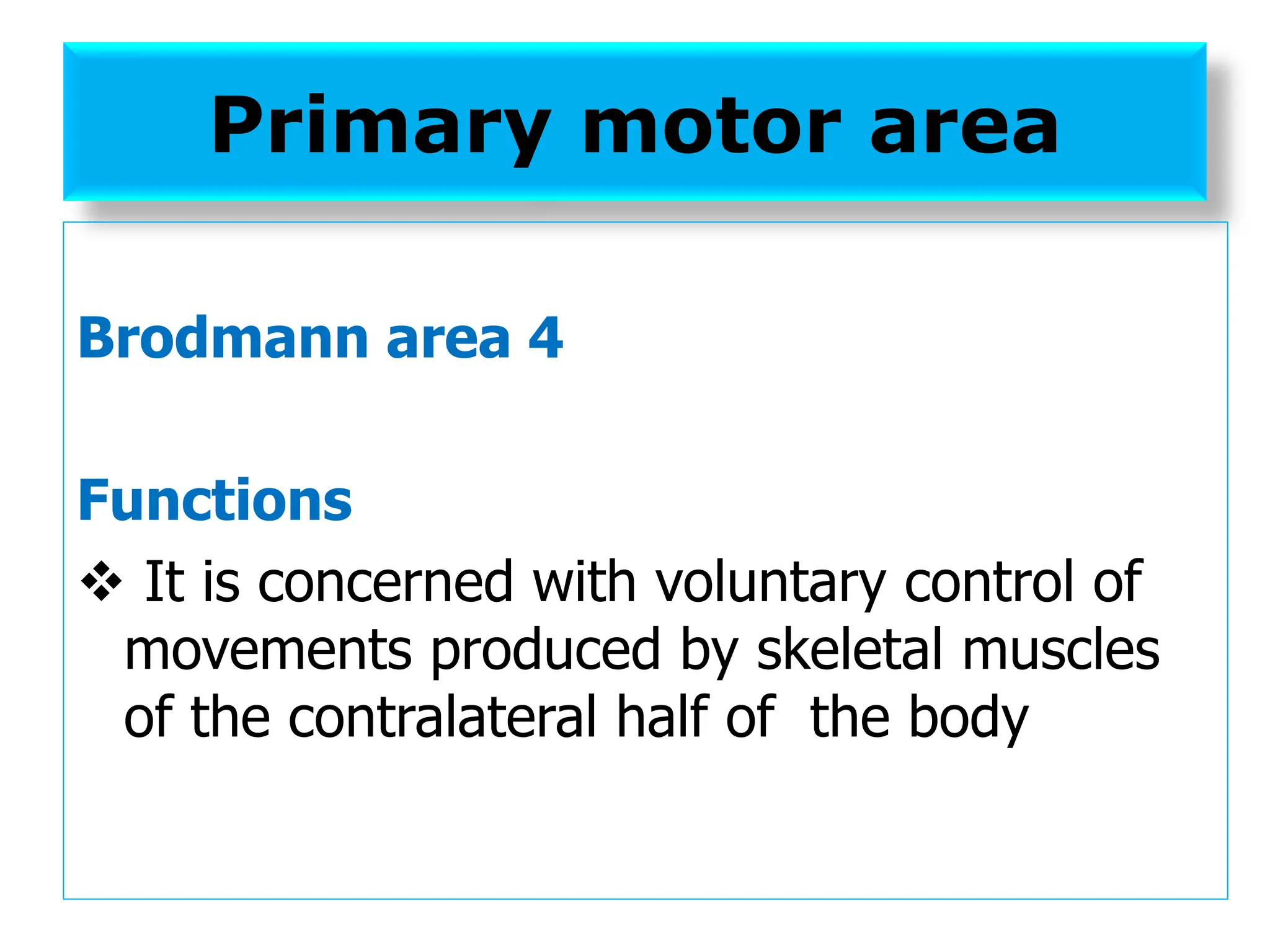
❖ Primary motor area [area 4]
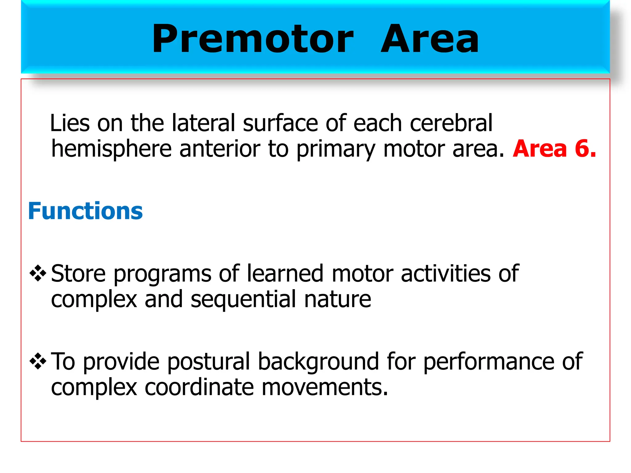
❖ Premotor area [area 6]
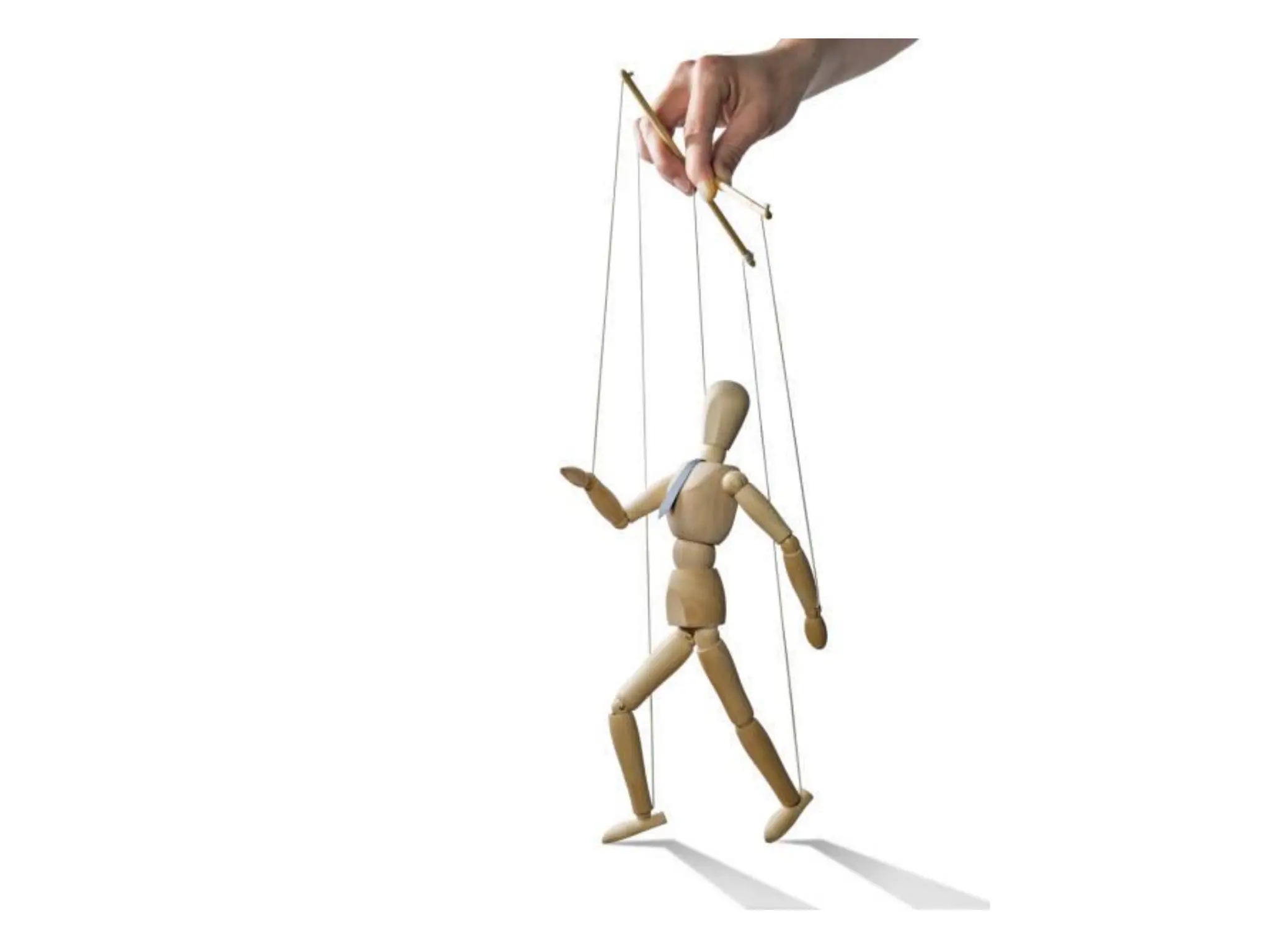
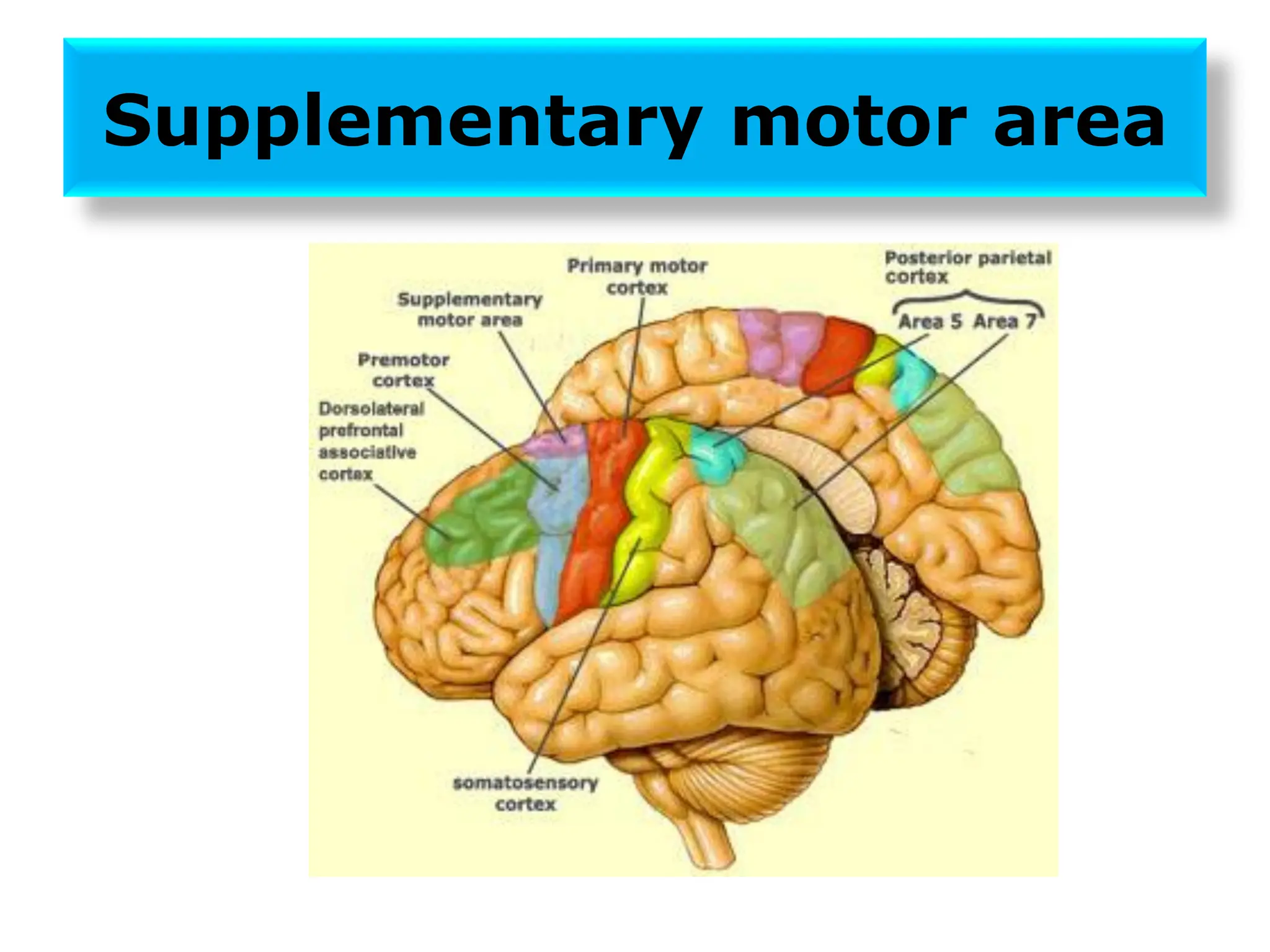
❖ Supplementary motor area
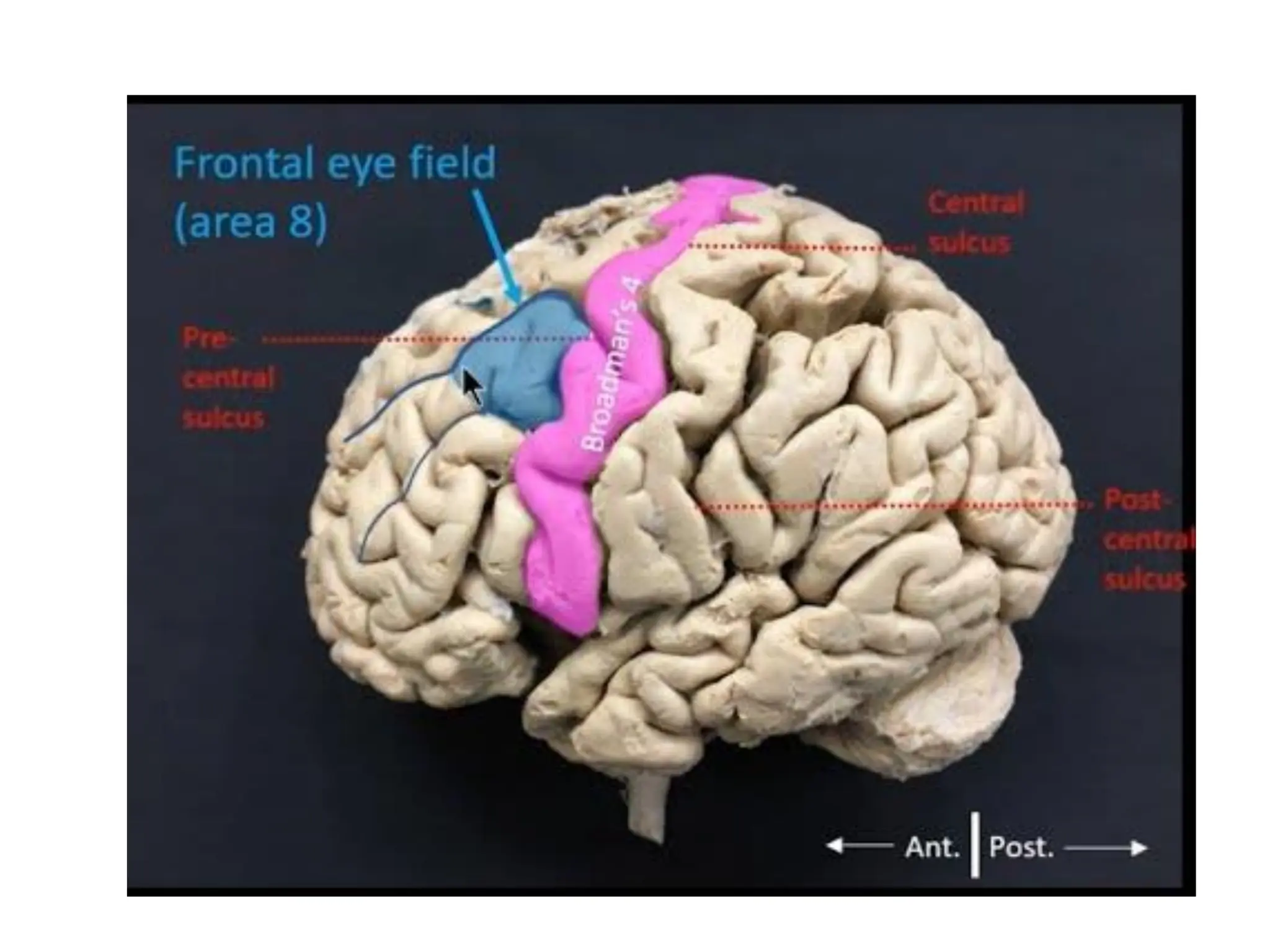
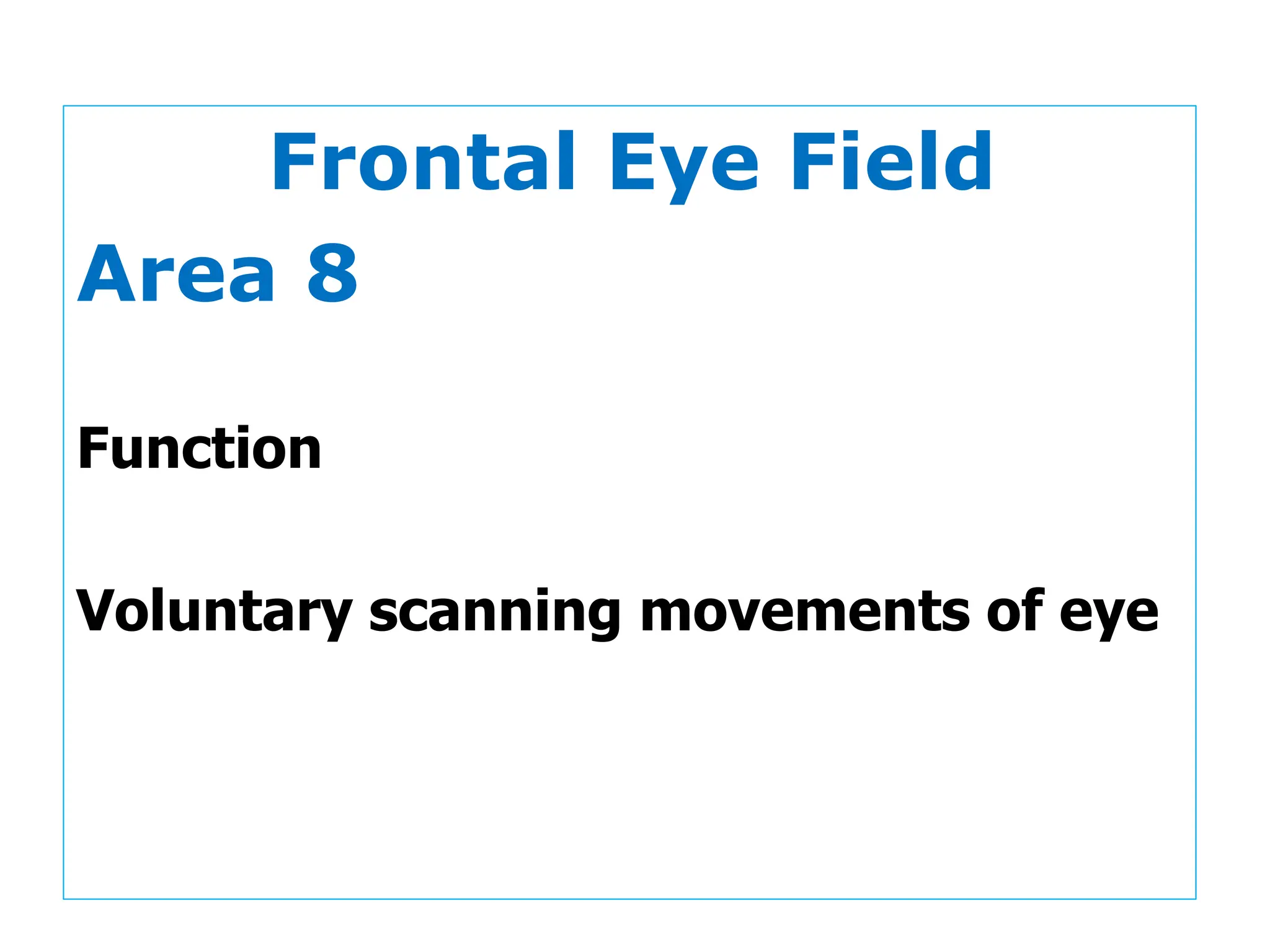
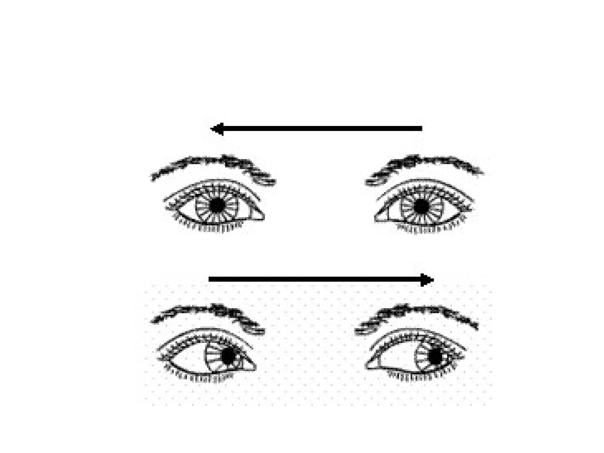
❖ Frontal eye field [area 8]
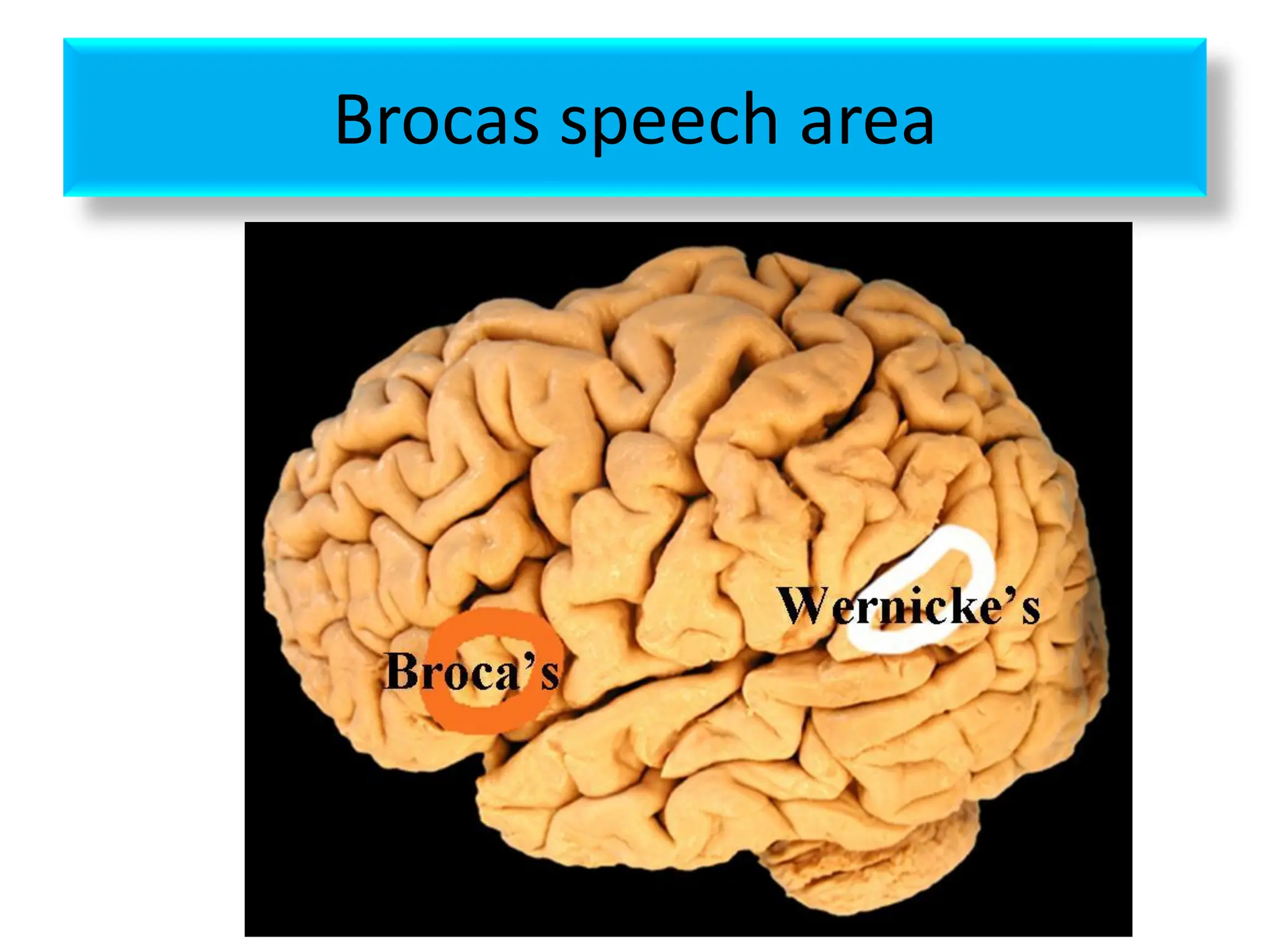
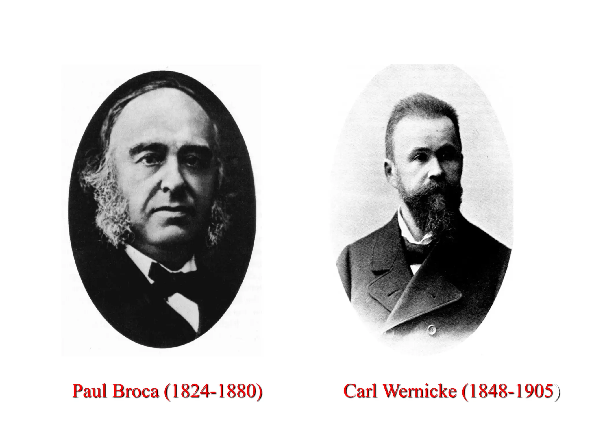
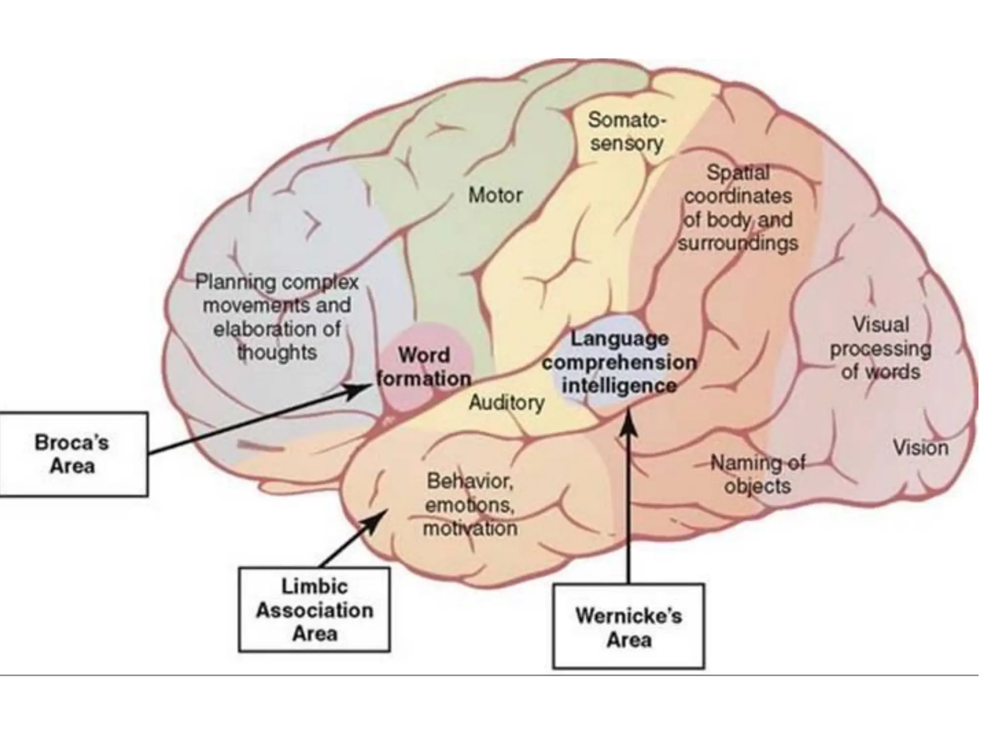
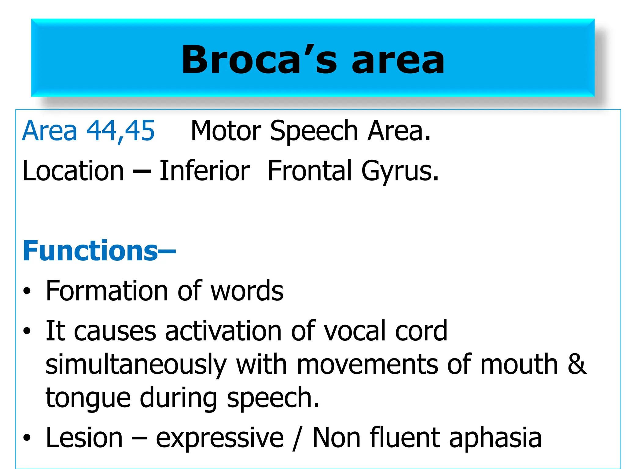
❖ Broca’s area (44,45)](https://image.slidesharecdn.com/cerebralcortex-241124075158-8ef9e2d9/75/CEREBRAL-CORTEX-FOR-BRAIN-THE-ORGAN-OF-pdf-23-2048.jpg)
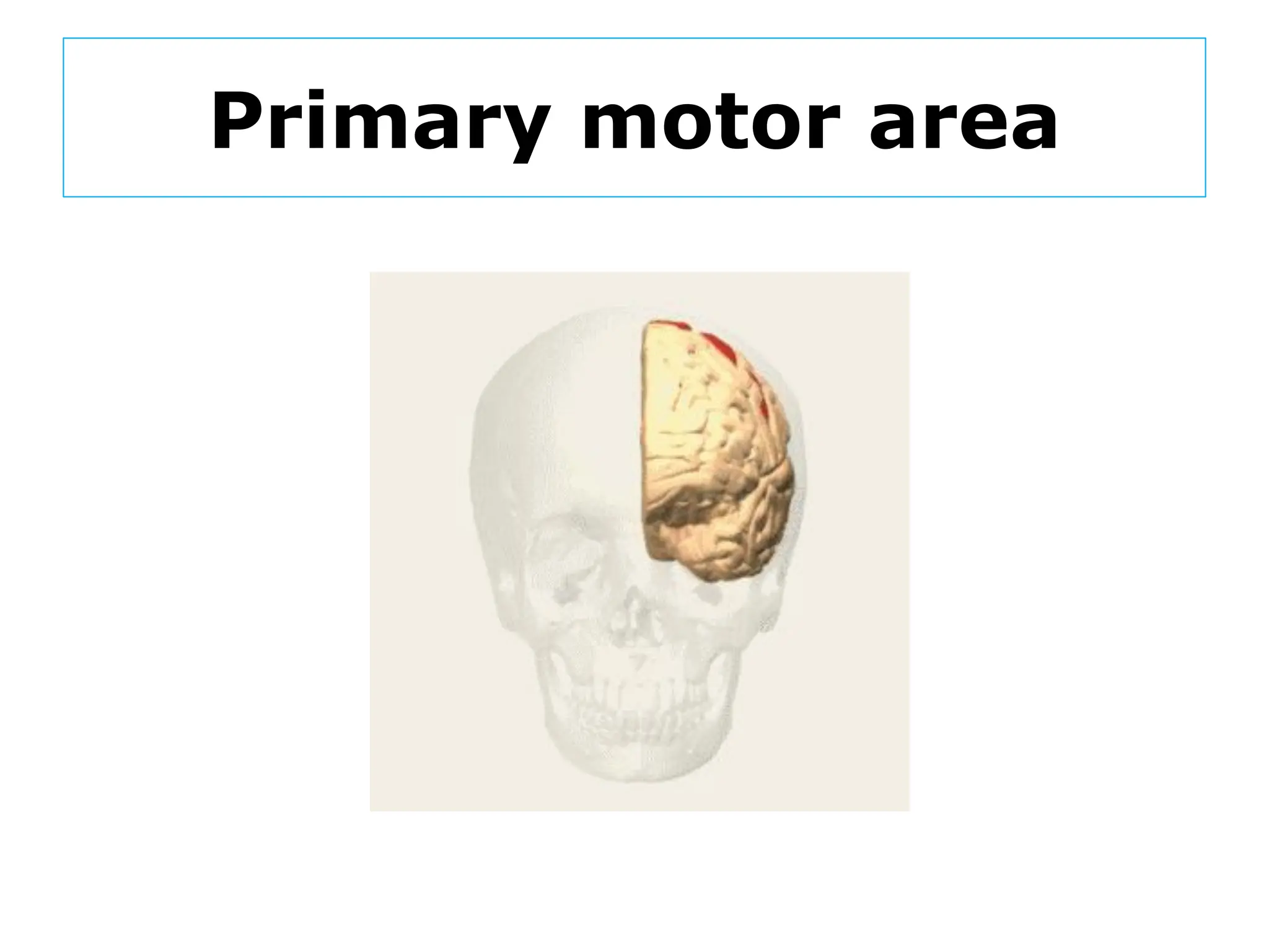
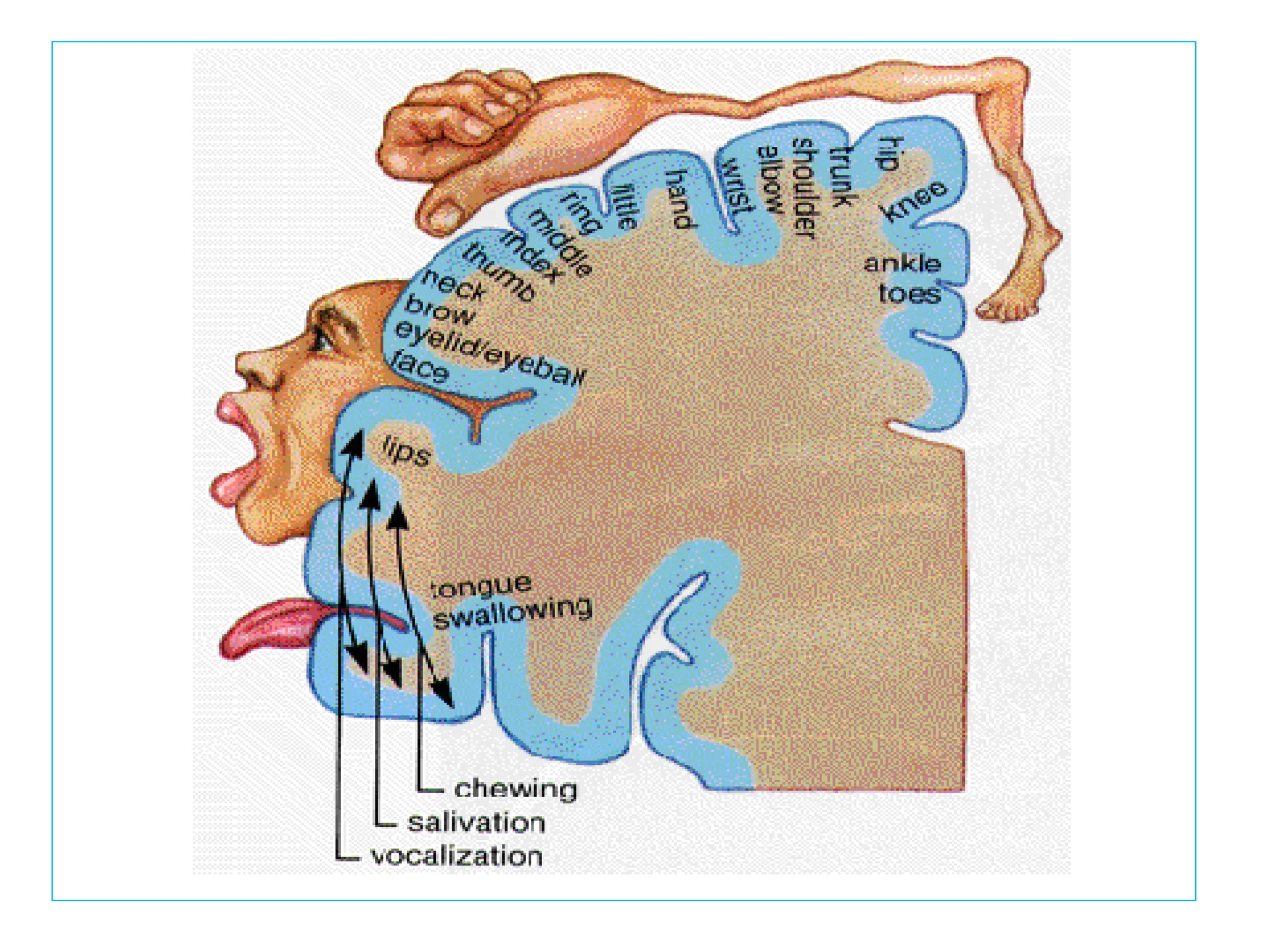
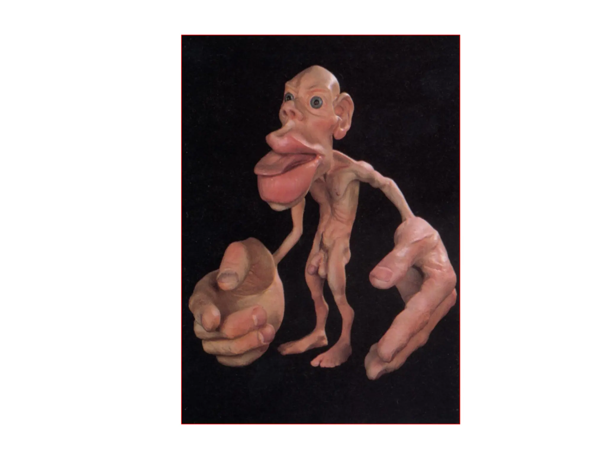
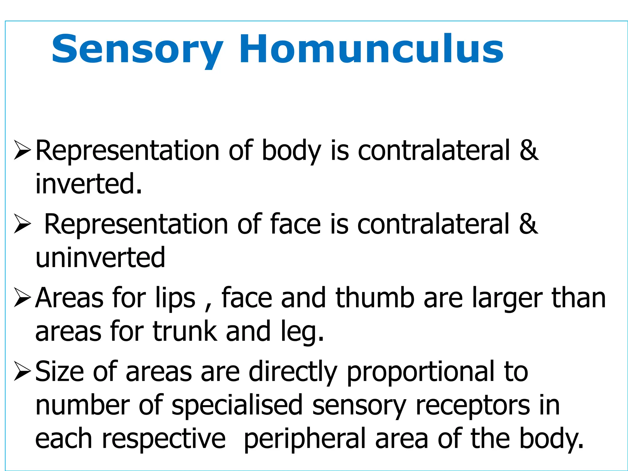
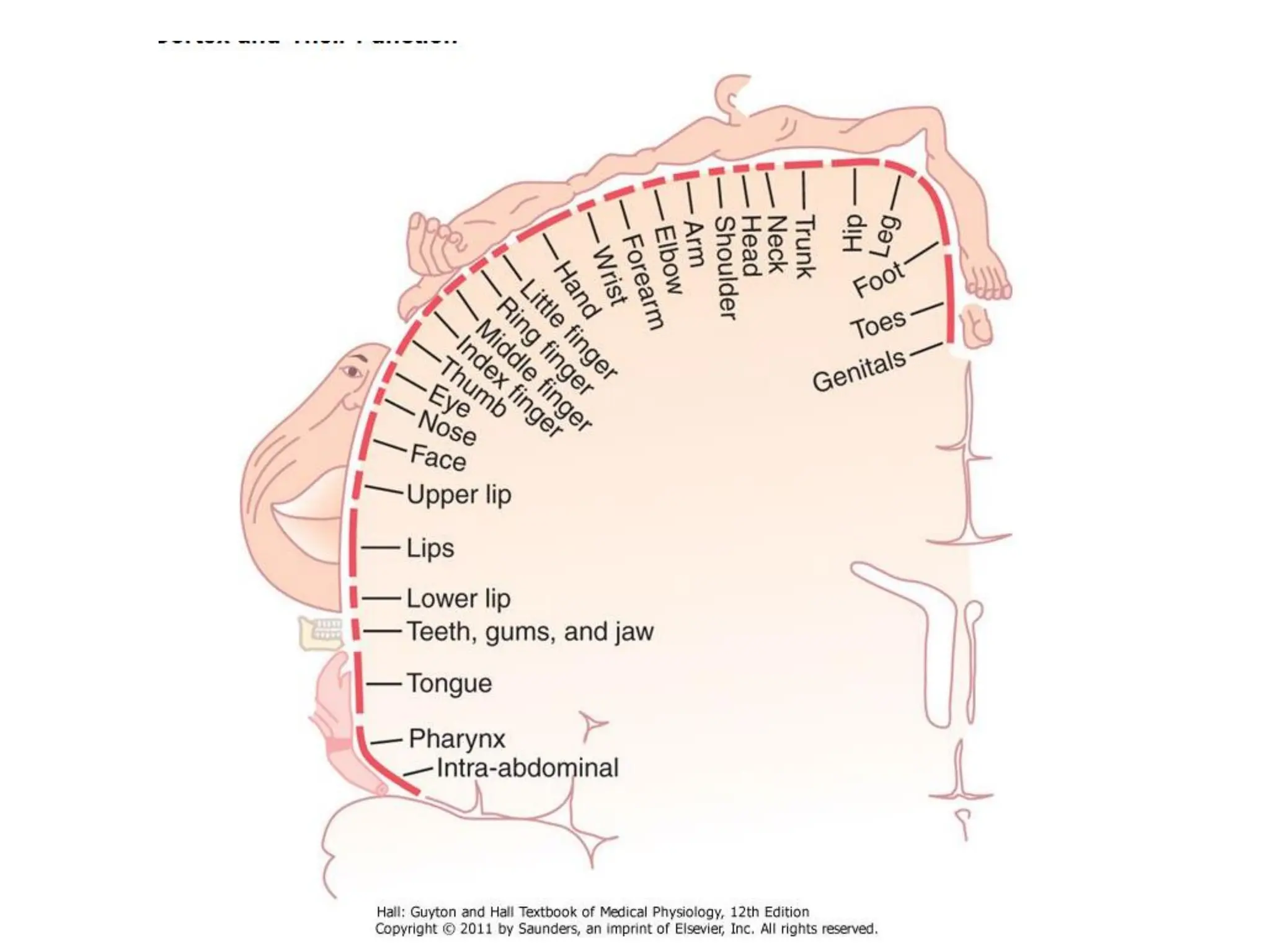
![❑Topographic representation [ Motor
Homunculus]
❑ Different parts of contralateral half of
body are represented separately in more
or less inverted orders except for the face.](https://image.slidesharecdn.com/cerebralcortex-241124075158-8ef9e2d9/75/CEREBRAL-CORTEX-FOR-BRAIN-THE-ORGAN-OF-pdf-27-2048.jpg)
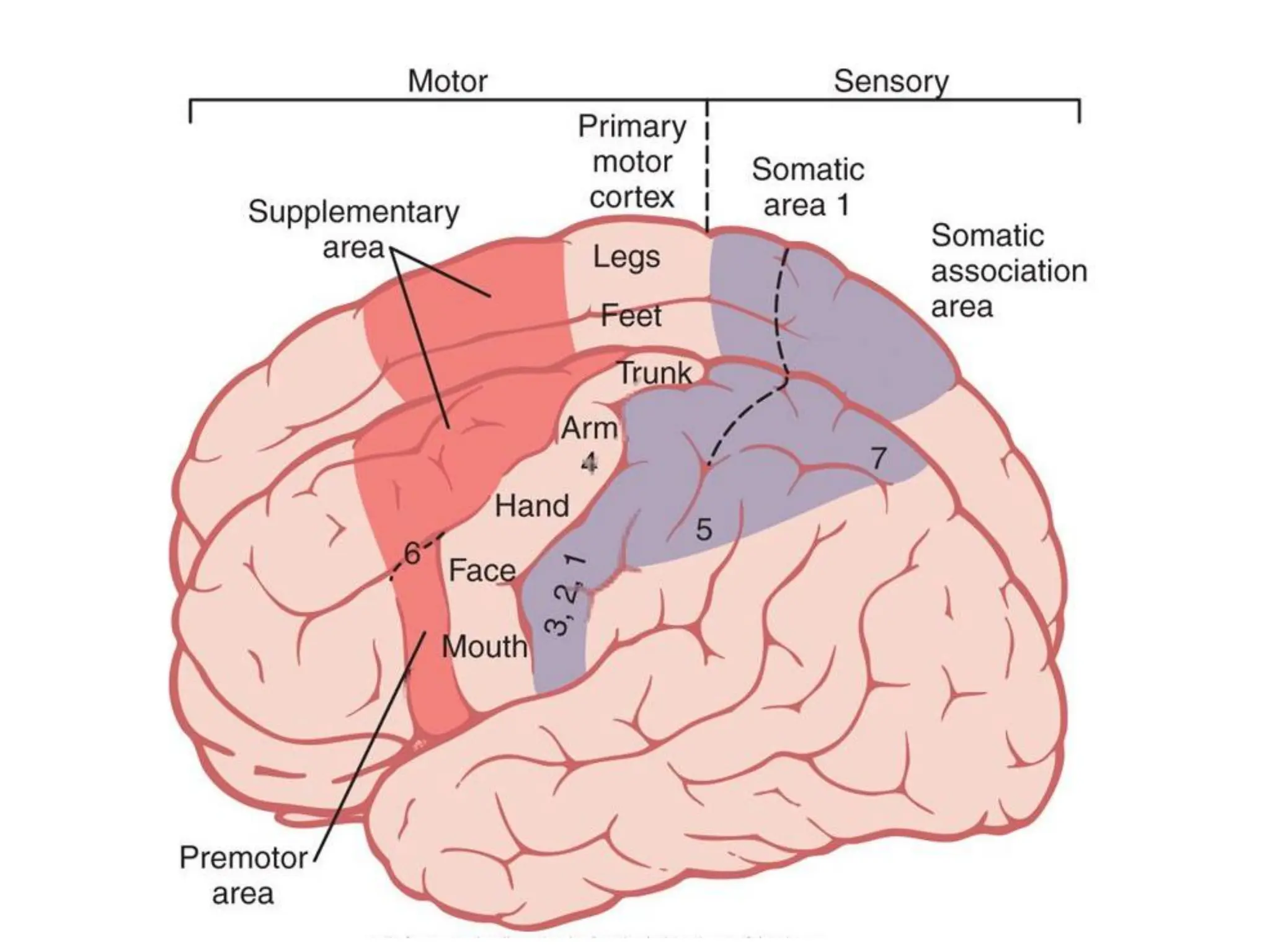

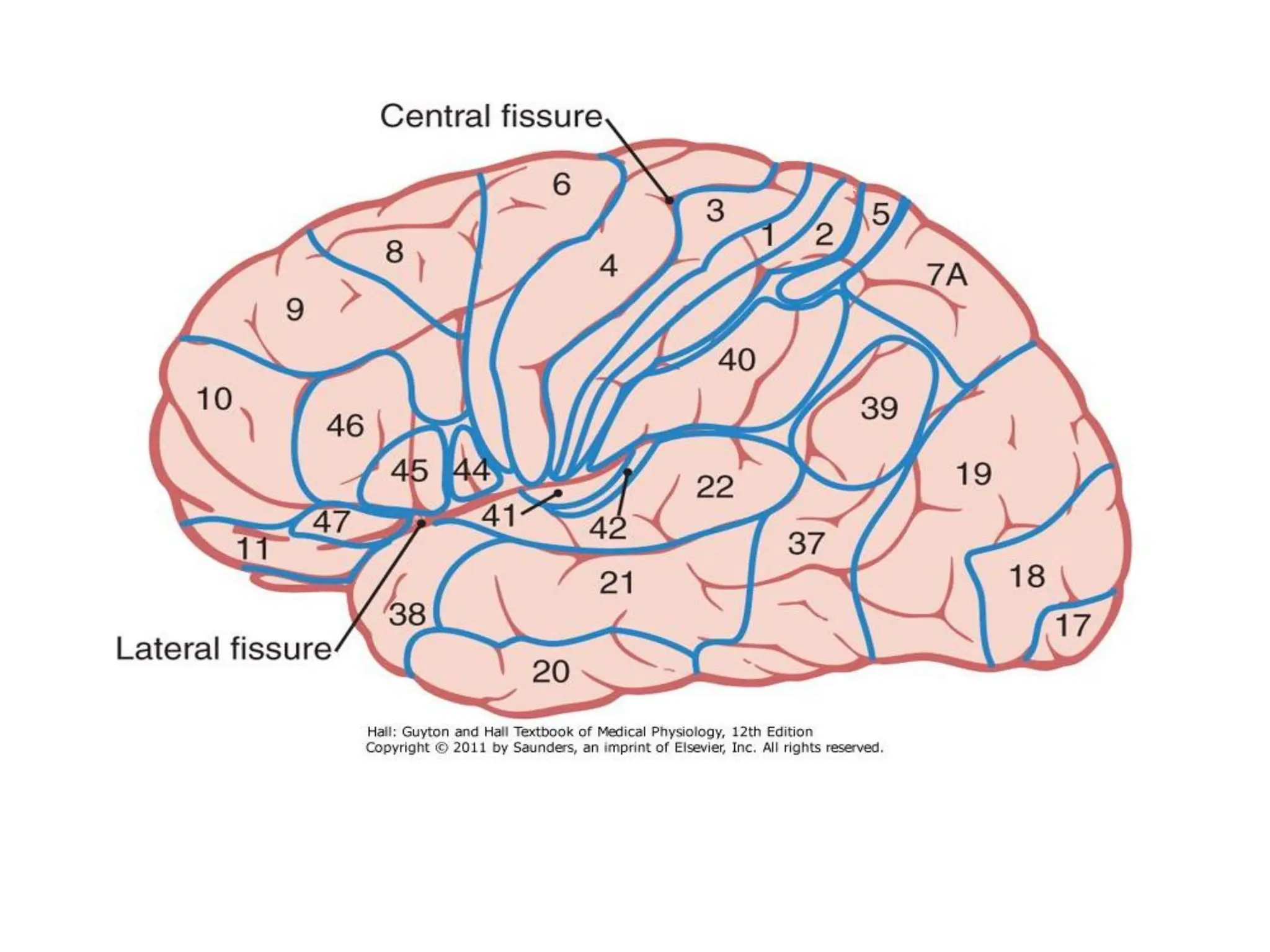
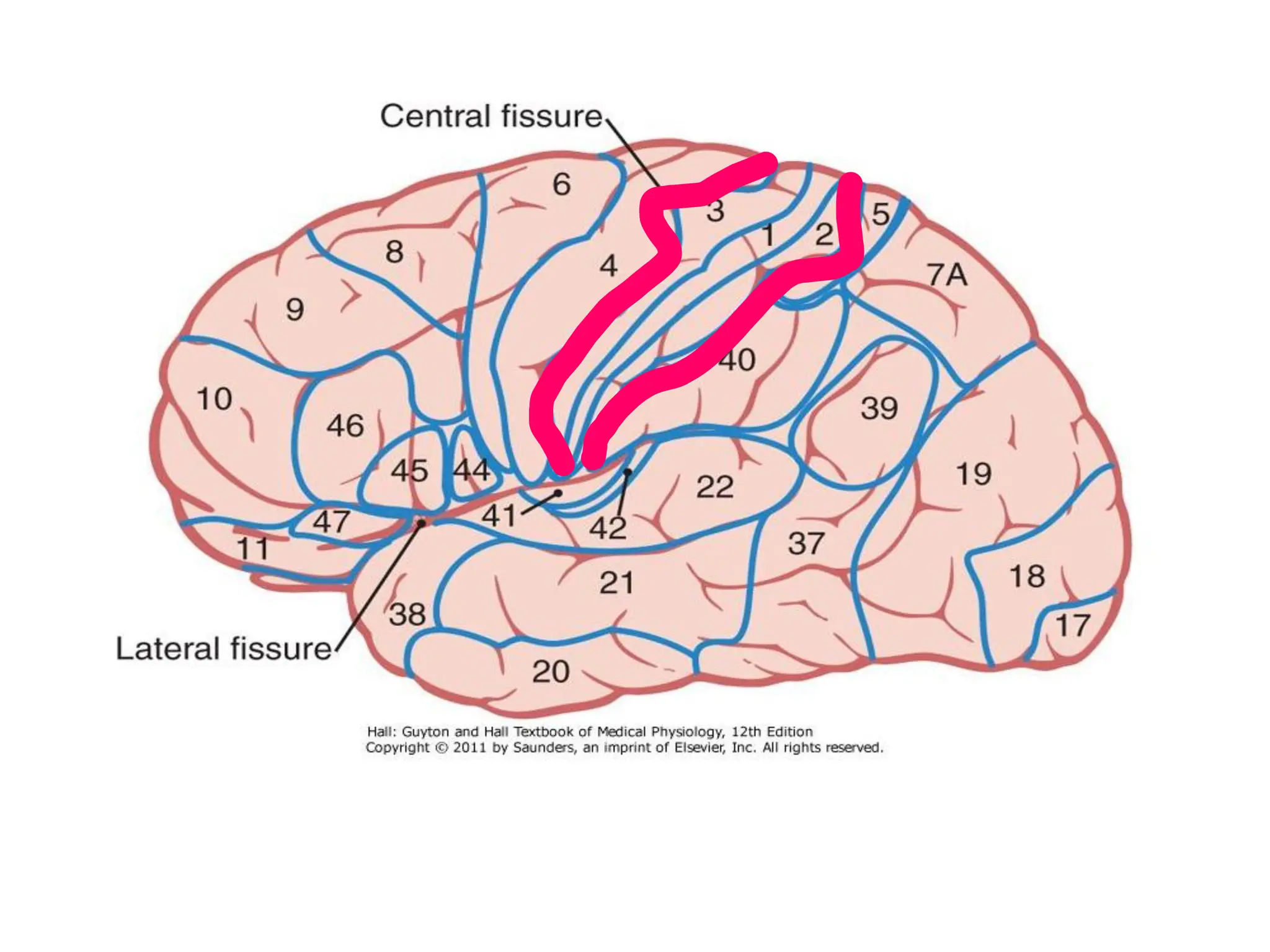
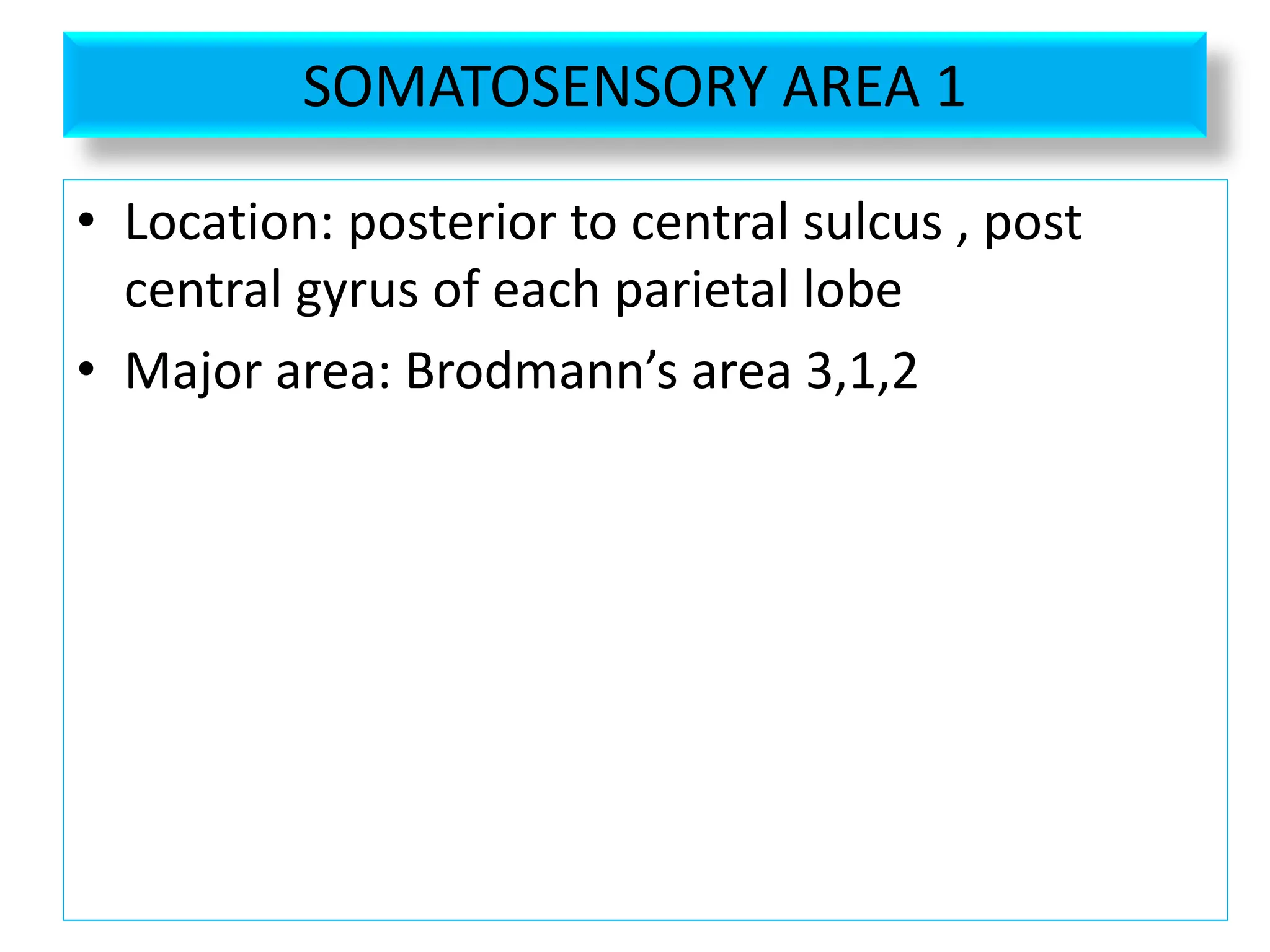
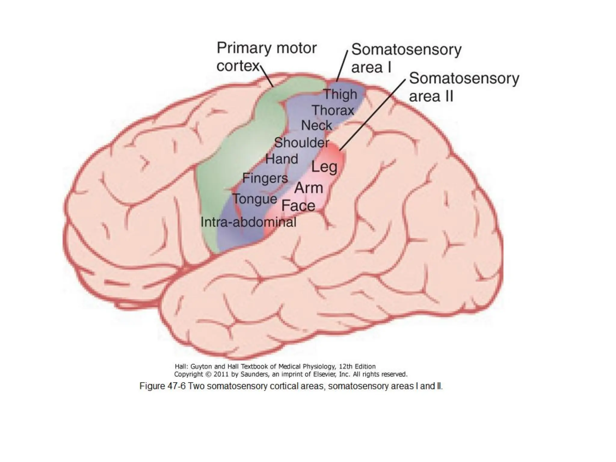
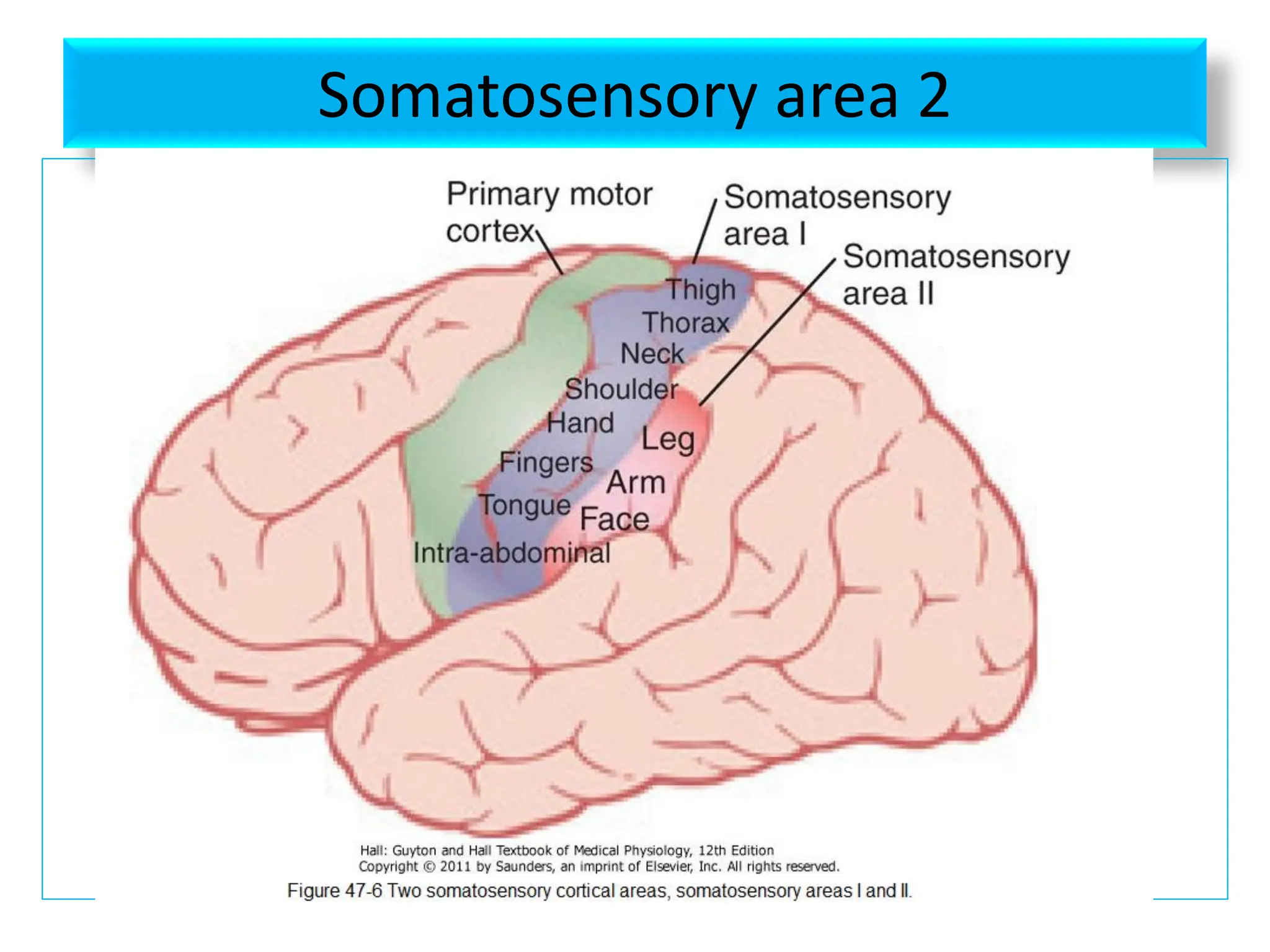
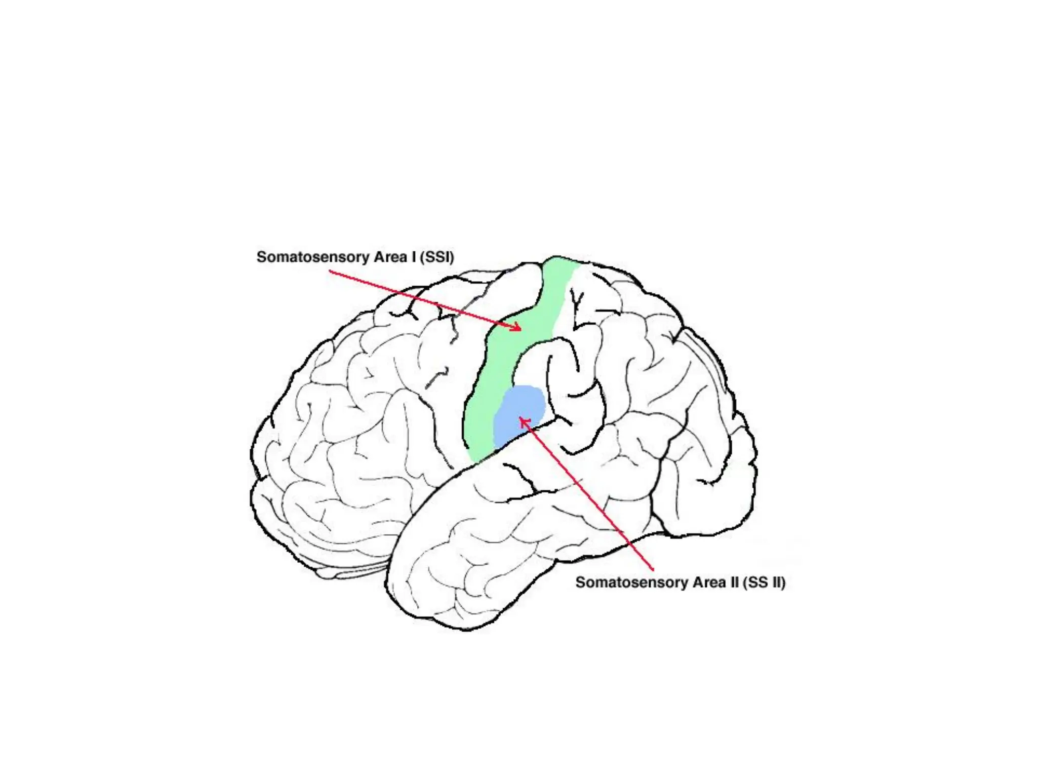
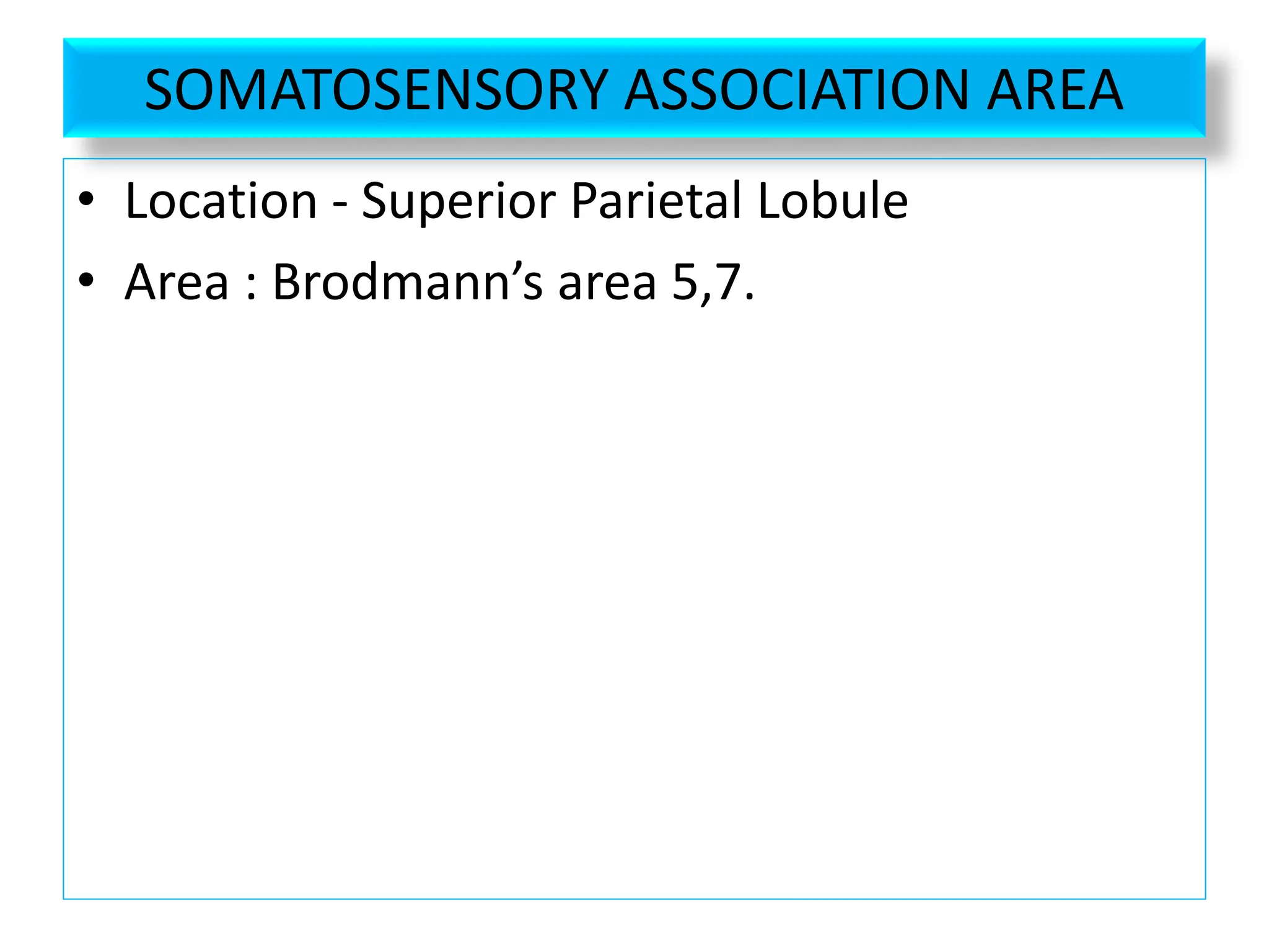
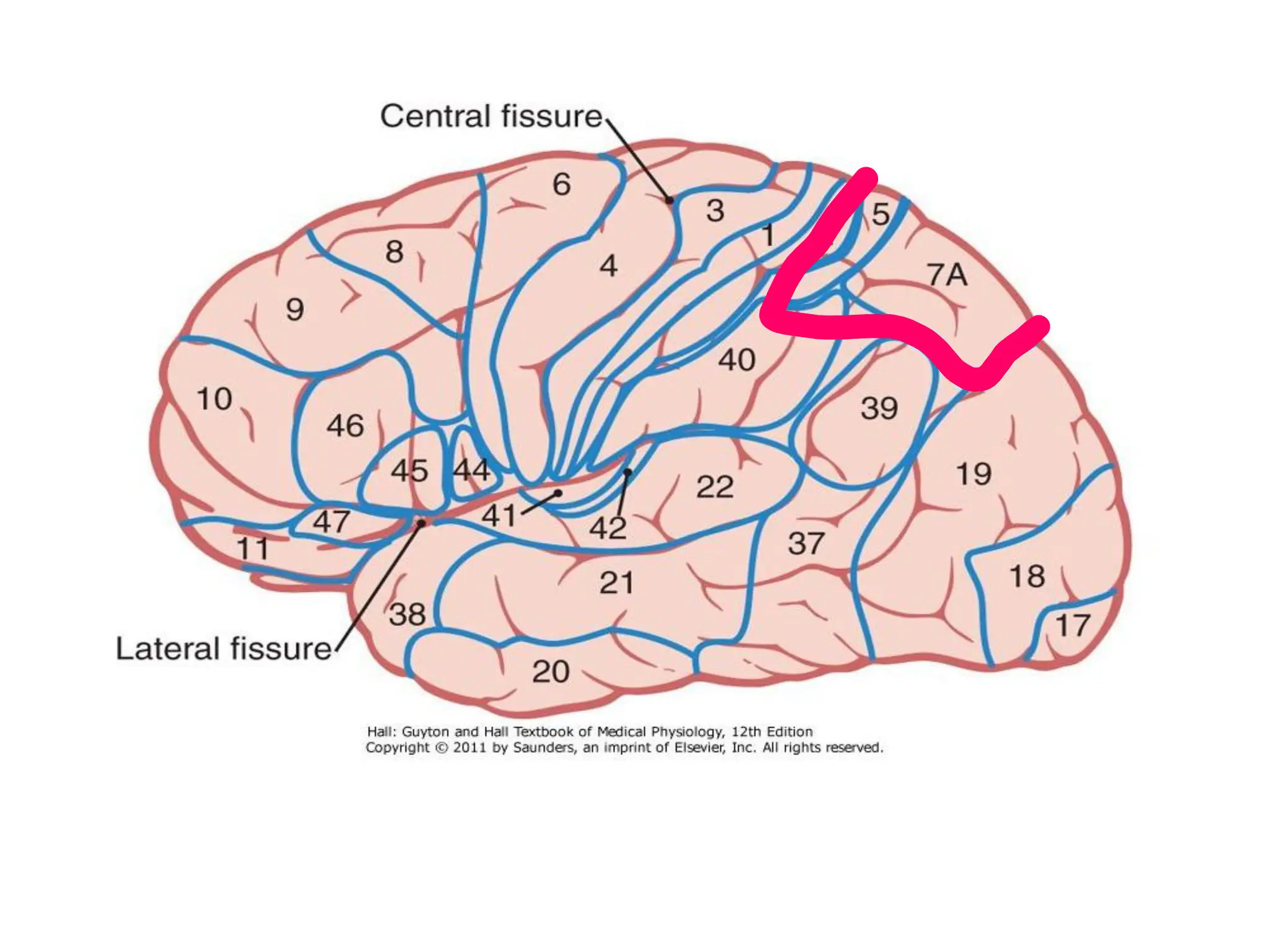
![SENSORY AREAS ARE
• Somatosensory area I [area 3,1,2]
• Somatosensory area II
• Sensory association area[area 5,7 and higher
association area 40]](https://image.slidesharecdn.com/cerebralcortex-241124075158-8ef9e2d9/75/CEREBRAL-CORTEX-FOR-BRAIN-THE-ORGAN-OF-pdf-51-2048.jpg)


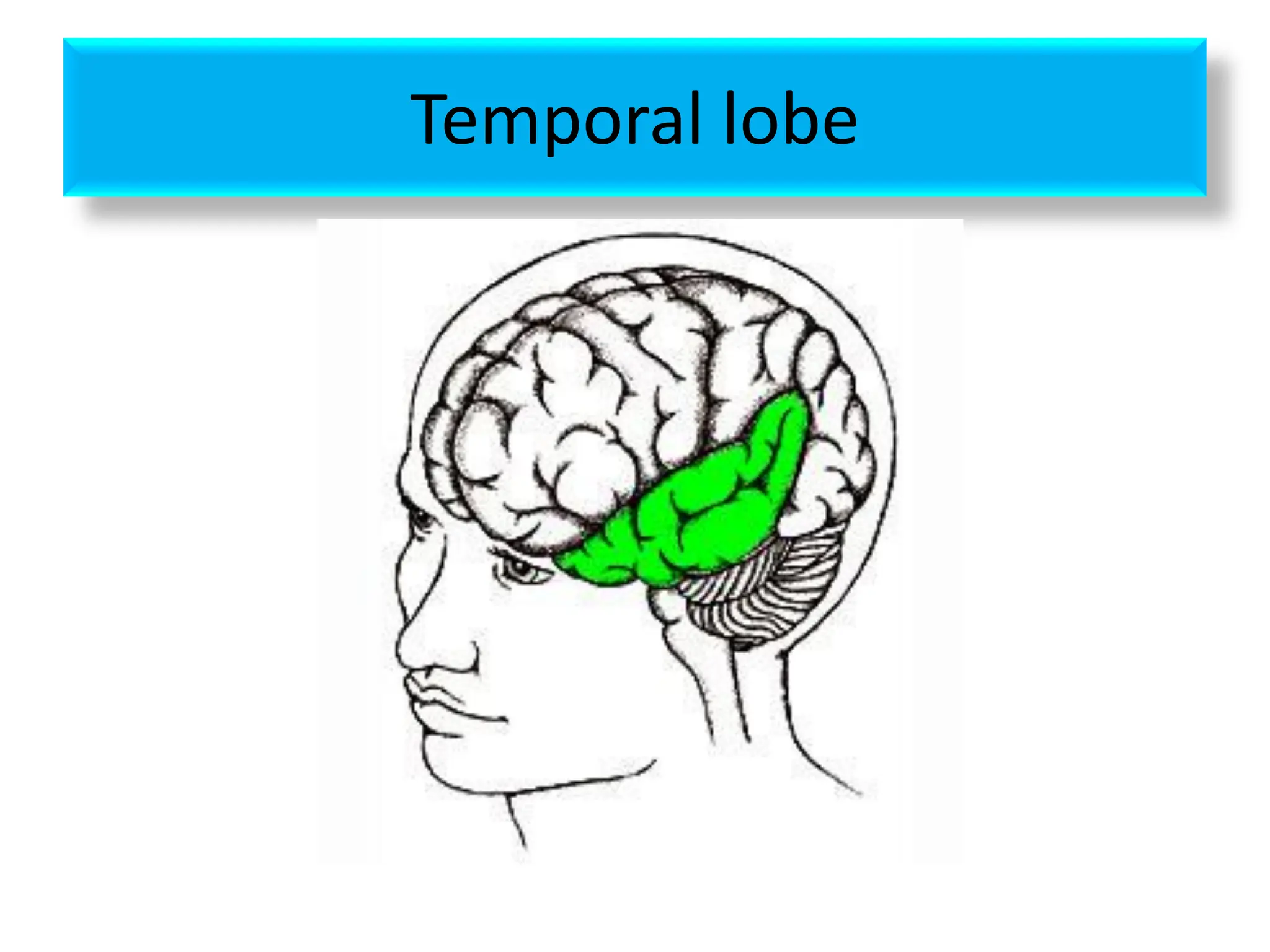
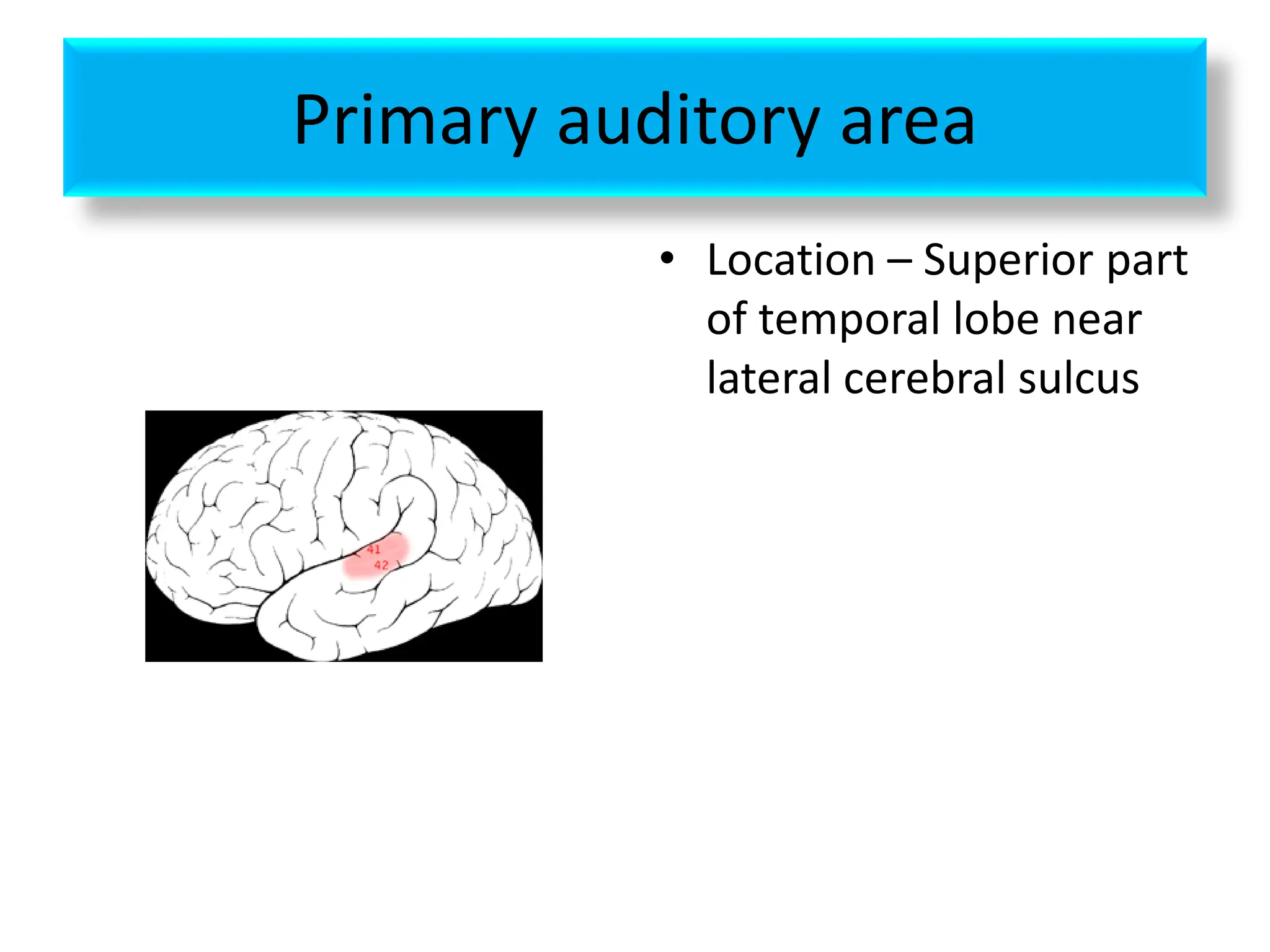
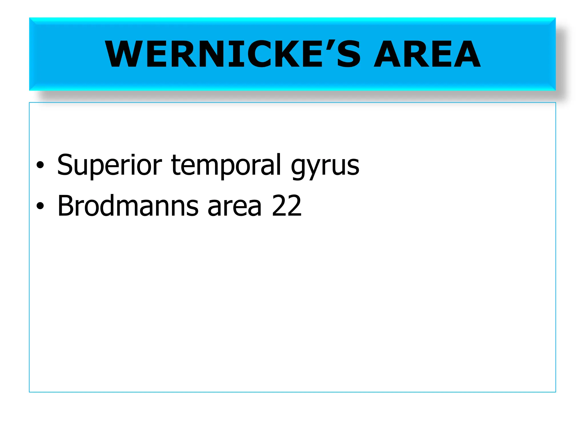
![TEMPORAL LOBE
Location – area inferior to lateral sulcus
AREAS
• Primary auditory area [area 41,42]
• Auditory association area [area 22]](https://image.slidesharecdn.com/cerebralcortex-241124075158-8ef9e2d9/75/CEREBRAL-CORTEX-FOR-BRAIN-THE-ORGAN-OF-pdf-65-2048.jpg)