Defination
• A lumbarpuncture (also called a spinal tap) is the removal of spinal
fluid (called the cerebrospinal fluid, or CSF) from the spinal canal; the
fluid is withdrawn through a needle and examined in a laboratory
3.
Indications
• To obtainC S F for analysis & diagnosis of:
• Meningitis
• Meningoencephalitis
• Subarachnoid hemorrhage
• Malignancy – diagnosis and treatment
• Pseudotumor Cerebri
• Other neurologic syndromes
• To drain C S F & reduce intracranial space
• To instill medications for spinal anaesthesia
4.
Spinal anesthesia -Itis the regional anesthesia obtained by temporary
interruption of nerve transmission of spinal nerves by injecting
anesthetic agents in to the subarachnoid space.
5.
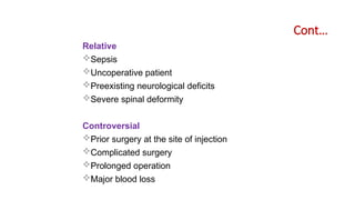
Contraindications of Spinal
ABSOLUTE
Infectionat the site of injection
Patient refusal
Coagulopathy and other bleeding disorders
Severe hypovolemia
Increased intracranial pressure
Severe MS & AS
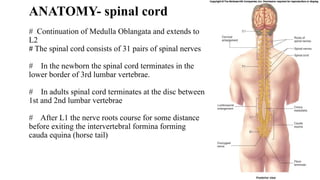
ANATOMY- spinal cord
#Continuation of Medulla Oblangata and extends to
L2
# The spinal cord consists of 31 pairs of spinal nerves
# In the newborn the spinal cord terminates in the
lower border of 3rd lumbar vertebrae.
# In adults spinal cord terminates at the disc between
1st and 2nd lumbar vertebrae
# After L1 the nerve roots course for some distance
before exiting the intervertebral formina forming
cauda equina (horse tail)
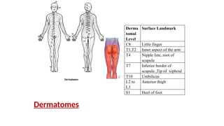
Derma
tomal
Level
Surface Landmark
C8 Littlefinger
T1,T2 Inner aspect of the arm
T4 Nipple line, root of
scapula
T7 Inferior border of
scapula ,Tip of xiphoid
T10 Umbilicus
L2 to
L3
Anterior thigh
S1 Heel of foot
Dermatomes
10.
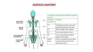
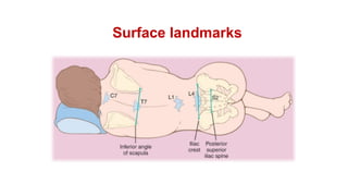
SURFACE ANATOMY
Anatomic Landmarksto Identify Vertebral
Levels
Anatomic
Landmark
Features
C7 Vertebral prominence, the most
prominent process in the neck
T7 Inferior angle of the scapula
L4 Line connecting iliac crests
S2 Line connecting the posterior
superior iliac spines
Sacral
hiatus
Groove or depression just above
or between the gluteal clefts
above the coccyx
SITE
Adult :L3-L4 or L4-L5 ( or even
L2-L3)
Infant : L4-L5
A line drawn b/w the highest pt. of
iliac crests (Tuffier’s line) usually
cross either body of L4 or the L4-
L5 interspace
Position
Sitting
lateral
Prone(anorectal procedure,
hypobaric solution, jackknife position)
15.
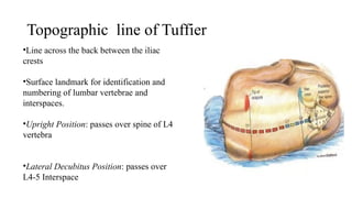
Topographic line ofTuffier
•Line across the back between the iliac
crests
•Surface landmark for identification and
numbering of lumbar vertebrae and
interspaces.
•Upright Position: passes over spine of L4
vertebra
•Lateral Decubitus Position: passes over
L4-5 Interspace
16.
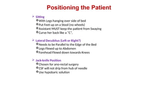
Positioning the Patient
Sitting
With Legs hanging over side of bed
Put Feet up on a Stool (no wheels)
Assistant MUST keep the patient from Swaying
Curve her back like a “C”,
Lateral Decubitus (Left or Right?)
Needs to be Parallel to the Edge of the Bed
Legs Flexed up to Abdomen
Forehead Flexed down towards Knees
Jack-knife Position
Chosen for ano-rectal surgery
CSF will not drip from hub of needle
Use hypobaric solution
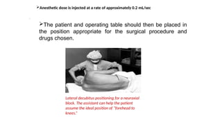
.
The patient andoperating table should then be placed in
the position appropriate for the surgical procedure and
drugs chosen.
Lateral decubitus positioning for a neuraxial
block. The assistant can help the patient
assume the ideal position of “forehead to
knees.”
Anesthetic dose is injected at a rate of approximately 0.2 mL/sec
20.
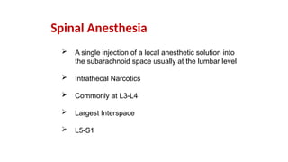
Spinal Anesthesia
Asingle injection of a local anesthetic solution into
the subarachnoid space usually at the lumbar level
Intrathecal Narcotics
Commonly at L3-L4
Largest Interspace
L5-S1
21.
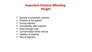
Important Factors Affecting
Height
Baricity of anesthetic solution
Position of the patient
During injection
Immediately after injection
Drug Dosage (mg)
Concentration times volume
Addition of Opioids
Site of Injection
22.
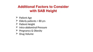
Additional Factors toConsider
with SAB Height
Patient Age
Elderly patients > 80 yrs
Patient Height
Intra-abdominal Pressure
Pregnancy & Obesity
Drug Volume
23.
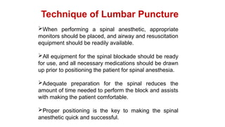
When performing aspinal anesthetic, appropriate
monitors should be placed, and airway and resuscitation
equipment should be readily available.
All equipment for the spinal blockade should be ready
for use, and all necessary medications should be drawn
up prior to positioning the patient for spinal anesthesia.
Adequate preparation for the spinal reduces the
amount of time needed to perform the block and assists
with making the patient comfortable.
Proper positioning is the key to making the spinal
anesthetic quick and successful.
Technique of Lumbar Puncture
24.
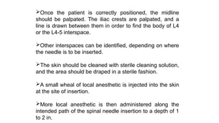
Once the patientis correctly positioned, the midline
should be palpated. The iliac crests are palpated, and a
line is drawn between them in order to find the body of L4
or the L4-5 interspace.
Other interspaces can be identified, depending on where
the needle is to be inserted.
The skin should be cleaned with sterile cleaning solution,
and the area should be draped in a sterile fashion.
A small wheal of local anesthetic is injected into the skin
at the site of insertion.
More local anesthetic is then administered along the
intended path of the spinal needle insertion to a depth of 1
to 2 in.
25.
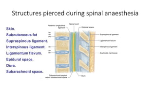
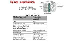
1. MIDLINE APPROACH
2.PARAMEDIAN APPROACH
Midline Approach Paramedian
approach
Skin Skin
Subcutaneous fat Subcutaneous fat
Supraspinous ligament
Interspinous ligament
Ligamentum flavum Ligmentum flavum
Dura mater Dura mater
Subdural space Subdural space
Arachnoid mater Arachnoid mater
Subarachnoid space Subarachnoid space
Spinal : approaches
Structure Pierced
26.
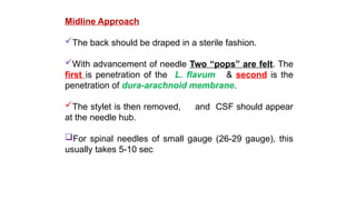
Midline Approach
The backshould be draped in a sterile fashion.
With advancement of needle Two “pops” are felt. The
first is penetration of the L. flavum & second is the
penetration of dura-arachnoid membrane.
The stylet is then removed, and CSF should appear
at the needle hub.
For spinal needles of small gauge (26-29 gauge), this
usually takes 5-10 sec
27.
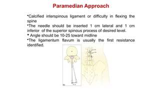
Paramedian Approach
•Calcified interspinousligament or difficulty in flexing the
spine
•The needle should be inserted 1 cm lateral and 1 cm
inferior of the superior spinous process of desired level.
Angle should be 10-25 toward midline
•The ligamentum flavum is usually the first resistance
identified.
28.
Paramedian Approach
•Calcified interspinousligament or difficulty in flexing the
spine
•The needle should be inserted 1 cm lateral and 1 cm
inferior of the superior spinous process of desired level.
Angle should be 10-25 toward midline
•The ligamentum flavum is usually the first resistance
identified.
29.
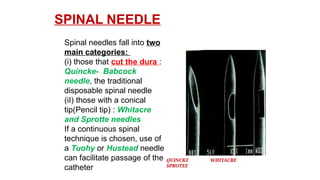
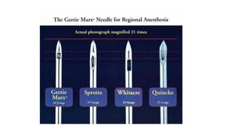
SPINAL NEEDLE
QUINCKE WHITACRE
SPROTEE
Spinalneedles fall into two
main categories:
(i) those that cut the dura :
Quincke- Babcock
needle, the traditional
disposable spinal needle
(iI) those with a conical
tip(Pencil tip) : Whitacre
and Sprotte needles
If a continuous spinal
technique is chosen, use of
a Tuohy or Hustead needle
can facilitate passage of the
catheter
31.
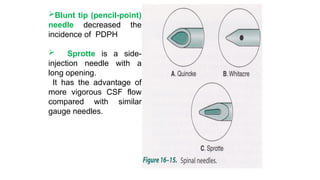
Blunt tip (pencil-point)
needledecreased the
incidence of PDPH
Sprotte is a side-
injection needle with a
long opening.
It has the advantage of
more vigorous CSF flow
compared with similar
gauge needles.
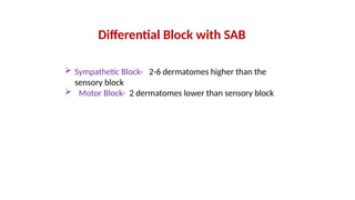
Differential Block withSAB
Sympathetic Block- 2-6 dermatomes higher than the
sensory block
Motor Block- 2 dermatomes lower than sensory block
35.
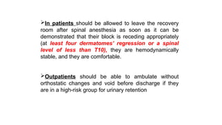
In patients shouldbe allowed to leave the recovery
room after spinal anesthesia as soon as it can be
demonstrated that their block is receding appropriately
(at least four dermatomes’ regression or a spinal
level of less than T10), they are hemodynamically
stable, and they are comfortable.
Outpatients should be able to ambulate without
orthostatic changes and void before discharge if they
are in a high-risk group for urinary retention
36.
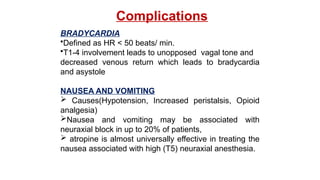
BRADYCARDIA
•Defined as HR< 50 beats/ min.
•T1-4 involvement leads to unopposed vagal tone and
decreased venous return which leads to bradycardia
and asystole
NAUSEA AND VOMITING
Causes(Hypotension, Increased peristalsis, Opioid
analgesia)
Nausea and vomiting may be associated with
neuraxial block in up to 20% of patients,
atropine is almost universally effective in treating the
nausea associated with high (T5) neuraxial anesthesia.
Complications
37.
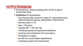
HYPOTENSION
Prevented by:Volume loading with 10-20 mL/kg of
intravenous fluid
Predictors of hypotension
low intravascular volume in case of hypovolemia due
external loss by trauma, dehydration, internal loss
sensory block ≥ T5
age > 40 years
systolic BP < 120 mm Hg
combined spinal and general anesthesia
dural puncture between L2-3 and above
emergency surgery
pt with h/o uncontrolled hypertension
underlying autonomic dysfunction
38.
Post Dural PunctureHeadache:
Due to leak of CSF from dural defect leads to traction in
supporting structure especially in dura and tentorium &
vasodialatation of cerebral blood vessels.
Usually bifrontal and or occipital, usually worse in
upright , coughing , straining
Causes nausea, photophobia, tinnitus, diplopia[6th
nerve],
cranial nerve palsy
Treatment plan include keeping patient supine,
adequate hydration, NSAIDS with without caffeine
[increases production of csf and causes vasoconstriction
of intracranial vessels], if not relieved within 12-24 hr
then epidural blood patch.
Epidural blood patch consists of giving 20 ml
39.
Post Dural PunctureHeadache:
Due to leak of CSF from dural defect leads to traction in
supporting structure especially in dura and tentorium &
vasodialatation of cerebral blood vessels.
Usually bifrontal and or occipital, usually worse in
upright , coughing , straining
Causes nausea, photophobia, tinnitus, diplopia[6th
nerve],
cranial nerve palsy
Treatment plan include keeping patient supine,
adequate hydration, NSAIDS with without caffeine
[increases production of csf and causes vasoconstriction
of intracranial vessels], if not relieved within 12-24 hr
then epidural blood patch.
Epidural blood patch consists of giving 20 ml


![Post Dural Puncture Headache:
Due to leak of CSF from dural defect leads to traction in
supporting structure especially in dura and tentorium &
vasodialatation of cerebral blood vessels.
Usually bifrontal and or occipital, usually worse in
upright , coughing , straining
Causes nausea, photophobia, tinnitus, diplopia[6th
nerve],
cranial nerve palsy
Treatment plan include keeping patient supine,
adequate hydration, NSAIDS with without caffeine
[increases production of csf and causes vasoconstriction
of intracranial vessels], if not relieved within 12-24 hr
then epidural blood patch.
Epidural blood patch consists of giving 20 ml](https://image.slidesharecdn.com/lumbarpunctureforundergraduate-260112095704-3ff05eaf/85/Lumbar-puncture-for-undergraduate-students-38-320.jpg)
![Post Dural Puncture Headache:
Due to leak of CSF from dural defect leads to traction in
supporting structure especially in dura and tentorium &
vasodialatation of cerebral blood vessels.
Usually bifrontal and or occipital, usually worse in
upright , coughing , straining
Causes nausea, photophobia, tinnitus, diplopia[6th
nerve],
cranial nerve palsy
Treatment plan include keeping patient supine,
adequate hydration, NSAIDS with without caffeine
[increases production of csf and causes vasoconstriction
of intracranial vessels], if not relieved within 12-24 hr
then epidural blood patch.
Epidural blood patch consists of giving 20 ml](https://image.slidesharecdn.com/lumbarpunctureforundergraduate-260112095704-3ff05eaf/85/Lumbar-puncture-for-undergraduate-students-39-320.jpg)


![CTEV [ clubfoot] DR ARUN LAL ,DR MOHAMED ASHRAF travancore medical college k...](https://cdn.slidesharecdn.com/ss_thumbnails/ctevclubfootdrarunlaldrmohamedashraftravancoremedicalcollegekollamkeralaindia-260208063247-18fc466c-thumbnail.jpg?width=640&height=640&fit=bounds)


