This document summarizes the relationship between cell cycle regulation and cancer. It discusses how cancer originates from the abnormal expression or activation of cell cycle regulators that promote progression and the suppression of regulators that act as brakes. The cell cycle involves multiple checkpoints controlled by cyclin-dependent kinases (CDKs) and their cyclin partners, which induce progression, and CDK inhibitors (CKIs), which act as brakes. Deregulation of these regulators, through mutations in tumors, underscores their importance in preventing cancer. Understanding these molecular mechanisms could provide insights into tumor development and new treatment strategies.
The cell cycle consists of interphase and the mitotic phase. Interphase includes the G1, S, and G2 phases where the cell grows and prepares for division. The mitotic phase includes mitosis where the cell divides into two daughter cells. Cyclin-dependent kinases (CDKs) and cyclins control progression through the cell cycle by phosphorylating proteins. CDK-cyclin complexes activate at different phases, such as cyclin D-CDK4 in G1 and cyclin B-CDK1 in mitosis. Checkpoints in G1/S and G2/M ensure DNA is intact before the cell divides.
1) The study examines how the telomere-associated protein TIN2 is reorganized in the nuclei of human mammary epithelial cells undergoing growth arrest and differentiation.
2) The researchers find that TIN2 forms one to three large nuclear subdomains that do not contain telomeres and occur simultaneously with TIN2 remaining at telomeres.
3) Expression of truncated forms of TIN2 prevents formation of the large TIN2 domains and relaxation of growth arrest in the cells, suggesting TIN2 organization regulates proliferation and differentiation.
The cell cycle is the regulated process by which cells grow and divide to produce two daughter cells. It has two main phases: interphase and mitosis. Interphase consists of G1, S, and G2 phases where the cell grows and duplicates its DNA. Mitosis is when the cell divides. The cell cycle is regulated by cyclins, cyclin-dependent kinases (CDKs), and CDK inhibitors. Checkpoints ensure DNA quality and control cell cycle progression. Cell cycle dysregulation can lead to uncontrolled cell division and cancer.
Molecular Pathogenesis and mechanisms of carcinogenesis involve several key hallmarks and changes in cell physiology that drive cancer development and progression. The hallmarks include self-sufficiency in growth signals, evasion of growth suppression, resistance to cell death, replication immortality, induction of angiogenesis, activation of invasion and metastasis, immune evasion, microRNA involvement, and epigenetic alterations. Cancer results from dysregulation of oncogenes and tumor suppressor genes that control the normal cell cycle and proliferation.
The cell cycle is regulated by internal and external factors that control when a cell divides. External factors include cell-cell contact and growth factors, while internal factors include cyclins and kinases that help control the cell cycle. Uncontrolled cell division can lead to cancer as cancer cells divide without regard for growth signals and contact inhibition. Cancer occurs due to mutations in genes that regulate the cell cycle, which can be caused by environmental exposures like radiation, viruses, and carcinogens. Treatment options aim to stop rapidly dividing cells like chemotherapy but may also harm healthy cells.
This research article examines the role of sphingosine-1-phosphate (S1P) in regulating the proliferative and stem-like properties of glioblastoma stem cells (GSCs). The results showed that GSCs rapidly consume ceramide and export S1P into the extracellular environment. Extracellular S1P levels reached nanomolar concentrations in response to increased sphingosine. S1P was also found to act as an autocrine factor that promotes GSC proliferation and maintains their stem-like phenotype. This suggests that microenvironmental S1P critically modulates the GSC population by acting as an autocrine signal to support stemness and favor proliferation, survival, and therapeutic resistance of GSCs.
1. The document discusses the role of cyclin D1 in regulating cell cycle progression and its implications in tumor initiation, maintenance, progression and metastasis through altering cell adhesion, migration, redox balance and drug resistance.
2. Cyclin D1 disrupts redox balance by producing reactive oxygen species and regulates cell cycle progression from G1 to S phase.
3. It also regulates cell adhesion and migration by stabilizing F-actin fibers and enhancing chemotaxis and inflammation.
Mutations are changes in genetic material that can be caused by errors in DNA replication or by exposure to mutagens. There are several types of mutations including substitutions, insertions, deletions, and chromosomal mutations. Mutations can have varying effects, from being harmless to causing genetic disorders or cancer. Carcinogenesis is the process by which normal cells are transformed into cancer cells through a series of mutations that disrupt the balance between cell proliferation and cell death.
The document discusses the relationship between cancer and cellular metabolism. It provides a brief history of how Otto Warburg first observed that cancer cells metabolize glucose differently than normal cells through aerobic glycolysis. More recently, there has been a renaissance in studying how oncogenes and tumor suppressor genes can alter cellular metabolism in cancer. The document then overviews key metabolic pathways and regulators that are altered in cancer, such as mutations in the TCA cycle enzymes IDH1 and IDH2. It describes techniques that can be used to study metabolism and cancer, such as analyzing mutations, gene expression, epigenetic changes, and cellular pathways. Throughout, it provides examples of studies investigating specific metabolic changes in cancer, such as how miRNAs
Dr Zahid Azeem, working as Assistant Professor of Biochemistry at Azad Jammu and Kashmir Medical College, Muzaffarabad since 2012.
email; paym_zahid@live.com
The document discusses checkpoints in the cell cycle that control progression from one phase to the next. It describes the G1, G2, and M phase checkpoints, noting that the G1 checkpoint determines whether a cell will continue through the cell cycle or become quiescent. It also explains that the G2 checkpoint is driven by maturation promoting factor, a protein complex of cyclins and cyclin-dependent kinases, and that the M phase checkpoint ensures proper chromosome attachment before anaphase.
Carcinogenesis
Theories of carcinogenesis
Hallmarks of cancer
Important Oncogenes
RB & p53 genes
Metastasis
Aetiology and Pathogenesis of cancer
Tests for carcinogenicity
How to repair damaged DNA?
Basic DNA repair mechanism
Repair of double stranded break
CELL CYCLE
CELL CYCLE CHECK POINT
PHASES IN CELL CYCLE CHECK POINT
ROLE OF CYLINE AND CDKS
MUTURATIONAL PROMOTING FACTOR
FUNCTION OF MPR
CONCLUSION
REFRENCE
Ribavirin shows promise as a novel therapeutic for certain cancers by targeting the translation factor eIF4E. In clinical trials, ribavirin monotherapy resulted in complete remissions in some acute myeloid leukemia patients and partial responses in some metastatic breast cancer patients. Ribavirin treatment led to relocalization of eIF4E from the nucleus to the cytoplasm in patients, correlating with clinical response. Ribavirin's activity was potentiated when combined with other chemotherapeutics, and it inhibited the growth and colony formation of cancer cell lines while sparing normal cells.
The cell cycle is regulated by cyclin-dependent kinases (Cdks) whose activity oscillates throughout the cycle. Cdks form complexes with cyclins, which activate the Cdks and determine which phase of the cycle they control. The cyclin-Cdk complexes phosphorylate target proteins to promote replication and mitosis. Progression through the cell cycle is also controlled by ubiquitin ligases and phosphorylation/dephosphorylation events. The cycle operates through a series of switches that trigger irreversible events, keeping it tightly regulated and coordinated.
The cell cycle involves an interphase of growth and DNA replication followed by mitosis, where the cell divides. The cell cycle is regulated by cyclins and cyclin-dependent kinases (CDKs) that drive progression between phases. CDK activity increases upon binding to cyclins and decreases when cyclins are degraded. Growth hormones like auxins and cytokinins promote cell cycle progression by increasing cyclin and CDK expression, while abscisic acid inhibits the cell cycle. Together, these regulatory mechanisms precisely control cell division.
We sequenced 45 independent clonal organoid cultures derived from 19 donors ranging in age from 3 to 87 years to assess somatic mutation accumulation in adult stem cells (ASCs) from the small intestine, colon and liver. We found that ASCs from all organs gradually accumulate mutations at a rate of around 36 mutations per year. While the mutation rates were similar, the mutation spectra differed between tissues. Notably, ASCs from the colon and small intestine showed very similar age-related mutation characteristics, although cancer incidence is much higher in the colon.
The document discusses carcinogenesis and cancer. It begins with an overview of cell anatomy and the cell cycle, including the phases of interphase and mitosis. It then discusses how abnormalities in cell cycle regulation and checkpoints can lead to uncontrolled cell growth and carcinogenesis. Factors involved in carcinogenesis include oncogenes, tumor suppressor genes, and DNA damage. The document outlines several theories of carcinogenesis and describes the multistage process. It also covers the characteristics, metastatic process, and classification of malignant tumors.
This document discusses the cell cycle and its relevance to cancer. It begins by describing the basic components and organelles of the cell. It then explains the different phases of the cell cycle, including interphase (G1, S, G2 phases) and mitosis. Key control mechanisms like cyclin-dependent kinases and checkpoints are described. Alterations in cell cycle pathways and genes can lead to uncontrolled cell proliferation and cancer. Understanding the cell cycle provides opportunities to target specific phases with chemotherapy or radiotherapy to treat cancer.
Molecular event during Cell cycle By KK Sahu SirKAUSHAL SAHU
WHAT IS CELL?
WHAT IS CELL DIVISION OR CELL CYCLE?
WHY DO CELL DIVIDE?
HISTORY
CELL CYCLE
INTERPHASE
M-PHASE
MOLECULAR EVENT DURING CELL CYCLE AND CELL REGULATION
TYPES OF CELL DIVISION
IMPORTANCE OF CELL DIVISION
ABNORMALTIES OF CELL CYCLE
REFRENCES
1) NF-kB is constitutively activated in pancreatic cancer cells but not normal pancreatic cells, suggesting it plays a role in pancreatic tumorigenesis.
2) Expressing a mutant IkBa (IkBaM) that inhibits NF-kB suppressed the tumorigenicity of pancreatic cancer cells in a mouse model.
3) Inhibiting NF-kB reduced expression of pro-survival genes like Bcl-xL and Bcl-2, and reduced VEGF and IL-8 expression which are involved in angiogenesis. This suggests NF-kB inhibition can suppress tumorigenesis by reducing pro-survival and angiogenic factors.
Brock biology-of-microorganisms-(13th-edition)Shahab Pour
This document provides an overview of microbiology. It begins with an introduction to microbiology as a science and discusses the historical discoveries that contributed to the field. It then describes techniques used to study microbes like microscopy. It explores the diversity of microbial life including bacteria, archaea, and microbial eukaryotes. The document also examines microbial cell structure, metabolism, growth, genetics, and genomics. It discusses the roles of microbes in areas like photosynthesis, biogeochemical cycles, and commercial applications. Finally, it considers microbial evolution and systematics.
This document reviews the role of microRNAs (miRNAs) in the occurrence and development of esophageal squamous cell carcinoma (ESCC). It summarizes that various miRNAs have been found to be dysregulated in ESCC and can function either as tumor suppressors or oncogenes by targeting different genes involved in processes like cell proliferation, apoptosis and invasion. Specifically, it provides tables listing miRNAs that have been identified as tumor suppressors and oncogenes in ESCC, along with their biological effects and target genes. The document aims to enhance understanding of the molecular mechanisms by which miRNAs contribute to the progression of ESCC.
Cell division in yeast involves four main processes - cell growth, DNA replication, chromosome separation, and cell division. These occur in distinct phases: interphase (G1, S, G2) and mitosis (M). Progression between phases is controlled by protein kinases like MPF and CDK1. Checkpoints like G1/S and G2 ensure replication is complete before progression. Factors like nutrients and cell size regulate the G1-S transition through START. DNA replicates only once per cycle due to licensing/de-licensing of MCM proteins.
The document outlines six hallmarks of cancer cells:
1. Self-sufficiency in growth signals through autocrine or paracrine signaling and expression of growth-promoting extracellular matrix receptors.
2. Insensitivity to antigrowth signals through mutations that block cell cycle inhibitors like Rb or induce differentiation.
3. Evading apoptosis by mutations in genes like p53 that derail cell suicide pathways and activate pro-survival signals.
4. Limitless replicative potential enabled by mechanisms like telomerase that maintain telomere length indefinitely.
5. Sustained angiogenesis driven by factors like VEGF that promote growth of blood vessels.
6. Tissue invasion and
Cell cycle- Dr Deenadayalan,Medical Oncologst,MauraiDr DEENADAYALAN T
This document provides information about the cell cycle and its regulation. It discusses the key phases of the cell cycle - prophase, metaphase, anaphase and telophase. It describes the cell cycle regulators including cyclins, cyclin dependent kinases, and cyclin dependent kinase inhibitors. It also discusses cell cycle checkpoints and alterations in cell cycle control proteins that can lead to cancer. Finally, it briefly mentions cell cycle phase specific and non-specific agents used in cancer treatment.
A Bioinformatic Analysis of Oral Cancer Proteomics.pptx . Dr.Erabah IlyasERABAHILYAS1
This work focuses on the role of cyclins in oral squamous cell carcinoma, it's degradation and it's clinical applications. It also unveils proteomics of oral cancer using bioinformatic tools
The document summarizes key aspects of the cell cycle. It discusses that the cell cycle is a series of events that a cell passes through from the time it is formed until it replicates. There are two main periods - interphase and mitosis. Interphase consists of G1, S, and G2 phases where the cell grows and duplicates its DNA. Mitosis is where the cell nucleus and cytoplasm divide. The cell cycle is tightly regulated by checkpoints and cyclins/CDKs to ensure DNA is properly replicated and divided between daughter cells. Dysregulation of cell cycle controls can lead to cancer if cells continue dividing uncontrollably.
The document summarizes the cell cycle and its checkpoints. It describes the different phases of the cell cycle including interphase consisting of G1, S, and G2 phases and the mitotic phase. Checkpoints ensure the cell cycle processes are accurately completed before progression including the G1, G2, and metaphase checkpoints. The cell cycle is regulated by cyclins, cyclin-dependent kinases, and cyclin-dependent kinase inhibitors that act as positive and negative regulators.
The cell cycle is tightly regulated by cyclins and cyclin-dependent kinases (Cdks). Cyclins bind to and activate Cdks at specific points in the cell cycle to promote progression. Cyclins fluctuate in concentration throughout the cycle while Cdk levels remain constant. Rb protein and p53 tumor suppressor proteins also regulate the cell cycle by inhibiting progression from G1 to S phase when DNA damage is detected. Checkpoints at G1/S and G2/M ensure conditions are suitable before allowing division to proceed and prevent errors from being passed on. Dysregulation of the cell cycle, as seen in cancer, results from defects in these regulatory proteins and checkpoints.
The document discusses regulation of the cell cycle. It explains that the cell cycle is regulated through checkpoints and cyclin-dependent protein kinases (Cdks) and cyclins. There are three main checkpoints - at the ends of G1 and G2 phases, and during metaphase. These checkpoints ensure DNA integrity and replication, and proper chromosome attachment before progression to the next phase. Cdks drive progression when activated by binding with cyclins, which are synthesized and degraded throughout the cycle in response to cellular signals.
The Role of Cyclin-Dependent Kinases on the Metastasis of Breast Cancer-Crims...CrimsonpublishersCancer
Cyclin-Dependent kinases (CDKs) function in mitosis by allowing the cycle to progress from one stage to another due to their properties as a family of protein kinases. Because of this function, abnormalities with CDKs can lead to uncontrolled cell division, leading to diseases such as cancer. Breast cancer is one form of cancer in which CDKs are a prevalent area of study. The role CDKs play in controlling and coordinating cell division makes it an important process to understand in breast cancer and, specifically, the metastasis of breast cancer. Lack of controlled CDK function could allow the cancer to spread to other parts of the body, leading to metastasis. Inhibiting CDK activity is an area of interest in searching for ways to treat breast cancer, especially once it has spread to the point where tumors cannot be surgically removed. Investigating these pathways and the effects of CDK inhibition on breast cancer cells has revealed much on the reestablishment of cell cycle control, which consequently leads to control of the cancer. This could be an effective form of non-localized treatment against metastatic cancer that is able to target specific cells throughout the body.
Cell cycle regulation presentation by me and my colleagues. Not the Best work but still it will give a general idea about DNA damage checkpoints, roles of Cdk-Cyclin complexes, Rb proteins, ATM&ATR kinases, p51, etc.
Reference : Nature reviews & The Cell a molecular approach. (cooper)
The document discusses chromosome organization and packaging, as well as the cell cycle and its regulation. It describes the different phases of the cell cycle (G1, S, G2, M) and checkpoints that ensure DNA replication is complete and any damage repaired before the cell divides. Key regulators of the cell cycle include cyclins, CDKs, and CDKIs, which promote or inhibit phase transitions in response to internal and external signals. DNA damage checkpoints in particular arrest the cell cycle to allow repair or induce apoptosis if damage is too severe.
New Microsoft Office PowerPoint Presentation-1.pptxShounakKamat1
The cell cycle is a precisely programmed series of events that enables a cell to duplicate its contents and divide into two daughter cells. It consists of interphase (G1, S, G2 phases) and mitosis (M phase). Progression through the cell cycle is regulated by cyclins and cyclin-dependent kinases (CDKs). CDK activity is controlled by association with cyclins, CDK inhibitors, and phosphorylation. Checkpoint pathways like the G1/S, G2, and spindle assembly checkpoints ensure replication and division errors are corrected before progression. Deregulation of these checkpoint pathways can lead to genomic instability and carcinogenesis.
Cyclin Dependent Kinases: Old Target with New Challenges for Anti-Cancer DrugsCrimsonPublishersMAPP
Cyclin Dependent Kinases: Old Target with New Challenges for Anti-Cancer Drugs by Shashank Shekhar Mishra in Modern Applications in Pharmacy & Pharmacology
The cell cycle and molecular basis of cancer can be summarized as follows:
The cell cycle is tightly regulated by cyclins, cyclin-dependent kinases (CDKs), and their inhibitors to ensure proper cell division and growth. Defects in cell cycle regulation can lead to uncontrolled cell growth and cancer. Cancer develops through the accumulation of genetic mutations in oncogenes and tumor suppressor genes, which disrupt the normal cell cycle checkpoints and allow cells to proliferate uncontrollably. Key cellular capabilities acquired during cancer progression include self-sufficiency in growth signals, insensitivity to anti-growth signals, evasion of apoptosis, limitless replicative potential, sustained angiogenesis, and tissue invasion and metastasis.
Cellular mechanisms of development in animals involve processes like cell division, differentiation, signaling, morphogenesis, and apoptosis to orchestrate the growth and organization of tissues and organs. Gametogenesis is the process by which germ cells undergo meiosis and differentiation into haploid gametes. In males, spermatogenesis yields sperm through spermatogonia undergoing meiosis and spermiogenesis. In females, oogenesis arrests oocytes in meiosis I until puberty, when oocytes resume meiosis to form eggs if fertilized.
The document summarizes the cell cycle and its regulation. It describes the main stages of the cell cycle - interphase consisting of G1, S, and G2 phases and the M phase. Key regulators of the cell cycle include cyclins, cyclin-dependent kinases, and checkpoints like G1, G2, and M that ensure fidelity of DNA replication and chromosome segregation. Dysregulation of these processes can lead to genomic instability and cancer.
The cell cycle consists of four main phases - G1, S, G2, and M phase. The cell cycle is tightly regulated by cyclins and cyclin-dependent kinases (CDKs) that drive progression between phases. CDK activity is controlled by association with cyclins, phosphorylation, and degradation. Checkpoints between phases like G1/S and G2/M ensure DNA replication fidelity before division. Apoptosis, or programmed cell death, can be triggered intrinsically via DNA damage or extrinsically through ligand-receptor interactions and leads to cell death. The cell cycle and apoptosis are assessed using techniques like propidium iodide staining to measure DNA content and annexin assays to detect early apoptotic
The document summarizes key aspects of cell cycle regulation in plants. It discusses the cell cycle phases of interphase (G1, S, G2) and mitosis. Key regulators of the plant cell cycle include cyclin-dependent kinases (CDKs) and cyclins. CDKs activate during specific cell cycle phases upon binding to cyclins. Plant hormones also influence the cell cycle through effects on CDKs, cyclins and other regulators. Precise control of the cell cycle is important for plant growth and development.
The phenomenon of signal transduction, also known as cell signaling, pertains to the intricate mechanisms that facilitate the transfer of biological information between cells. The effective coordination of diverse specialized cell types in various tissues and organs is a prerequisite for the proper functioning of complex multicellular organisms, necessitating intercellular communication. This communication must be continuous and dynamic to maintain coordination. Additionally, cell signaling pathways play a crucial role in the mechanisms of action of numerous drugs, including both local and general anesthetics. Consequently, a fundamental understanding of cell signaling mechanisms is imperative for comprehending various pathophysiologic and pharmacologic mechanisms.
The document discusses the cell cycle, its phases and checkpoints. It describes that the cell cycle consists of interphase (G1, S, G2 phases) and the mitotic phase. Key checkpoints include G1, G2 and metaphase checkpoints, which verify processes are accurately completed before progression. Cyclins and CDKs regulate progression through phases. Disruptions to checkpoints can contribute to cancer by allowing mutations to propagate. Anticancer drugs target specific phases through cyclin/CDK inhibition or other mechanisms.
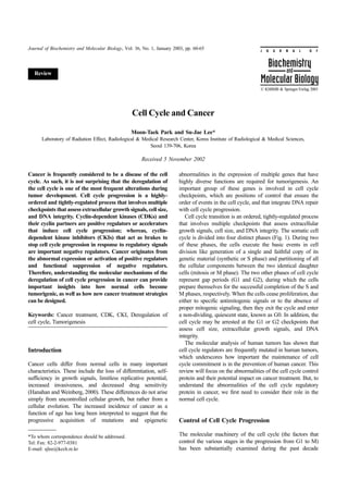
2. Cell Cycle and Cancer 61
Fig. 1. Progression of cell cycle. Somatic cell cycle consists of four distinct phases: initial growth (G1), DNA synthesis (S), a gap (G2),
and mitosis (M). The critical point of cell cycle control is the restriction point. After passing this point, the cell cycle is irreversibly
committed to the next cell division.
(Pardee et al., 1989; Xiong et al., 1991; Sherr et al., 1993; residues (T14/Y15 in CDC2) is mediated by dual specific
Morgan et al., 1995; Hwang et al., 1998; Zhan et al., 1999; kinases (Wee1 and MYT1). This inhibition is relieved when
Raleigh et al., 2000) the CDC25 phosphatases dephosphorylate these residues,
The heart of the regulatory apparatus during the cell cycle which triggers entry into mitosis (Morgan, 1997; Ekholm and
progression is a family of enzymes, called the cyclin- Reed, 2000). CDKs and their cyclin partners are positive
dependent kinases (CDKs). The active forms of CDKs are a regulators or accelerators that induce cell cycle progression;
complex of at least two proteins, a kinase and a cyclin (Table whereas, important negative regulators, such as cyclin-
1). They often contain other proteins with poorly understood dependent kinase inhibitors (CKIs), act as brakes to stop the
functions. These complexes undergo changes in the kinase cell cycle progression in response to regulatory signals (Fig.
and cyclin components that are believed to drive the cell from 2). By direct association with CDK, CKIs can negatively
one stage of the cell cycle to another (Pardee et al., 1989; regulate CDK activity. There are two types of CKIs. The four
Xiong et al., 1991; Sherr et al., 1993; Morgan et al., 1995; members of the INK family, INK4A (p16), INK4B (p15),
Hwang et al., 1998; Zhan et al., 1999; Raleigh et al., 2000). INK4C (p18), and INK4D (p19), exert their inhibitory activity
According to this paradigm, the cell cycle is determined by by binding to CDK4 and CDK6, and preventing their
the constellation of proteins that are activated or inactivated by association with D-type cyclins. The three members of the
phosphorylation, a result of the activity of the CDKs during CIP/KIP family, CIP1 (p21), KIP1 (p27), and KIP2 (p57),
that stage (Fig. 1). In mammalian cells, a succession of kinase form heterotrimeric complexes with the G1/S CDKs. CKIs
subunits (CDK4, CDK6, CDK2, and CDC2) is expressed are induced in response to different cellular processes (Sherr
along with a succession of cyclins (cyclin D, E, A, and B), as and Roberts, 1999; Sherr, 2000). For instance, in quiescent
the cells progress from G1 to mitosis. CDK4 and CDK6 cells, the KIP1 levels are generally high. CIP1 is one of the
complexed with one of several D-type cyclins functions early effectors of p53, a tumor suppressor that is important in the
in the G1 phase, probably in response to growth factors. DNA damage checkpoint.
CDK2 that complexed with cyclin E, cyclin A, or both is The critical point of cell cycle control is the restriction
essential for the G1 S transition and DNA replication,
respectively. CDC2 that complexed with cyclin A and cyclin
B is essential for mitosis. Additional CDKs and cyclins will Table 1. Mammalian cyclin-dependent kinases (CDKs) and their
regulatory cyclins
be added to this list (Table 1).
The passage of cells from one stage of the cell cycle to CDKs Cyclins
another is tightly regulated by a wealth of controls that act on
Cdc2 Cyclin A&B
the transcription of cyclin genes, the degradation of cyclins,
Cdk2 Cyclin A ,E & D
and modification of the kinase subunits by phosphorylation. A
Cdk3 Cyclin E
number of positive or negative feedback loops also contribute
Cdk4 Cyclin D1,D2,D3
to the cell cycle progression (Fig. 2). CDK activity is
Cdk5 p35
positively regulated by the association with the cyclins, and by
Cdk6 Cyclin D1,D2,D3
phosphorylation of the T-loop threonine by the CDK-
Cdk7 Cyclin H
activating kinase (CAK), a serine/threonine kinase that is also
Cdk8 Cyclin C
involved in transcription and DNA repair (Nigg, 1996).
Cdk9 Cyclin T
Inhibitory phosphorylation of adjacent threonine and tyrosine
3. 62 Moon-Taek Park and Su-Jae Lee
Fig. 2. Regulation of G1/S cell cycle progression. The kinase activity of cyclin D-CDK4/6 and cyclin E-CDK2 complexes are
negatively-regulated by two cyclin-dependent kinase inhibitor (CKIs) families the INK4 (inhibitor of CDK4) and CIP/KIP (CDK
interacting protein/CDK inhibitor) proteins.
point. After passing this point, the cell is irreversibly become apparent that tumorigenesis is frequently associated
committed to the next phase of the cell cycle (Fig. 1). The with mutations or abnormalities in the expression of various
restriction point control is mediated by the cyclin D and cyclin cyclins, CDKs and CKIs in several types of human cancers
E-dependent kinases. The primary substrates of CDK4/6 and (Weinstein and Zhou, 1997; Sgambato et al., 1998). In 1991, a
CDK2 in G1 progression are the members of the presumptive oncogene that is composed of the parathyroid
retinoblastoma protein family pRB, p107, and p130 (Fig. 2) hormone gene (PRAD1) that fused to the gene that encodes
(Morgan, 1997; Adams, 2001). These molecules function as cyclin D1 was identified in a human parathyroid adenoma
negative regulators at the restriction point. One important (Heichman and Roberts, 1994; King et al., 1994). This
target of pRB for regulating the early G1 cell cycle observation provided the first clue that cyclins might be
progression is the E2F family of transcription factors directly involved in some human cancers. Subsequently,
(Harbour and Dean, 2000). E2Fs regulate the expression of a evidence was presented for the involvement of cyclins in other
host of genes that mediates both restriction point transversals, human cancer cells. These included B cell lymphoma and
such as cyclin E, and the S phase progression, such as breast, gastric, colon and esophageal carcinomas, as well as
dihydrofolate reductase and thymidylate synthase (Fig. 2). several other types of cancers (Hunter and Pines, 1994).
Moreover, pRb has some CDK-independent functions, such as Indeed, the increased expression of cyclin D1 is one of the
the repression of some promoters and RNA pol III activity most frequent abnormalities in human cancer, since it occurs
(Adnane et al., 1995; Sellers et al., 1995; Weintraub et al., in ~60% of breast cancers, 40% of colorectal cancers, 40% of
1995; Chow and Dean, 1996; Chow et al., 1996; White et al., squamous carcinoma of the head and neck, and 20% of
1996). Therefore, the re-introduction and expression of pRb prostate cancers (Weinstein et al., 1997; Weinstein and Zhou,
into tumor cells may arrest growth by mechanisms that are 1997; Han et al., 1998; Sgambato et al., 1998). Consistant
independent of the CDK complex formation. with these results, cyclin D1 substitutes or partially substitutes
for certain oncogenes in the cellular transformation assay
(Hinds et al., 1994; Lovec et al., 1994). The cyclin E gene,
Abnormalities in the Cell Cycle Control Proteins in which acts in late G1, is also overexpressed and dysregulated
Cancer in a variety of human cancers (Malumbres and Barbacid,
2001). The amplification and overexpression of CDKs and its
Abnormalities in Cyclins and CDKs: In recent years, it has regulators have rarely been described in human cancers. Some
4. Cell Cycle and Cancer 63
Table 2. Alterations of INK4A (p16) in human cancers (Rocco and Sindransky, 2001). Specific deletions of INK4B
Tumor Type of alteration Frequency (%) have been found in only a few cases of leukemia and
lymphomas (Nguyen et al., 2000; Vidal and Koff, 2000;
Melanoma Mutation 25 Rocco and Sindransky, 2001). In contrast, the
Adult T cell leukemia Deletion 28 hypermethylation of INK4B seems to be frequent in several
T cell acute lymphocytic cancers (Cameron et al., 1999; Nguyen et al., 2000; Wong et
Deletion 59
leukemia al., 2000; Chim et al., 2001). This suggests that the silencing
Oral squamous cell of the INK4B promoter by methylation plays an important
Mutation 9
carcinoma
role in tumor development.
Gliosarcoma Deletion 37
The second family of CKIs is the CIP/KIP family, which
Bladder carcinoma Deletion 20
shares homology at the N-terminal CDK inhibitory domain.
Upper track urotherial
Deletion 31 This includes p21(CIP1/WAF1), p27 (KIP1), and p57 (KIP2).
carcoinoma
The p21CIP1 binds and inhibits several CDK-cyclin complexes,
Head and neck Mutation 13
including CDK2-cyclin E and CDK4-cyclin D1. The p21CIP1
expression is directly induced by p53 (Sherr, 1994). This
immediately focuses attention on p21CIP1 as a potential
reports demonstrated that the CDK4 gene is overexpressed in mediator of p53-dependent tumor suppression. Furthermore,
certain tumor cell lines (Hunter and Pines, 1994). As with the overexpression of p21CIP1 can cause G1 arrest. So far,
cyclins, however, a general involvement of CDKs in cancer however, there have been no reports of p21CIP1 alterations in
has yet to be demonstrated. tumors or cell lines. If p21CIP1 is an important mediator in the
p53-dependent tumor suppression, then the mutations of
p21CIP1 might be expected in some fraction of tumors and cell
Abnormalities in CKIs lines. Therefore, the genetic evidence of p21CIP1 as a general
tumor suppressor is still undetermined.
Two families of CKIs have been characterized, based on their The p27KIP1 was identified as CDK-binding proteins that are
specificity. The first is the INK4 family, which includes p15 activated by TGF-β, contact inhibition of cell growth (Polyak et
(INK4B), p16 (INK4A), p18 (INK4C), and p19 (INK4D) al., 1994; Toyoshima and Hunter, 1994). The p27KIP1 protein
(Sherr and Roberts, 1995). The INK4 family of proteins sequence bears some similarities to p21, and also inhibits
specifically targets the cyclin D-dependent kinases (CDK4 several CDKs. No mutations in the p27KIP1 gene have been
and CDK6) (Sherr and Roberts, 1995; Ruas and Peters, 1998). reported in tumor and cell lines. However, the reduced
The INK4A (p16) protein, which was originally identified as a expression of p27KIP1 is frequently detected in human cancers.
CDK4-interacting protein that inhibits CDK4 kinase activity These include the breast, prostate, gastric, lung, skin, colon, and
(Serrano et al., 1993), has been mapped to chromosome 9p21 ovarian cancers (Catzavelos et al., 1997; Esposito et al., 1997;
(Kamb et al., 1994). Due to a hot spot of genomic alterations Mori et al., 1997; Loda et al., 1997; Porter et al., 1997; Cordon-
in cancers, intense studies have focused on the role of INK4A Cardo et al., 1998; Florenes et al., 1998). Surprisingly, however,
in tumorigenesis. Mutations and deletions of INK4A genes a relatively high expression of p27KIP1 is found in a series of
are frequently found in a variety of human malignancies and human esophageal cancer cell lines (Doki et al., 1997).
transformed cells (Table 2) (Kamb et al., 1994; Nobori et al., Furthermore, several human colon and breast cancer cell lines
1994). These include melanoma, acute lymphocytic leukemia, also express high levels of p27KIP1, but low levels in three normal
osteosarcoma, lung, brain, breast, head and neck, bladder, and human mammary cell lines. It is also overexpressed in the
ovarian cancers. The frequent inactivation of INK4A in these small-cell carcinomas of the lung, despite their high degree of
tumors suggests that the loss of INK4A provides a selective malignancy (Yatabe et al., 1997). The increased expression of
cellular growth advantage. p27KIP1 in cancer cells seems paradoxical, because mutations of
INK4B is encoded immediately adjacent to INK4A at the this gene have not been found or are extremely rare in various
INK4 locus, 9p21. Its expression is induced in response to cancers (Sgambato et al., 2000). A possible expression for the
transforming-growth factor beta (TGF-β) treatment (Hannon increase of p27KIP1 in some cancer cells is that they have become
and Beach, 1994). The INK4B sequence is very similar to refractory to the inhibitory effects of this protein. Further studies
INK4A, about 70% at the amino acid level. Its nearly identical will be required to establish the role of p21CIP1 and p27KIP1 in
biochemical behavior, as an inhibitor of CDK4/6, supports the cancer development.
notion that INK4B might be a player in tumor suppression.
Although evidence for the tumor suppressor role of INK4B is
abundant, the INK4B role in tumor suppression is unclear. No, Cell Cycle Control and Cancer Treatment
or rare, point mutations have identified INK4B in tumor cell
lines. The majority of homozygous deletions affect either both The frequent loss of cell cycle regulation in human cancer has
the INK4A and INK4B loci, or INK4A alone in most tumors revealed targets for possible therapeutic intervention. Indeed,
5. 64 Moon-Taek Park and Su-Jae Lee
restoring proper restriction point control to cancer cells might Chim, C. S. M., Liang, R., Tam, C. Y. and Kwong, Y. L. (2001)
allow them to return to a quiescent state. Alternatively, it could Methylation of p15 and p16 genes in acute promyelocytic
take advantage of their uncontrolled proliferation to facilitate leukemia: potential diagnostic and prognostic significance. J.
apoptotic death, or to specifically exposure cancer cells to Clin. Oncol. 19, 2033-2040.
Chow, K. N. and Dean, D. C. (1996) (1996) Domains A and B in
cytotoxic treatments (Chen et al., 1999).
the Rb pocket interact to form a transcriptional repressor motif.
CDKs are actively being targeted, due to their central role
Mol. Cell. Biol. 16, 4862-4868.
in the control of cell cycle progression. Designing inhibitors Chow, K. N., Starostik, P. and Dean, D. C. (1996). The Rb family
that block CDK activity are the most direct and promising contains a conserved cyclin-dependent-kinase-regulated
strategy. Substantial efforts from many groups have led to the transcriptional repressor motif. Mol. Cell. Biol.,16, 7173-7181.
discovery, optimization, and characterization of potent CDK Cordon-Cardo, C., Koff, A., Drobnjak, M., Capodieci, P., Osman,
inhibitors. Three properties make CDK inhibitors attractive as I., and Millard, S. S. (1998) Distinct altered patterns of
anti-tumor agents. First, they are potent anti-proliferative p27KIP1 gene expression in benign prostatic hyperplasia and
agents, arresting cells in G1 or G2/M (Damiens et al., 2000; prostatic carcinoma. J. Natl. Cancer Inst. 90, 1284-1291.
Soni et al., 2001). Second, they trigger apoptosis, alone or in Damiens, E., Baratte, B., Marie, D., Eisenbrand, G. and Meijer, L.
combination with other treatments (Edamatsu et al., 2000). (2000) Anti-mitotic properties of indirubin-3'-monoxime, a
CDK/GSK-3 inhibitor: induction of endoreplication following
Third, in some instances, the inhibition of CDKs contributes
prophase arrest. Oncogene 20, 3786¯3797.
to the cell differentiation (Matushansky et al., 2000). Only
Doki, Y., Imoto, M., Han, E. K. -H., Sgambato, A. and Weinstein,
reports on the clinical trials of flavopiridol and UCN01 (7- I. B. (1997) Increased expression of the p27kip1 protein in
hydroxystaurosporine) are available, but several other CDK human esophageal cancer cell lines that over-express cyclin D1.
inhibitors are currently being researched. Carcinogenesis 18, 11391148.
Potential gene therapeutic strategies are also being Edamatsu, H., Gau, C. L., Nemoto, T., Guo, L. and Tamanoi, F.
established, based on the negative regulators of cell cycle (2000) Cdk inhibitors, roscovitine and olomoucine, synergize
progression (such as INK4A, p21CIP1, and p27KIP1) to inhibit with farnesyl transferase inhibitor (FTI) to induce efficient
cell transformation and cancer growth. The use of gene apoptosis of human cancer cell lines. Oncogene 19, 3059¯3068.
therapy continues to be a promising, yet elusive, alternative Ekholm, S. V. and Reed, S. I. (2000) Regulation of G1 cyclin-
for the treatment of cancer. The origins of cancer must be well dependent kinases in the mammalian cell cycle. Curr. Opin.
Cell Biol. 12, 676-684.
understood so that the therapeutic gene can be chosen that has
Esposito, V., Baldi, A., De Luca, A., Groger, A. M., Loda, M.,
the highest chance of successful tumor regression. The gene
and Giordano, G. G. (1997) Prognostic role of the cyclin-
delivery system must be tailored for optimum transfer of the dependent kinase inhibitor p27 in non-small cell lung cancer.
therapeutic gene to the target tissue. In the near future, new Cancer Res. 57, 3381-3385.
drug compounds and gene therapy protocols will be available. Florenes, V. A., Maelandsmo, G. M., Kerbel, R. S., Slingerland, J.
They will help the fight against cancer, since they will broaden M., Nesland, J. M. and Holm, R. (1998) Protein expression of
our understanding of the cell cycle and cancer. the cell-cycle inhibitor p27Kip1 in malignant melanoma:
inverse correlation with disease-free survival. Am. J. Pathol.
153, 305-312.
References Han, E. K. -H., Rubin, M. A., Lim, J. T., Arber, N., Xing, W. -Q.
and Weinstein, I. B. (1998) Cyclin D1 expression in human
Adams, P. D. (2001) Regulation of the retinoblastoma tumor prostate cell lines and primary tumors. The Prostate, 35, 95101.
suppressor protein by cyclin/CDKs. Biochim. Biophys. Acta Hanahan, D. and Weinberg, R. A. (2000) The hallmarks of cancer.
1471, 123-133. Cell 100, 57-70.
Adnane, J., Shao, Z. and Robbins, P. D. (1995) The Hannon, G. J. and Beach, D. (1994) p151INK4B is a potential
retinoblastoma susceptibility gene product represses effector of TGF-beta-induced cell cycle arrest. Nature 371,
transcription when directly bound to the promoter. J. Biol. 257-261.
Chem. 270, 8837-8843. Harbour, J. and Dean, D. (2000) The Rb/E2F pathway: expanding
Cameron, E. E., Baylin, S. B. and Herman, J. G. (1999) roles and emerging paradigms. Genes Dev. 14, 2393¯2409.
p15(INK4B) CpG island methylation in primary acute Heichman, K. A. and Roberts, J. M. (1994) Rules to replicate by.
leukemia is heterogeneous and suggests density as a critical Cell 79, 557-562.
factor for transcriptional silencing. Blood 94, 2445-2451. Hinds, P. W., Dowdy, S. F., Eaton, E. N., Arnold, A. and
Catzavelos, C., Bhattacharya, N., Ung, Y. C., Wilson, J. A., Weinberg, R. A. (1994) Function of a human cyclin gene as an
Roncari, L. and Sandhu, C. (1997) Decreased levels of the cell oncogene. Proc. Natl. Acad. Sci. USA 91, 709-713.
cycle inhibitor p27Kip1 protein: prognostic implications in Hunter, T. and Pines, J. (1994) Cyclins and cancer. II: Cyclin D
primary breast cancer. Nature Med. 3, 227-230. and CDK inhibitors come of age. Cell 79, 573-582.
Chen, Y. N., Sharma, S. K., Ramsey, T. M., Jiang, L., Martin, M. Hwang, A. and Muschel, R. J. (1998) Radiation and the G2 phase
S., Baker, K., Adams, P. D., Bair, K. W. and Kaelin, W. G. Jr. of the cell cycle. Radiat. Res. 150 (Suppl.), S52-S59.
(1999) Selective killing of transformed cells by cyclin/cyclin- Kamb, A., Gruis, N. A., Weaver-Feldhaus, J., Liu Q., Harshman,
dependent kinase 2 antagonists. Proc. Natl. Acad. Sci. USA 96, K., and Tavigian, S. V. (1994) A cell cycle regulator potentially
4325-4329. involved in genesis of many tumor types. Science 264, 436-
6. Cell Cycle and Cancer 65
440. transrepression domain in the retinoblastoma protein induces a
Kamb, A., Shattuck-Eidens, D., Eeles, R., Liu, Q., Gruis, N. A., cell cycle arrest when bound to E2F sites. Proc. Nat. Acad.
and Ding, W. (1994) Analysis of the p16 gene (CDKN2) as a Sci. USA 92, 11544-11548.
candidate for the chromosome 9p melanoma susceptibility Serrano, M., Hannon, G. J. and Beach, D. (1993) A new
locus. Nature Genet. 8, 23-26. regulatory motif in cell-cycle control causing specific inhibition
King, R. W., Jackson, P. K. and Kirschner, M. W. (1994) Mitosis of cyclin D/CDK4. Nature 366, 704-707.
in transition. Cell 79, 563-571. Sgambato, A., Flamini, G., Cittadini, A. and Weinstein, I.B.
Loda, M., Cukor, B., Tam, S. W., Lavin, P., Fiorentino, M., (1998) Abnormalities in cell cycle control in cancer and their
Draetta, G. F., Jessup, J. M. and Pagano, M. (1997) Increased clinical implications. Tumori 84, 421433.
proteasome-dependent degradation of the cyclin-dependent Sgambato, A. A., Cittadini, A. and Weinstein, I. B. (2000)
kinase inhibitor p27 in aggressive colorectal carcinomas. Nature Multiple functions of p27Kip1 and its alterations in tumor cells.
Med. 3, 231-234. A review. J. Cell. Physiol. 183, 18-27.
Lovec, H., Sewing, A., Lucibello, F. C., Muller, R. and Moroy, T. Sherr, C. J. (1993) Mammalian G1 cyclins. Cell 73, 1059-1065..
(1994) Oncogenic activity of cyclin D1 revealed through Sherr, C. J. and Roberts, J. M. (1995) Inhibitions of mammalian
cooperation with Ha-ras: link between cell cycle control and G1 cyclin-dependent kinases. Genes Dev. 9, 114-1163.
malignant transformation. Oncogene 9, 323-326. Sherr, C. J. and Roberts, J. M. (1999) Cdk inhibitors: positive and
Malumbres, M. and Barbacid, M. (2001) To cycle or not to cycle: negative regulators of G1-phase progression. Genes Dev. 13,
a critical decision in cancer. Nat Rev Cancer 3, 222-31. 1501-1512.
Matushansky, I., Radparvar, F. and Skoultchi, A. I. (2000) Sherr, C. J. (1994) G1 phase progression: cycling on cue. Cell 79,
Reprogramming leukemic cells to terminal differentiation by 551-555.
inhibiting specific cyclin-dependent kinases in G1. Proc. Natl. Sherr, C. J. (2000) Cancer cell cycle revisited. Cancer Res. 60,
Acad. Sci. USA 97, 14317¯14322. 3689-3695.
Morgan, D. O. (1995) Principles of CDK regulation. Nature 374, Soni, R., O’Reilly, T., Furet. P., Muller, L., Stephan, C., Zumstein-
131-134. Mecker, S., Fretz, H., Fabbro, D. and Chaudhuri, B. (2001)
Morgan, D. O. (1997) Cyclin-dependent kinases: engines, clocks, Selective in vivo and in vitro effects of a small molecule
and microprocessors. Annu. Rev. Cell Dev. Biol. 13, 261-291. inhibitor of cyclin-dependent kinase 4. J. Natl. Cancer Inst. 21,
Mori, M., Mimori, K., Shiraishi, T., Tanaka, S., Ueo, H. and 436¯446.
Sugimachi, K. (1997) p27 expression and gastric carcinoma. Toyoshima, H. and Hunter, T. (1994) p27, a novel inhibitor of G1
Nature Med. 3, 593. cyclin-Cdk protein kinase activity, is related to p21. Cell 78,
Nguyen, T. T., Mohrbacher, A. F., Tsai, Y. C., Groffen, J., 67-74.
Heisterkamp, N. and Nichols, P. W. (2000) Quantitative Vidal, A. and Koff, A. (2000) Cell-cycle inhibitors: three families
measure of c-abl and p15 methylation in chronic myelogenous untied by a common cause. Gene 247, 1-15.
leukemia: biological implications. Blood 95, 2990-2992. Weinstein, I. B., Begemann, M., Zhou, P., Han, E. K. -H.,
Nigg, E. A. (1996) Cyclin-dependent kinase 7: at the cross-roads Sgambato, A., Doki, Y., Arber, N., Ciaparrone, M. and
of transcription, DNA repair and cell cycle control? Curr. Opin. Yamamoto, H. (1997) Disorders in cell circuitry associated
Cell Biol. 8, 312-317. with multistage carcinogenesis: exploitable targets for cancer
Nobori, T., Miura, K., Wu, D. J., Lois, A., Takabayashi, K, and prevention and therapy. Clin. Cancer Res. 3, 2696-2702.
Carson, D. A. (1994) Deletions of the cyclin-dependent kinase- Weinstein, I. B. and Zhou, P. (1997) Defects in cell cycle control
4 inhibitor gene in multiple human cancers. Nature 368, 753- genes in human cancer: in Encyclopedia of Cancer, Bertino, J.
756. R. (ed.), pp. 256-267, Academic Press, New York, New York.
Pardee, A. B. (1989) G1 events and regulation of cell Weintraub, S. J., Chow, K. N., Luo, R. X., Zhang, S. H., He, S.
proliferation. Science 246, 603-608. and Dean, D. C. (1995) Mechanism of active transcriptional
Polyak, K., Lee, M. H., Erdjument-Bromage, H., Koff, A., repression by the retinoblastoma protein. Nature 375, 812-815.
Roberts, J. M., Tempst, P. and Massague, J. (1994) Cloning of White, R. J., Trouche, D., Martin, K., Jackson, S. P. and
p27Kip1, a cyclin-dependent kinase inhibitor and a potential Kouzarides, T. (1996) Repression of RNA polymerase III
mediator of extracellular antimitogenic signals. Cell 78, 59-66. transcription by the retinoblastoma protein. Nature 382, 88-90.
Porter, P. L., Malone, K. E., Heagerty, P. J., Alexander, G. M., Wong, I. H., Ng, M. H., Huang, D. P. and Lee, J. C. (2000)
Gatti, L. A. and Pirpo, E. J. (1997) Expression of cell-cycle Aberant p15 promoter methylation in adult and childhood acute
regulators p27Kip1 and cyclin E, alone and in combination, leukemia of nearly all morphologic subtypes: potential
correlate with survival in young breast cancer patients. Nature prognostic implications. Blood 95, 1942-1949.
Med. 3, 222-225. Xiong, Y., Connolly, T., Futcher, B., and Beach, D. (1991) Human
Raleigh, J. M. and O’Connell, M. J. (2000) The G2 DNA damage D-type cyclin. Cell 65, 691-699.
checkpoint targets both Wee1 and Cdc25. J. Cell Sci. 113, Yatabe, Y., Masuda, A., and Koshikawa, T. (1997) Increased
1727-1736. proteasome-dependent degradation of the cyclin-dependent
Rocco, J. W. and Sindransky, D. (2001) p16(MTS-1/CDKN2/ kinase inhibitor p27 in aggressive colorectal carcinomas. Nature
INK4a) in cancer progression. Exp. Cell Res. 264, 42-55. Med. 3, 231-234.
Ruas, M. and Peters, G. (1998) The p16INK4a/CDKN2A tumor Zhan, Q., Antinore, M. J., Wang, X. W., Carrier, F., Smith, M. L.,
suppressor and its relatives. Biochim. Biophys. Acta 1378, 115- Harris, C. C. and Fornace, A. J. Jr. (1999) Association with
177. Cdc2 and inhibition of Cdc2/cyclinB1 kinase activity by the
Sellers, W. R., Rodgers, J. W. and Kaelin, W. G. (1995). A potent P53-regulated protein Gadd45. Oncogene 18, 2892-2900.