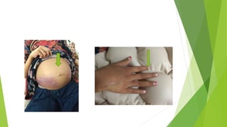
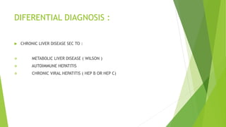
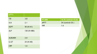
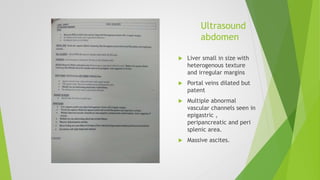
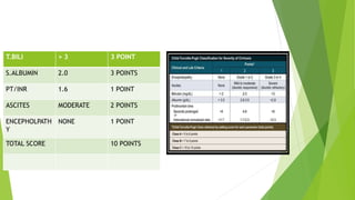
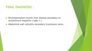
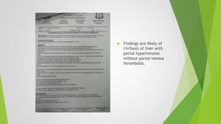
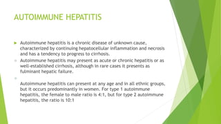
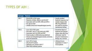
MORNING MEETING Dr.Hadia nasir
Unit 3
Presenting complain
 11-year-old female child, vaccinated, weighing 20 kg resident of Larkana
admitted with c/o :
Abdominal distention – 1 year
blood in stool– 2 episodes
HOPC
 BIRTH HISTORY : Non significant.
 IMMUNIZATION HISTORY :Completely vaccinated according to EPI schedule.
 NUTRITION HISTORY : Consumes up to 1200 kcal per day including dairy,
vegetables and meat. No history of fava bean consumption in diet.
 DEVELOPMENTAL HISTORY : Developmentally normal
 PAST MEDICAL HISTORY : No significant history of hospital admissions and IV
medications before this illness.
 PAST SURGICAL HISTORY : Non significant.
 FAMILY HISTORY : 6th product of consanguineous marriage. All elder siblings
are alive and healthy. Mother had history of hepB which was treated before
her birth.
 DRUG AND TRANSFUSION HISTORY : No history of blood transfusions. No
significant history of prolonged use of painkillers , anti epileptics or any other
drug.
 PERSONAL HISTORY : sleep and eating habits were normal before this illness.
Normal bowel and bladder habits. Drinks unboiled tap water.
 SOCIOECONOMIC HISTORY : belongs to middle class family, lives in well
ventilated house.
GENERAL PHYSICALEXAMINATION
 Young girl lying on bed well oriented with following
vitals:
 HR = 84b/m
 RR= 24b/m
 BP = 100/60mmhg ( 50th centile)
 SO2= 96% at room air
Patient has A+, CL+, E+ at the time of admission while
No signs of J-,CY-, LN-,D-
 Weight = 30kg (-1.23 SDS)
 Height =130cm ( -2.22 SDS)
HEADTO TOE EXAMINATION
 Clubbing positive
 Nail & palmer crease pale
 Purplish discoloration of skin at both right lumber
regions
 No cervical lymphadenopathy.
 No gum bleeding or oral ulcer.
 No jaundice ,Palmar erythema, Asterixis,
koilonychias, leukonychia, Spider navi, Caput
medusa ,Telangiectasia
 BCG scar present.
 All joints normal with no tenderness & swelling
 No bruising or bleeding seen With no spider nevi
seen
ABDOMINAL EXAMINATION
 on inspection abdomen is grossly distended with slit like
centrally placed everted umbilicus ,abdomen is moving with
respiration. Purplish discoloration of both sides of lower abdomen
is present extending to back , cough reflex is -ve
 on palpation abdomen is soft distended mildly tender and
abdominal grith is 40cm
 visceromegaly not appreciated due to massive ascites
 on percussion : fluid thrill and shifting dullness is positive
 on auscultation gut sounds audible with no abdominal bruits
 perineum and genital exam is unremarkable with no pubic hair
.
 CNSEXAMINATION
 CVSEXAMINATION
 CHESTEXAMINATION
6/4/2023 11
UNREMARKABLE
DIFERENTIAL DIAGNOSIS :
 CHRONIC LIVER DISEASE SEC TO :
 METABOLIC LIVER DISEASE ( WILSON )
 AUTOIMMUNE HEPATITIS
 CHRONIC VIRAL HEPATITIS ( HEP B OR HEP C)
INVESTIGATIONS
CBC
Hb 7.6
MCV 60.6
TLC 11.6
L 06
N 91
PLT 199
SUCE
Ur 26
Cr 0.4
Na 135
K 4.0
Cl 100
Ca 6.8
LFT’s
T.B 3.8
D.B 2.1
SGPT 50 (N=41)
ALP 130 (N=480)
ALBUMIN 2.0
G.GT 24 (N=40)
CRP 1.0
PT/INR 16.9( control=10.8)
APTT 34 (control=35 )
INR 1.6
Ultrasound
abdomen
 Liver small in size with
heterogenous texture
and irregular margins
 Portal veins dilated but
patent
 Multiple abnormal
vascular channels seen in
epigastric ,
peripancreatic and peri
splenic area.
 Massive ascites.
T.BILI > 3 3 POINT
S.ALBUMIN 2.0 3 POINTS
PT/INR 1.6 1 POINT
ASCITES MODERATE 2 POINTS
ENCEPHOLPATH
Y
NONE 1 POINT
TOTAL SCORE 10 POINTS
Workup for Wilson disease :
 Serum ceruloplasmin : 0.29 age 4-12yrs ( 0.25-0.45
)
 Urinary copper : 19.08 ug/Day normal : (less than
60ug/day )
Viral markers
 Anti HCV = non reactive
 HbsAg = non reactive
ANA PROFILE :
 ANA = POSITIVE
 ASMA = POSITIVE
COOMB’S TEST
Direct coombs test : positive.
IMMUNOGLOBULIN :
SERUM IgG : 24.7 (6.5-15 g/dl )
FINAL DIAGNOSIS :
 Decompensated chronic liver disease secondary to
Autoimmune hepatitis ( type 1 ) .
 Abdominal wall cellulitis secondary to pressure sores.
GASTRO OPINION
 Gastroenterology was taken on board , they advised to
continue conservative management .
 Add tablet rifaximine and tab Aldactone.
 Liver biopsy in plan.
MANAGEMENT IN WARD
 Admit in unit 3
 IV line maintained
 IV fluids started
 IV antibiotics started
 Inj vit K and omeprazole started.
 Syp duphalac 30ml HS
 Tab Aldactone 25mg 1+1+1
 Tab rifixamine 500mg 1 OD
 Findings are likely of
cirrhosis of liver with
portal hypertension
without portal venous
thrombosis.
AUTOIMMUNE HEPATITIS
AUTOIMMUNE HEPATITIS
 Autoimmune hepatitis is a chronic disease of unknown cause,
characterized by continuing hepatocellular inflammation and necrosis
and has a tendency to progress to cirrhosis.
 Autoimmune hepatitis may present as acute or chronic hepatitis or as
well-established cirrhosis, although in rare cases it presents as
fulminant hepatic failure.

Autoimmune hepatitis can present at any age and in all ethnic groups,
but it occurs predominantly in women. For type 1 autoimmune
hepatitis, the female to male ratio is 4:1, but for type 2 autoimmune
hepatitis, the ratio is 10:1
TYPES OF AIH :
DIAGNOSIS :
 Evidence for Autoimmune hepatitis include:
 Elevation in transaminases.
 Association with hypergammaglobulinemia and the
presence of a rheumatoid factor
 Circulating autoantibodies (ie, nuclear, smooth muscle,
thyroid, liver-kidney microsomal, soluble liver antigen,
hepatic lectin)
 Hepatic histopathologic lesions composed predominantly
of cytotoxic T cells and plasma cells
 Association with other autoimmune diseases
 Response to steroid and/or immunosuppressive therapy
DIAGNOSTIC SCORING SYSTEM FOR AIH
NON INVASIVE FIBROSIS ASSESMENT :
 FibroTest (APRI) : AST/Platelet ratio index
 Fibroscan (VCTE )
 MRE
THERAPEUTIC STRATEGY :
TREATMENT END-POINTS :
Patient may achieve 1 of 4 end points
 Remission: Remission is indicated by the absence of symptoms,
normalization of aminotransferases, and histologic improvement to
normal or minimal inflammatory activity on liver biopsy.
 Treatment failure: Treatment failure is defined as deterioration in a
patient's clinical condition, laboratory tests, or histologic features
during therapy.
 Incomplete response: Incomplete response is defined as an
improvement that is insufficient to satisfy remission criteria. It is
estimated to occur in 13% of patients.
 Drug toxicity: Drug toxicity may occur. Patients must be tapered off
from the culprit medication. Some patients successfully achieve
treatment goals on alternative medications.
 RELAPSE
Relapse occurs in 50% of patients within 6 months of
treatment withdrawal and in 80% of patients within 3 years
of treatment. Reinstitution of the original treatment
regimen usually induces another remission; however, relapse
commonly recurs after a second attempt at terminating
therapy. The major consequence of relapse and re-treatment
is the development of drug-related complications, which
occurs in 70% of patients
PROGNOSIS :
 Without treatment, approximately 40% to 50% of the individuals
with severe disease will die within 6 months to 5 years.
Treatment with steroids has dramatically changed the course of
the disease. Most patients respond to therapy and the 10-year
survival rate is approximately 83.8% to 94%.
 The long-term outlook after liver transplantation is excellent,
with 10-year survival rates reported as greater than
70%.Positive autoantibodies and hypergammaglobulinemia tend
to disappear within 2 years of transplantation.
 Treatment failure, Relapse and need for long term Maintenance
is common in type 2 AIH. So, compared to type 1 AIH it’s
prognosis is poor.
Case presentation on AUTOIMMUNE HEP final.pptx

Case presentation on AUTOIMMUNE HEP final.pptx

  • 1.
  • 2.
    Presenting complain  11-year-oldfemale child, vaccinated, weighing 20 kg resident of Larkana admitted with c/o : Abdominal distention – 1 year blood in stool– 2 episodes
  • 3.
  • 4.
     BIRTH HISTORY: Non significant.  IMMUNIZATION HISTORY :Completely vaccinated according to EPI schedule.  NUTRITION HISTORY : Consumes up to 1200 kcal per day including dairy, vegetables and meat. No history of fava bean consumption in diet.  DEVELOPMENTAL HISTORY : Developmentally normal
  • 5.
     PAST MEDICALHISTORY : No significant history of hospital admissions and IV medications before this illness.  PAST SURGICAL HISTORY : Non significant.  FAMILY HISTORY : 6th product of consanguineous marriage. All elder siblings are alive and healthy. Mother had history of hepB which was treated before her birth.  DRUG AND TRANSFUSION HISTORY : No history of blood transfusions. No significant history of prolonged use of painkillers , anti epileptics or any other drug.
  • 6.
     PERSONAL HISTORY: sleep and eating habits were normal before this illness. Normal bowel and bladder habits. Drinks unboiled tap water.  SOCIOECONOMIC HISTORY : belongs to middle class family, lives in well ventilated house.
  • 7.
    GENERAL PHYSICALEXAMINATION  Younggirl lying on bed well oriented with following vitals:  HR = 84b/m  RR= 24b/m  BP = 100/60mmhg ( 50th centile)  SO2= 96% at room air Patient has A+, CL+, E+ at the time of admission while No signs of J-,CY-, LN-,D-  Weight = 30kg (-1.23 SDS)  Height =130cm ( -2.22 SDS)
  • 8.
    HEADTO TOE EXAMINATION Clubbing positive  Nail & palmer crease pale  Purplish discoloration of skin at both right lumber regions  No cervical lymphadenopathy.  No gum bleeding or oral ulcer.  No jaundice ,Palmar erythema, Asterixis, koilonychias, leukonychia, Spider navi, Caput medusa ,Telangiectasia  BCG scar present.  All joints normal with no tenderness & swelling  No bruising or bleeding seen With no spider nevi seen
  • 10.
    ABDOMINAL EXAMINATION  oninspection abdomen is grossly distended with slit like centrally placed everted umbilicus ,abdomen is moving with respiration. Purplish discoloration of both sides of lower abdomen is present extending to back , cough reflex is -ve  on palpation abdomen is soft distended mildly tender and abdominal grith is 40cm  visceromegaly not appreciated due to massive ascites  on percussion : fluid thrill and shifting dullness is positive  on auscultation gut sounds audible with no abdominal bruits  perineum and genital exam is unremarkable with no pubic hair
  • 11.
    .  CNSEXAMINATION  CVSEXAMINATION CHESTEXAMINATION 6/4/2023 11 UNREMARKABLE
  • 12.
    DIFERENTIAL DIAGNOSIS : CHRONIC LIVER DISEASE SEC TO :  METABOLIC LIVER DISEASE ( WILSON )  AUTOIMMUNE HEPATITIS  CHRONIC VIRAL HEPATITIS ( HEP B OR HEP C)
  • 13.
  • 14.
    CBC Hb 7.6 MCV 60.6 TLC11.6 L 06 N 91 PLT 199
  • 15.
    SUCE Ur 26 Cr 0.4 Na135 K 4.0 Cl 100 Ca 6.8
  • 16.
    LFT’s T.B 3.8 D.B 2.1 SGPT50 (N=41) ALP 130 (N=480) ALBUMIN 2.0 G.GT 24 (N=40) CRP 1.0 PT/INR 16.9( control=10.8) APTT 34 (control=35 ) INR 1.6
  • 17.
    Ultrasound abdomen  Liver smallin size with heterogenous texture and irregular margins  Portal veins dilated but patent  Multiple abnormal vascular channels seen in epigastric , peripancreatic and peri splenic area.  Massive ascites.
  • 18.
    T.BILI > 33 POINT S.ALBUMIN 2.0 3 POINTS PT/INR 1.6 1 POINT ASCITES MODERATE 2 POINTS ENCEPHOLPATH Y NONE 1 POINT TOTAL SCORE 10 POINTS
  • 19.
    Workup for Wilsondisease :  Serum ceruloplasmin : 0.29 age 4-12yrs ( 0.25-0.45 )  Urinary copper : 19.08 ug/Day normal : (less than 60ug/day )
  • 20.
    Viral markers  AntiHCV = non reactive  HbsAg = non reactive
  • 21.
    ANA PROFILE : ANA = POSITIVE  ASMA = POSITIVE
  • 22.
  • 23.
    IMMUNOGLOBULIN : SERUM IgG: 24.7 (6.5-15 g/dl )
  • 24.
    FINAL DIAGNOSIS : Decompensated chronic liver disease secondary to Autoimmune hepatitis ( type 1 ) .  Abdominal wall cellulitis secondary to pressure sores.
  • 25.
    GASTRO OPINION  Gastroenterologywas taken on board , they advised to continue conservative management .  Add tablet rifaximine and tab Aldactone.  Liver biopsy in plan.
  • 26.
    MANAGEMENT IN WARD Admit in unit 3  IV line maintained  IV fluids started  IV antibiotics started  Inj vit K and omeprazole started.  Syp duphalac 30ml HS  Tab Aldactone 25mg 1+1+1  Tab rifixamine 500mg 1 OD
  • 27.
     Findings arelikely of cirrhosis of liver with portal hypertension without portal venous thrombosis.
  • 28.
  • 29.
    AUTOIMMUNE HEPATITIS  Autoimmunehepatitis is a chronic disease of unknown cause, characterized by continuing hepatocellular inflammation and necrosis and has a tendency to progress to cirrhosis.  Autoimmune hepatitis may present as acute or chronic hepatitis or as well-established cirrhosis, although in rare cases it presents as fulminant hepatic failure.  Autoimmune hepatitis can present at any age and in all ethnic groups, but it occurs predominantly in women. For type 1 autoimmune hepatitis, the female to male ratio is 4:1, but for type 2 autoimmune hepatitis, the ratio is 10:1
  • 30.
  • 32.
    DIAGNOSIS :  Evidencefor Autoimmune hepatitis include:  Elevation in transaminases.  Association with hypergammaglobulinemia and the presence of a rheumatoid factor  Circulating autoantibodies (ie, nuclear, smooth muscle, thyroid, liver-kidney microsomal, soluble liver antigen, hepatic lectin)  Hepatic histopathologic lesions composed predominantly of cytotoxic T cells and plasma cells  Association with other autoimmune diseases  Response to steroid and/or immunosuppressive therapy
  • 34.
  • 35.
    NON INVASIVE FIBROSISASSESMENT :  FibroTest (APRI) : AST/Platelet ratio index  Fibroscan (VCTE )  MRE
  • 36.
  • 39.
    TREATMENT END-POINTS : Patientmay achieve 1 of 4 end points  Remission: Remission is indicated by the absence of symptoms, normalization of aminotransferases, and histologic improvement to normal or minimal inflammatory activity on liver biopsy.  Treatment failure: Treatment failure is defined as deterioration in a patient's clinical condition, laboratory tests, or histologic features during therapy.  Incomplete response: Incomplete response is defined as an improvement that is insufficient to satisfy remission criteria. It is estimated to occur in 13% of patients.  Drug toxicity: Drug toxicity may occur. Patients must be tapered off from the culprit medication. Some patients successfully achieve treatment goals on alternative medications.
  • 40.
     RELAPSE Relapse occursin 50% of patients within 6 months of treatment withdrawal and in 80% of patients within 3 years of treatment. Reinstitution of the original treatment regimen usually induces another remission; however, relapse commonly recurs after a second attempt at terminating therapy. The major consequence of relapse and re-treatment is the development of drug-related complications, which occurs in 70% of patients
  • 41.
    PROGNOSIS :  Withouttreatment, approximately 40% to 50% of the individuals with severe disease will die within 6 months to 5 years. Treatment with steroids has dramatically changed the course of the disease. Most patients respond to therapy and the 10-year survival rate is approximately 83.8% to 94%.  The long-term outlook after liver transplantation is excellent, with 10-year survival rates reported as greater than 70%.Positive autoantibodies and hypergammaglobulinemia tend to disappear within 2 years of transplantation.  Treatment failure, Relapse and need for long term Maintenance is common in type 2 AIH. So, compared to type 1 AIH it’s prognosis is poor.