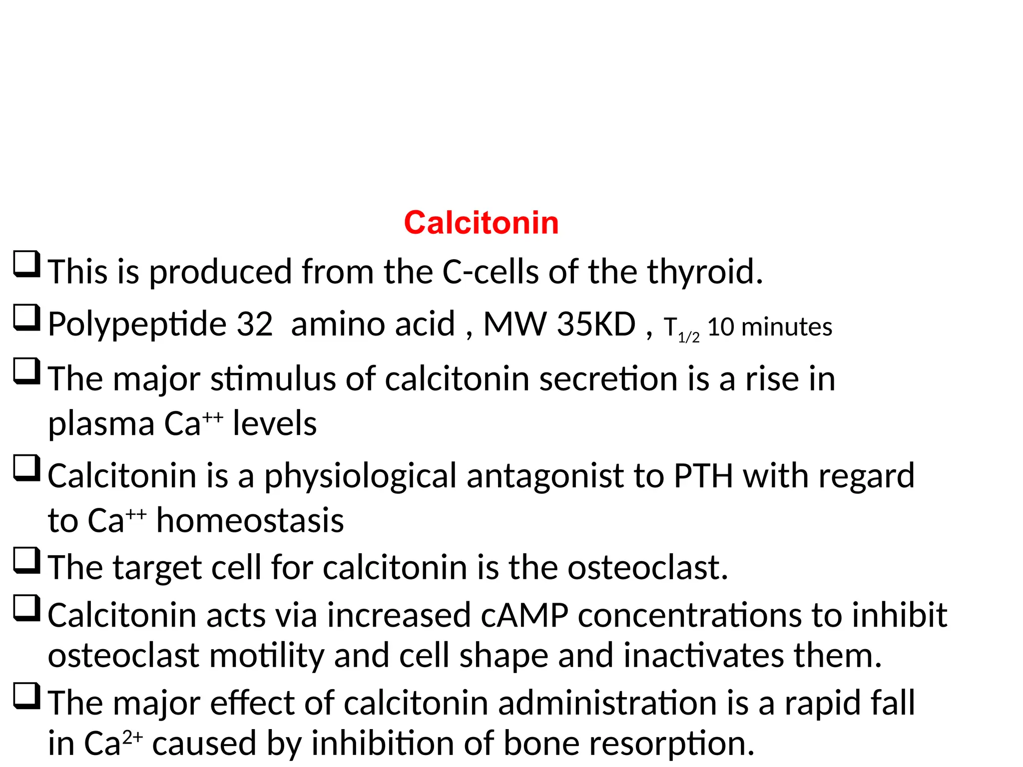
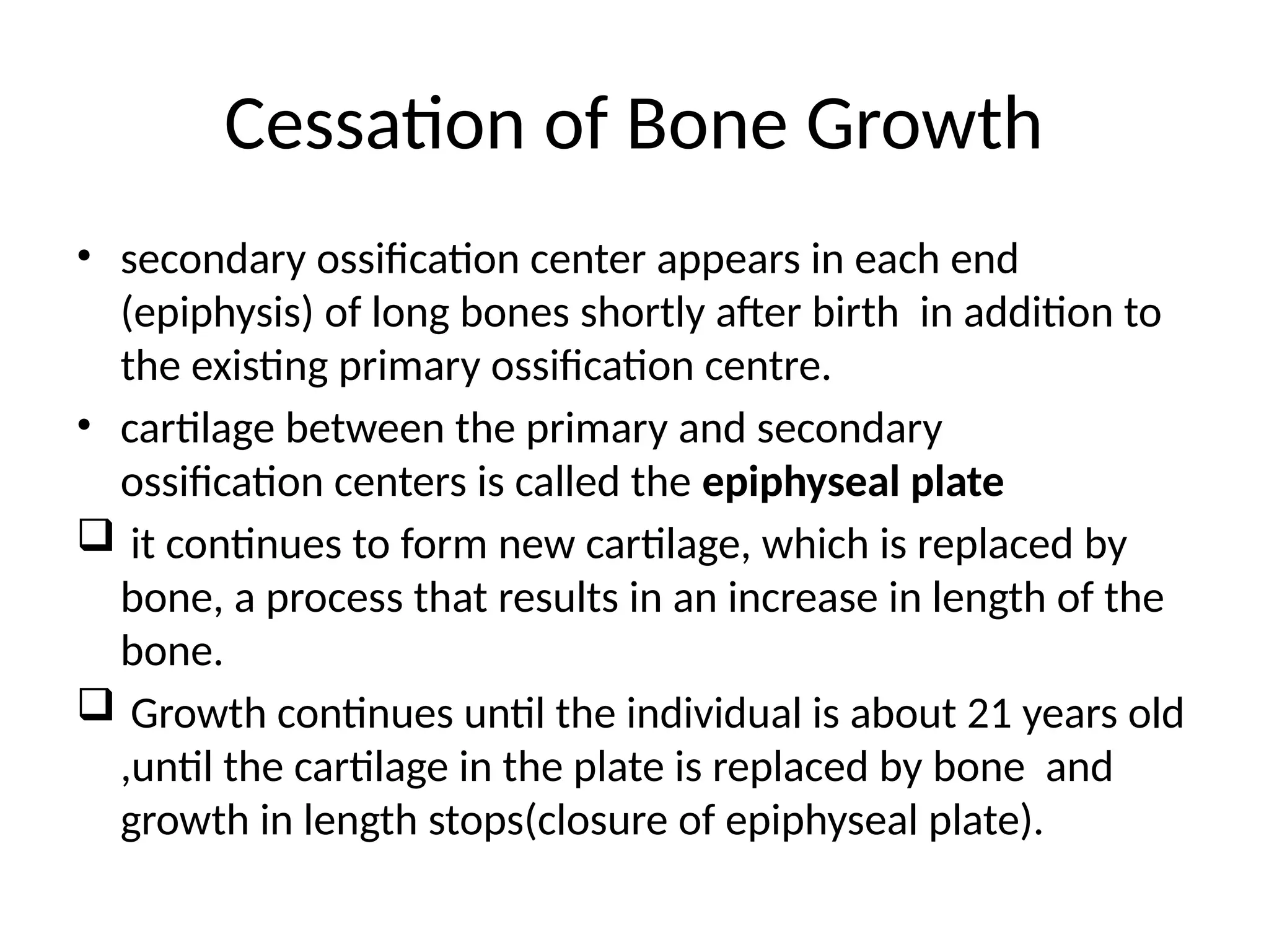
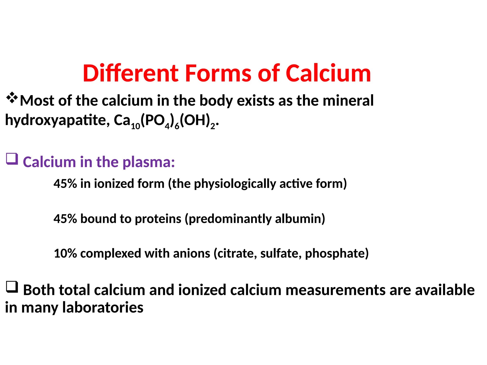
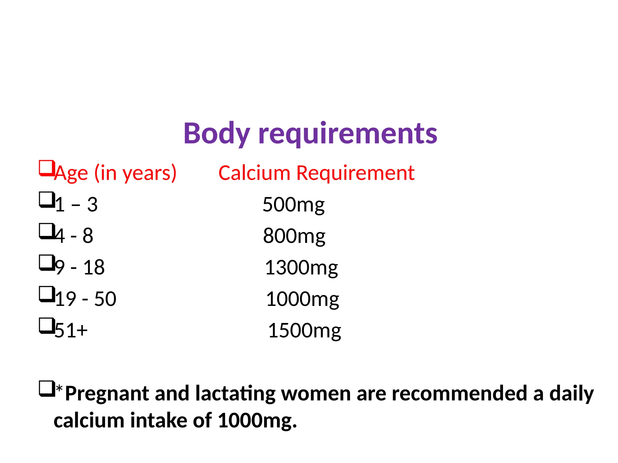
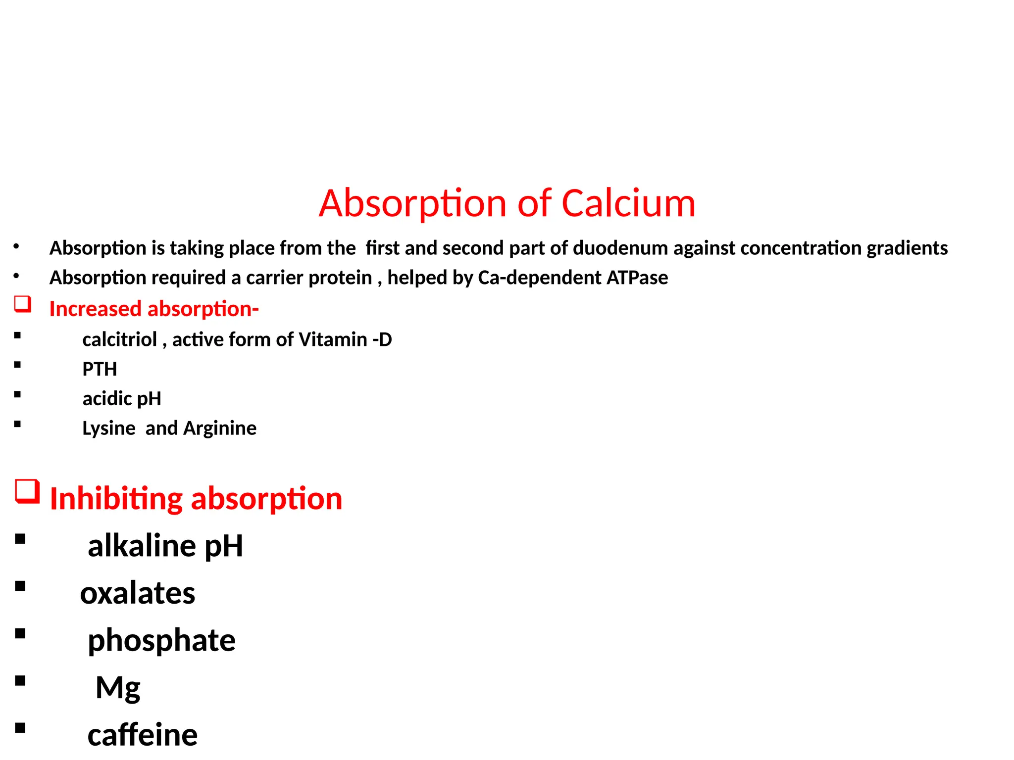
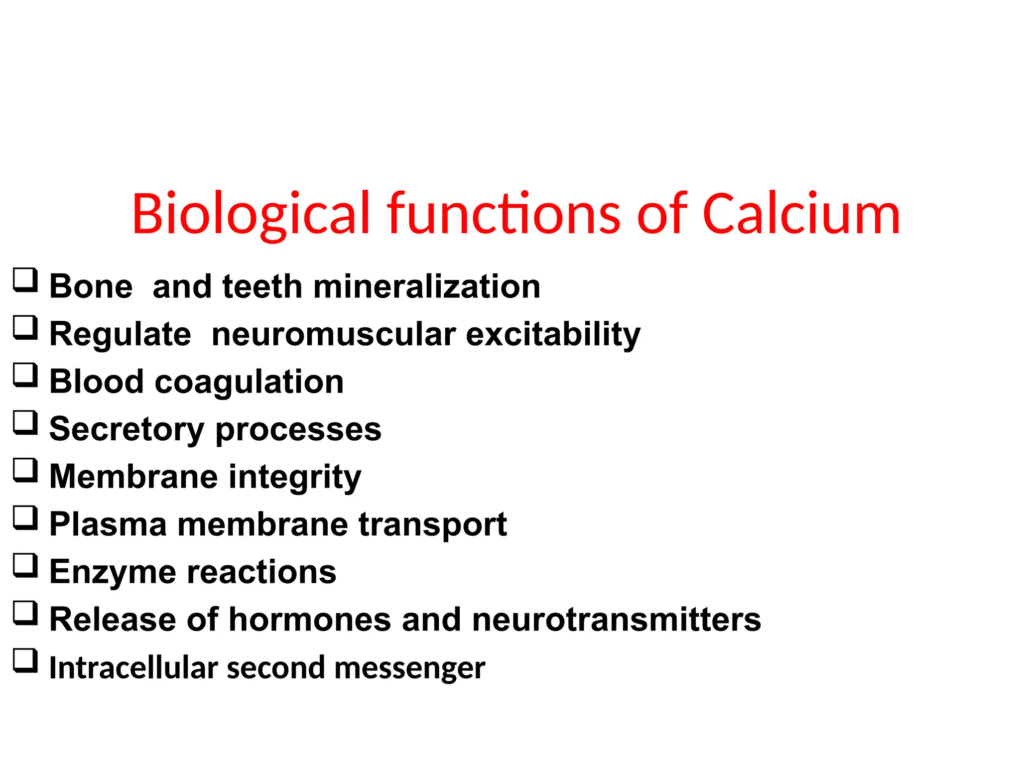
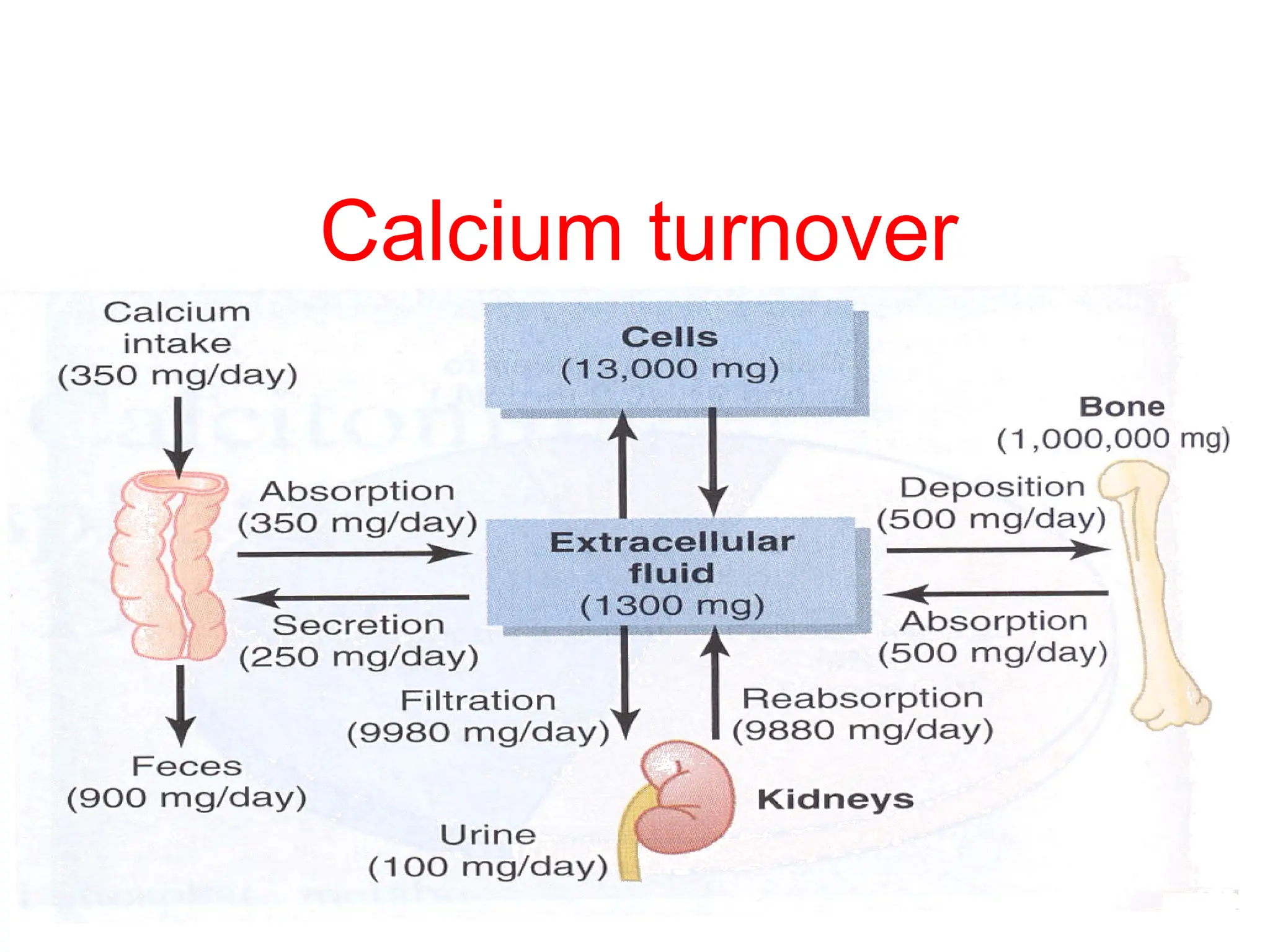
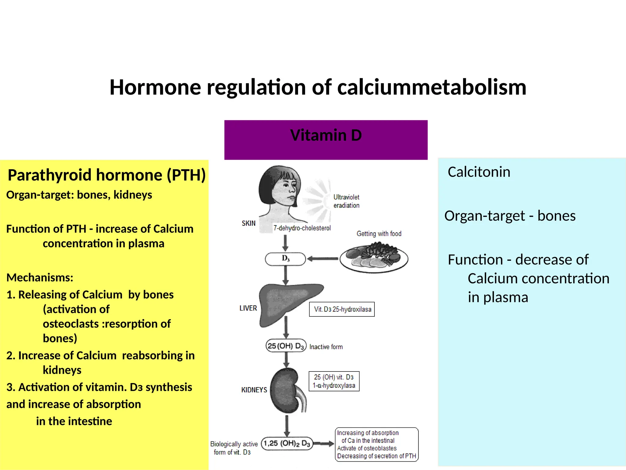
Calcium metabolism
Distribution
Different Forms of Calcium
Most of the calcium in the body exists as the mineral
hydroxyapatite, Ca10(PO4)6(OH)2.
 Calcium in the plasma:
45% in ionized form (the physiologically active form)
45% bound to proteins (predominantly albumin)
10% complexed with anions (citrate, sulfate, phosphate)
 Both total calcium and ionized calcium measurements are available
in many laboratories
Body requirements
Age (in years) Calcium Requirement
1 – 3 500mg
4 - 8 800mg
9 - 18 1300mg
19 - 50 1000mg
51+ 1500mg
*Pregnant and lactating women are recommended a daily
calcium intake of 1000mg.
source
 Calcium is found in milk and dairy products,
 Green leafy vegetables,
seafood,
almonds,
blackstrap molasses,
broccoli,
enriched soy and rice milk products, figs,
 soybeans
Absorption of Calcium
• Absorption is taking place from the first and second part of duodenum against concentration gradients
• Absorption required a carrier protein , helped by Ca-dependent ATPase
 Increased absorption-
 calcitriol , active form of Vitamin -D
 PTH
 acidic pH
 Lysine and Arginine
 Inhibiting absorption
 alkaline pH
 oxalates
 phosphate
 Mg
 caffeine
Biological functions of Calcium
 Bone and teeth mineralization
 Regulate neuromuscular excitability
 Blood coagulation
 Secretory processes
 Membrane integrity
 Plasma membrane transport
 Enzyme reactions
 Release of hormones and neurotransmitters
 Intracellular second messenger
Calcium turnover
Hormone regulation of calciummetabolism
Parathyroid hormone (PTH)
Organ-target: bones, kidneys
Function of PTH - increase of Calcium
concentration in plasma
Mechanisms:
1. Releasing of Саlcium by bones
(activation of
osteoclasts :resorption of
bones)
2. Increase of Саlcium reabsorbing in
kidneys
3. Activation of vitamin. Dз synthesis
and increase of absorption
in the intestine
Vitamin D
Calcitonin
Organ-target - bones
Function - decrease of
Calcium concentration
in plasma
Vitamin D3
 Dietary cholesterol is converted into 7-dehydrocholesterol and transported to
skin
 UV sunlight (290-320nm) penetrates the skin to break provitamine 7-
dehydrocholesterol which is converted to Cholecalciferol by the process of
isomerisation
 In the liver, cholecalciferol undergoes 25-hydroxylation to yield 25(OH) Vitamine
-D ( calcidiol)
 In the kidney , calcidiol undergoes further 1α-hydroxylation to produce 1,25 –
dihydroxy Vit-D (Calcitriol). Its production in the kidney is catalyzed by 1α -
hydroxylase
Vitamin D3
α -hydroxylase activity is increased by :
Decreased serum Ca2+
Increased PTH level
Decreased serum phosphate
Action of 1,25-dihydroxycholecalcififerol(Calcitriol)
• Increases intestinal Ca2+ absorption
• Increases intestinal phosphate absorption
• Increase renal reabsorption of Ca2+ and phosphate
• Increases osteoclast activity
Vitamin D3 and Calcium Control
Vitamin D3 promotes intestinal calcium absorption
• Vitamin D3 acts via steroid hormone like receptor to
increase transcriptional and translational activity
• One gene product is calcium-binding protein (Ca BP)
• CaBP facilitates calcium uptake by intestinal cells
• Estrogen, prolactin and growth hormone also
stimulate 1α -hydroxylase thus increasing Ca absorption
during pregnancy, lactation and growth
Vitamin D3 Actions on Bones
 Another important target for 1,25-(OH)2
-D3 is the bone.
 Osteoblasts, but not osteoclasts have vitamin D3 receptors.
 1,25-(OH)2
-D3 acts on osteoblasts which produce a paracrine signal
that activates osteoclasts to resorb Ca++
from the bone matrix.
 1,25-(OH)2
-D3 also stimulates osteocytic osteolysis.
 In its absence, excess osteoid accumulates from lack of 1,25-(OH)2
-
D3 repression of osteoblastic collagen synthesis.
 Inadequate supply of vitamin D3 results in rickets, a disease of
bone deformation
):
 It is synthesised as pre-pro-PTH(115aa) and is
cleaved to pro-PTH(90aa) with cleavage before
secretion of PTH(84aa).
 Intact PTH T1/2 3-4 mins
 Normal levels 1.3 – 6.8 pmol/L
 Secreted from the chief cells of the parathyroid glands.
Function:
Increase renal phosphate excretion , and increases plasma calcium by:
Increasing osteoclastic resorption of bone (occurring rapidly).
Increasing intestinal absorption of calcium (a slower response).
Increasing synthesis of 1,25-(OH)2D3 (stimulating GIT absorption).
Increasing renal tubular reabsorption of calcium
Parathyroid hormone (PTH
PTH action
The overall action of PTH is to increase plasma Ca++
levels and decrease plasma phosphate levels.
PTH acts directly on the bones to stimulate Ca++
resorption and kidney to stimulate Ca++ reabsorption in
the distal tubule of the kidney and to inhibit
reabosorptioin of phosphate (thereby stimulating its
excretion).
PTH also acts indirectly on intestine by stimulating 1,25-
(OH)2
-D synthesis.
PTH indirectly increases Calcium absorption from GIT
Regulation of PTH
 The dominant regulator of PTH is plasma Ca2+
.
 Secretion of PTH is inversely related to [Ca2+
].
 Maximum secretion of PTH occurs at plasma Ca2+
below 3.5 mg/dL.
 At Ca2+
above 5.5 mg/dL, PTH secretion is maximally inhibited.
 PTH secretion responds to small alterations in plasma Ca2+
within
seconds
 A unique calcium receptor within the parathyroid cell plasma membrane
senses changes in the extracellular fluid concentration of Ca2+
.
 A unique calcium receptor within the parathyroid cell plasma membrane senses
changes in the extracellular fluid concentration of Ca2+
 This is a typical G-protein coupled receptor that activates phospholipase C and
adenylate cyclase:result is increase in intracellular Ca2+
via generation of inositol
phosphates and decrease in cAMP which prevents exocytosis of PTH from
secretory granules.
 When Ca2+
falls, cAMP rises and PTH is secreted.
 1,25-(OH)2
-D inhibits PTH gene expression, providing another level of feedback
control of PTH.
 Despite close connection between Ca2+
and PO4
, no direct control of PTH is exerted
by phosphate levels.
Calcitonin
This is produced from the C-cells of the thyroid.
Polypeptide 32 amino acid , MW 35KD , T1/2 10 minutes
The major stimulus of calcitonin secretion is a rise in
plasma Ca++
levels
Calcitonin is a physiological antagonist to PTH with regard
to Ca++
homeostasis
The target cell for calcitonin is the osteoclast.
Calcitonin acts via increased cAMP concentrations to inhibit
osteoclast motility and cell shape and inactivates them.
The major effect of calcitonin administration is a rapid fall
in Ca2+
caused by inhibition of bone resorption.
Cessation of Bone Growth
• secondary ossification center appears in each end
(epiphysis) of long bones shortly after birth in addition to
the existing primary ossification centre.
• cartilage between the primary and secondary
ossification centers is called the epiphyseal plate
 it continues to form new cartilage, which is replaced by
bone, a process that results in an increase in length of the
bone.
 Growth continues until the individual is about 21 years old
,until the cartilage in the plate is replaced by bone and
growth in length stops(closure of epiphyseal plate).
END

calcium metabolism .pptx

  • 1.
  • 2.
  • 3.
    Different Forms ofCalcium Most of the calcium in the body exists as the mineral hydroxyapatite, Ca10(PO4)6(OH)2.  Calcium in the plasma: 45% in ionized form (the physiologically active form) 45% bound to proteins (predominantly albumin) 10% complexed with anions (citrate, sulfate, phosphate)  Both total calcium and ionized calcium measurements are available in many laboratories
  • 4.
    Body requirements Age (inyears) Calcium Requirement 1 – 3 500mg 4 - 8 800mg 9 - 18 1300mg 19 - 50 1000mg 51+ 1500mg *Pregnant and lactating women are recommended a daily calcium intake of 1000mg.
  • 5.
    source  Calcium isfound in milk and dairy products,  Green leafy vegetables, seafood, almonds, blackstrap molasses, broccoli, enriched soy and rice milk products, figs,  soybeans
  • 6.
    Absorption of Calcium •Absorption is taking place from the first and second part of duodenum against concentration gradients • Absorption required a carrier protein , helped by Ca-dependent ATPase  Increased absorption-  calcitriol , active form of Vitamin -D  PTH  acidic pH  Lysine and Arginine  Inhibiting absorption  alkaline pH  oxalates  phosphate  Mg  caffeine
  • 7.
    Biological functions ofCalcium  Bone and teeth mineralization  Regulate neuromuscular excitability  Blood coagulation  Secretory processes  Membrane integrity  Plasma membrane transport  Enzyme reactions  Release of hormones and neurotransmitters  Intracellular second messenger
  • 8.
  • 9.
    Hormone regulation ofcalciummetabolism Parathyroid hormone (PTH) Organ-target: bones, kidneys Function of PTH - increase of Calcium concentration in plasma Mechanisms: 1. Releasing of Саlcium by bones (activation of osteoclasts :resorption of bones) 2. Increase of Саlcium reabsorbing in kidneys 3. Activation of vitamin. Dз synthesis and increase of absorption in the intestine Vitamin D Calcitonin Organ-target - bones Function - decrease of Calcium concentration in plasma
  • 10.
    Vitamin D3  Dietarycholesterol is converted into 7-dehydrocholesterol and transported to skin  UV sunlight (290-320nm) penetrates the skin to break provitamine 7- dehydrocholesterol which is converted to Cholecalciferol by the process of isomerisation  In the liver, cholecalciferol undergoes 25-hydroxylation to yield 25(OH) Vitamine -D ( calcidiol)  In the kidney , calcidiol undergoes further 1α-hydroxylation to produce 1,25 – dihydroxy Vit-D (Calcitriol). Its production in the kidney is catalyzed by 1α - hydroxylase
  • 11.
    Vitamin D3 α -hydroxylaseactivity is increased by : Decreased serum Ca2+ Increased PTH level Decreased serum phosphate Action of 1,25-dihydroxycholecalcififerol(Calcitriol) • Increases intestinal Ca2+ absorption • Increases intestinal phosphate absorption • Increase renal reabsorption of Ca2+ and phosphate • Increases osteoclast activity
  • 12.
    Vitamin D3 andCalcium Control
  • 13.
    Vitamin D3 promotesintestinal calcium absorption • Vitamin D3 acts via steroid hormone like receptor to increase transcriptional and translational activity • One gene product is calcium-binding protein (Ca BP) • CaBP facilitates calcium uptake by intestinal cells • Estrogen, prolactin and growth hormone also stimulate 1α -hydroxylase thus increasing Ca absorption during pregnancy, lactation and growth
  • 14.
    Vitamin D3 Actionson Bones  Another important target for 1,25-(OH)2 -D3 is the bone.  Osteoblasts, but not osteoclasts have vitamin D3 receptors.  1,25-(OH)2 -D3 acts on osteoblasts which produce a paracrine signal that activates osteoclasts to resorb Ca++ from the bone matrix.  1,25-(OH)2 -D3 also stimulates osteocytic osteolysis.  In its absence, excess osteoid accumulates from lack of 1,25-(OH)2 - D3 repression of osteoblastic collagen synthesis.  Inadequate supply of vitamin D3 results in rickets, a disease of bone deformation
  • 15.
    ):  It issynthesised as pre-pro-PTH(115aa) and is cleaved to pro-PTH(90aa) with cleavage before secretion of PTH(84aa).  Intact PTH T1/2 3-4 mins  Normal levels 1.3 – 6.8 pmol/L  Secreted from the chief cells of the parathyroid glands. Function: Increase renal phosphate excretion , and increases plasma calcium by: Increasing osteoclastic resorption of bone (occurring rapidly). Increasing intestinal absorption of calcium (a slower response). Increasing synthesis of 1,25-(OH)2D3 (stimulating GIT absorption). Increasing renal tubular reabsorption of calcium Parathyroid hormone (PTH
  • 16.
    PTH action The overallaction of PTH is to increase plasma Ca++ levels and decrease plasma phosphate levels. PTH acts directly on the bones to stimulate Ca++ resorption and kidney to stimulate Ca++ reabsorption in the distal tubule of the kidney and to inhibit reabosorptioin of phosphate (thereby stimulating its excretion). PTH also acts indirectly on intestine by stimulating 1,25- (OH)2 -D synthesis. PTH indirectly increases Calcium absorption from GIT
  • 17.
    Regulation of PTH The dominant regulator of PTH is plasma Ca2+ .  Secretion of PTH is inversely related to [Ca2+ ].  Maximum secretion of PTH occurs at plasma Ca2+ below 3.5 mg/dL.  At Ca2+ above 5.5 mg/dL, PTH secretion is maximally inhibited.  PTH secretion responds to small alterations in plasma Ca2+ within seconds  A unique calcium receptor within the parathyroid cell plasma membrane senses changes in the extracellular fluid concentration of Ca2+ .
  • 18.
     A uniquecalcium receptor within the parathyroid cell plasma membrane senses changes in the extracellular fluid concentration of Ca2+  This is a typical G-protein coupled receptor that activates phospholipase C and adenylate cyclase:result is increase in intracellular Ca2+ via generation of inositol phosphates and decrease in cAMP which prevents exocytosis of PTH from secretory granules.  When Ca2+ falls, cAMP rises and PTH is secreted.  1,25-(OH)2 -D inhibits PTH gene expression, providing another level of feedback control of PTH.  Despite close connection between Ca2+ and PO4 , no direct control of PTH is exerted by phosphate levels.
  • 19.
    Calcitonin This is producedfrom the C-cells of the thyroid. Polypeptide 32 amino acid , MW 35KD , T1/2 10 minutes The major stimulus of calcitonin secretion is a rise in plasma Ca++ levels Calcitonin is a physiological antagonist to PTH with regard to Ca++ homeostasis The target cell for calcitonin is the osteoclast. Calcitonin acts via increased cAMP concentrations to inhibit osteoclast motility and cell shape and inactivates them. The major effect of calcitonin administration is a rapid fall in Ca2+ caused by inhibition of bone resorption.
  • 21.
    Cessation of BoneGrowth • secondary ossification center appears in each end (epiphysis) of long bones shortly after birth in addition to the existing primary ossification centre. • cartilage between the primary and secondary ossification centers is called the epiphyseal plate  it continues to form new cartilage, which is replaced by bone, a process that results in an increase in length of the bone.  Growth continues until the individual is about 21 years old ,until the cartilage in the plate is replaced by bone and growth in length stops(closure of epiphyseal plate).
  • 22.

Editor's Notes

  • #10 Levels of 1,25 OHD do not decrease until defy is severe D2 - ergocalciferol