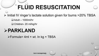
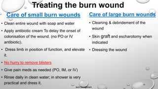
MANAGEMENT
OF BURN INJURY
Moderator : - Dr. Andinet Dessalegn
PRESENTER : - EYASU
OUTLINE
Pre hospital care of burn patient
Hospital care of burn patient
Indication for Escharotomy
Skin grafts and flaps treatment
burn management 2
OBJECTIVE
At the end of this presentation you are
expected to
Manage burn patient at the scene (pre hospital
care)
Manage burn patient in the hospital
Know indication of escharotomy
know Skin grafts and flaps treatment
burn management 3
PRE-HOSPITAL CARE /CARE AT THE
SCENE/
Stop the burning process :- stop, drop, roll sequence
Consider burn patient as a multiple trauma patient until determined otherwise
Check for ABC
Cold application
Remove constricting clothing and jewelry
Other injuries and transport
burn management 4
HOSPITAL CARE
 Can be divided in to two as any other trauma
management
Primary survey
Secondary survey
burn management 5
PRIMARY SURVEY
Airway
Early elective intubation is safest
Emergency cricothyroidotomy
Breathing
100% O2 via a non rebreathable mask
Circulation
Bleeding associated with trauma:- stop bleeding
Deformity
Due to trauma i.e. Fracture
Exposure
To poisonous chemicals e.g. CO poisoning
burn management
6
Secondary survey
Detailed history and
Head to toe examination with calculation of TBSA
And then managing the patient
burn management 7
Out come of treatment depends on
TBSA
Depth of burn
Presence of an inhalational injury
burn management 8
FLUID RESUSCITATION
 Initial IV ringer’s lactate solution given for burns >20% TBSA
 Adult – 1000ml/hr
 Children- 20 ml/kg/hr
PARKLAND
Formula= 4ml × wt. In kg × TBSA
burn management 9
LUND BROWDER CHART
burn management 10
MAINTENANCE FLUID
For the first 10 kg - 100ml/kg
For the next 10kg - 50ml/kg
For the rest kg - 20ml/kg
burn management 11
EXAMPLE
•Weight of a patient – 60 kg
•TBSA – 20%
Solution
PF- 4800ml per 24hr (4ml × 60 kg ×20)
MF = 1000ml+500ml+800ml =2300ml
TOTAL = 4800ml + 2300 ml=7100ml/24hrs
burn management 12
• In patients with large burns, do not initially spend much time
carefully calculating fluids
• Instead, start an IV and start giving fluids rather rapidly while exam is
being performed. Do not use bolus! 500 ml/hr. is a good rule.
• Later do the calculations
burn management 13
MONITORING OF RESUSCITATION
Urine output
 0.3 – 0.5ml/kg/hr in adults
 1.0ml/kg/hr in children
burn management 14
OTHERS
 Tetanus prophylaxis
Gastric decompression
Pain control
Psychological care
Care of burn wound, nutritional support
Escharotomy and fasciotomy
Skin graft
Amputation
burn management 15
Treating the burn wound
Care of small burn wounds
• Clean entire wound with soap and water
• Apply antibiotic cream To delay the onset of
colonisation of the wound. (no PO or IV
antibiotic).
• Dress limb in position of function, and elevate
it.
• No hurry to remove blisters
• Give pain meds as needed (PO, IM, or IV)
• Rinse daily in clean water; in shower is very
practical and dress it.
burn management
16
Care of large burn wounds
• Cleaning & debridement of the
wound
• Skin graft and escharotomy when
indicated
• Dressing the wound
Before and after debridement
• Removing the blister leaves a weeping, very tender wound, that requires
much care.
burn management
17
Silver sulfadiazene (1% is commonly used)
Options for topical
treatment
 0.5% silver nitrate solution
 Mafenide acetate cream
 Serum nitrate and cerium
nitrate
18
Escharotomy : - Circumferential full-thickness burns, in order to avoid the tourniquet
effect of this injury, is incised along the whole length of full-thickness burns.
Indication : - muscle compartment pressures > 30 mmHg, compartment syndrome
burn management 19
Inhalation injury
•Carbon monoxide poisoning – toxic=100% O2
•Upper airway thermal injury
•Lower airway burn injury
•Evaluate with bronchoscopy if uncertain
burn management 20
Criteria for intubation
• Changes in voice
• Wheezing / labored respirations
• Excessive, continuous coughing
• Altered mental status
• Singed facial or nasal hairs
• Facial burns, eyes swollen shut
• Oro-pharyngeal edema / stridor
• Assume inhalation injury in any patient confined in a fire environment
• Extensive burns of the face / neck
• Burns of 50% TBSA or greater
burn management 21
Chemical burns
• End the exposure
• Removing contaminated clothing can eliminate 85-90% of
trapped chemical substances
• Dry powder should be brushed off
• Initial treatment for acid or ALKALI:- irrigation with water
• ABCDE
• Empirical treatment
• Alkalis generally cause worse damage(more emphasis)
• Hydrofluoric acid causes hypocalcaemia (so we should give
IV Ca guluconate)
burn management 22
Summery of Hospital care of a burn patient
Skin grafts and flaps treatment
Graft
A tissue of epidermis
and varying amounts of
dermis that is detached from
its own blood supply and
placed in a new area with a
new blood supply.
Flap
Any tissue used for
reconstruction or wound
closure that retains all or part
of its original blood supply
after the tissue has been
moved to the recipient
location
burn management 24
Indications
Skin loss
- post traumatic (avulsion and degloving
injuries)
- post surgical (excision of burn wound)
Mucosal loss (excision lesion of oral cavity or
tongue)
Contraindication
Beta hemolytic streptococcal Infection
Infected wound with copious discharge
Avascular wound
Types of skin grafts
Definition
Split-
thickness
grafts
• superficial and some deep layers of skin
• Split-thickness grafts are used for non-weight-bearing parts of the body
Full-
thickness
grafts
• For weight-bearing portions of the body and friction prone areas such as, feet
and joints.
• contains all of the layers of the skin including blood vessels
Pinch grafts • quarter inch pieces of skin
• will then grow to cover injured sites (grow even in areas of poor blood supply
and resist infection)
Pedicle
grafts
• skin used from the donor site will remain attached to the donor area (remainder
is attached to the recipient site)
• blood supply remains intact at the donor location until the new blood supply has
completely developed.
• Mostly used for hands, face or neck areas of the body
GRAFT VS. FLAP
Graft
• Does not maintain original blood
supply
26
Flap
• Maintains original blood supply
• Flaps are chosen based on :-
 spatial relationship to the
defect
 Blood supply
Tissue type
Type of Graft Advantages Disadvantages
Thin Split
Thickness
-Best Survival
-Heals Rapidly
-Least resembles original skin.
-Least resistance to trauma.
-Poor Sensation
-Maximal Secondary Contraction
Thick Split
Thickness
-More qualities of normal skin.
-Less Contraction
-Looks better
-Fair Sensation
-Lower graft survival
-Slower healing
Full Thickness -Most resembles normal skin.
-Minimal Secondary contraction
-Resistant to trauma
-Good Sensation
-Poorest survival.
-Donor site must be closed surgically.
-Donor sites are limited.
burn management
27
References
• FAHC burn care manual
• Bailey & loves-short-practice-of-surgery-25th-edition
• Up to date online
• Google images
burn management 28
THANK YOU

burn eyasu.ppt

  • 1.
    MANAGEMENT OF BURN INJURY Moderator: - Dr. Andinet Dessalegn PRESENTER : - EYASU
  • 2.
    OUTLINE Pre hospital careof burn patient Hospital care of burn patient Indication for Escharotomy Skin grafts and flaps treatment burn management 2
  • 3.
    OBJECTIVE At the endof this presentation you are expected to Manage burn patient at the scene (pre hospital care) Manage burn patient in the hospital Know indication of escharotomy know Skin grafts and flaps treatment burn management 3
  • 4.
    PRE-HOSPITAL CARE /CAREAT THE SCENE/ Stop the burning process :- stop, drop, roll sequence Consider burn patient as a multiple trauma patient until determined otherwise Check for ABC Cold application Remove constricting clothing and jewelry Other injuries and transport burn management 4
  • 5.
    HOSPITAL CARE  Canbe divided in to two as any other trauma management Primary survey Secondary survey burn management 5
  • 6.
    PRIMARY SURVEY Airway Early electiveintubation is safest Emergency cricothyroidotomy Breathing 100% O2 via a non rebreathable mask Circulation Bleeding associated with trauma:- stop bleeding Deformity Due to trauma i.e. Fracture Exposure To poisonous chemicals e.g. CO poisoning burn management 6
  • 7.
    Secondary survey Detailed historyand Head to toe examination with calculation of TBSA And then managing the patient burn management 7
  • 8.
    Out come oftreatment depends on TBSA Depth of burn Presence of an inhalational injury burn management 8
  • 9.
    FLUID RESUSCITATION  InitialIV ringer’s lactate solution given for burns >20% TBSA  Adult – 1000ml/hr  Children- 20 ml/kg/hr PARKLAND Formula= 4ml × wt. In kg × TBSA burn management 9
  • 10.
  • 11.
    MAINTENANCE FLUID For thefirst 10 kg - 100ml/kg For the next 10kg - 50ml/kg For the rest kg - 20ml/kg burn management 11
  • 12.
    EXAMPLE •Weight of apatient – 60 kg •TBSA – 20% Solution PF- 4800ml per 24hr (4ml × 60 kg ×20) MF = 1000ml+500ml+800ml =2300ml TOTAL = 4800ml + 2300 ml=7100ml/24hrs burn management 12
  • 13.
    • In patientswith large burns, do not initially spend much time carefully calculating fluids • Instead, start an IV and start giving fluids rather rapidly while exam is being performed. Do not use bolus! 500 ml/hr. is a good rule. • Later do the calculations burn management 13
  • 14.
    MONITORING OF RESUSCITATION Urineoutput  0.3 – 0.5ml/kg/hr in adults  1.0ml/kg/hr in children burn management 14
  • 15.
    OTHERS  Tetanus prophylaxis Gastricdecompression Pain control Psychological care Care of burn wound, nutritional support Escharotomy and fasciotomy Skin graft Amputation burn management 15
  • 16.
    Treating the burnwound Care of small burn wounds • Clean entire wound with soap and water • Apply antibiotic cream To delay the onset of colonisation of the wound. (no PO or IV antibiotic). • Dress limb in position of function, and elevate it. • No hurry to remove blisters • Give pain meds as needed (PO, IM, or IV) • Rinse daily in clean water; in shower is very practical and dress it. burn management 16 Care of large burn wounds • Cleaning & debridement of the wound • Skin graft and escharotomy when indicated • Dressing the wound
  • 17.
    Before and afterdebridement • Removing the blister leaves a weeping, very tender wound, that requires much care. burn management 17
  • 18.
    Silver sulfadiazene (1%is commonly used) Options for topical treatment  0.5% silver nitrate solution  Mafenide acetate cream  Serum nitrate and cerium nitrate 18
  • 19.
    Escharotomy : -Circumferential full-thickness burns, in order to avoid the tourniquet effect of this injury, is incised along the whole length of full-thickness burns. Indication : - muscle compartment pressures > 30 mmHg, compartment syndrome burn management 19
  • 20.
    Inhalation injury •Carbon monoxidepoisoning – toxic=100% O2 •Upper airway thermal injury •Lower airway burn injury •Evaluate with bronchoscopy if uncertain burn management 20
  • 21.
    Criteria for intubation •Changes in voice • Wheezing / labored respirations • Excessive, continuous coughing • Altered mental status • Singed facial or nasal hairs • Facial burns, eyes swollen shut • Oro-pharyngeal edema / stridor • Assume inhalation injury in any patient confined in a fire environment • Extensive burns of the face / neck • Burns of 50% TBSA or greater burn management 21
  • 22.
    Chemical burns • Endthe exposure • Removing contaminated clothing can eliminate 85-90% of trapped chemical substances • Dry powder should be brushed off • Initial treatment for acid or ALKALI:- irrigation with water • ABCDE • Empirical treatment • Alkalis generally cause worse damage(more emphasis) • Hydrofluoric acid causes hypocalcaemia (so we should give IV Ca guluconate) burn management 22
  • 23.
    Summery of Hospitalcare of a burn patient
  • 24.
    Skin grafts andflaps treatment Graft A tissue of epidermis and varying amounts of dermis that is detached from its own blood supply and placed in a new area with a new blood supply. Flap Any tissue used for reconstruction or wound closure that retains all or part of its original blood supply after the tissue has been moved to the recipient location burn management 24 Indications Skin loss - post traumatic (avulsion and degloving injuries) - post surgical (excision of burn wound) Mucosal loss (excision lesion of oral cavity or tongue) Contraindication Beta hemolytic streptococcal Infection Infected wound with copious discharge Avascular wound
  • 25.
    Types of skingrafts Definition Split- thickness grafts • superficial and some deep layers of skin • Split-thickness grafts are used for non-weight-bearing parts of the body Full- thickness grafts • For weight-bearing portions of the body and friction prone areas such as, feet and joints. • contains all of the layers of the skin including blood vessels Pinch grafts • quarter inch pieces of skin • will then grow to cover injured sites (grow even in areas of poor blood supply and resist infection) Pedicle grafts • skin used from the donor site will remain attached to the donor area (remainder is attached to the recipient site) • blood supply remains intact at the donor location until the new blood supply has completely developed. • Mostly used for hands, face or neck areas of the body
  • 26.
    GRAFT VS. FLAP Graft •Does not maintain original blood supply 26 Flap • Maintains original blood supply • Flaps are chosen based on :-  spatial relationship to the defect  Blood supply Tissue type
  • 27.
    Type of GraftAdvantages Disadvantages Thin Split Thickness -Best Survival -Heals Rapidly -Least resembles original skin. -Least resistance to trauma. -Poor Sensation -Maximal Secondary Contraction Thick Split Thickness -More qualities of normal skin. -Less Contraction -Looks better -Fair Sensation -Lower graft survival -Slower healing Full Thickness -Most resembles normal skin. -Minimal Secondary contraction -Resistant to trauma -Good Sensation -Poorest survival. -Donor site must be closed surgically. -Donor sites are limited. burn management 27
  • 28.
    References • FAHC burncare manual • Bailey & loves-short-practice-of-surgery-25th-edition • Up to date online • Google images burn management 28
  • 29.

Editor's Notes

  • #20 Eschar = burned skin Escharotomy = cut burned skin to relieve underlying pressure Similar to bivalving a tight cast. Cut along inside and outside of limb from good skin to good skin Knife can be used, or cautery. Use local or no anesthesia.(Full-thickness burn should have no sensation, but underlying tissues do!) Indication of eschartomy when muscle compartment pressures > 30 mmHg(detacted best with ultrasound) Circulation to distal limb is in danger due to swelling. Progressive loss of sensation / motion in hand / foot. Progressive loss of pulses in the distal extremity by palpation or doppler. In circumferential chest burn, patient might not be able to expand his chest enough to ventilate, and might need escharotomy of the skin of the chest.
  • #21 CO: Dx: carboxyhemaglobin level. CO toxicity is easily treated with 100% inhaled oxygen, which rapidly accelerates CO dissociation from hemoglobin, +/- HBO Upper airway thermal injury: 2/2 hot air or chemical toxins. Dx: direct visualization of the posterior pharynx. The decision to intubate should be based on visual evidence of posterior pharyngeal swelling, mucosal sloughing, or carbonaceous sputum coming from below the level of the vocal cords. The heat absorptive capacity of the oropharynx is sufficiently efficient that thermal burns to the lower airway are rare; however, steam can cause a lower airway thermal burn. Lower airway burn injury: 2/2 smoke > steam. ARDS
  • #22 Signs and symptoms of CO poisoning Confused, irritable, restless Headache Tachycardia, arrhythmias or infarction Vomiting / incontinence Dilated pupils Bounding pulse Pale or cyanotic complexion Seizures Overall cherry red color – rarely seen
  • #23 Empirical treatment of the casualties of an acute chemical emergency is of paramount importance. Treatment begins with ending the exposure, which can be accomplished by evacuating or extricating the affected persons and then by thorough decontamination. Persons who suspect that they have sustained an exposure to a chemical contaminant should remove and bag their clothing and shower thoroughly with soap and water as soon as possible. Removing contaminated clothing can eliminate 85 to 90 percent of trapped chemical substances. After their clothing has been removed, injured persons should be irrigated with water, and then washed with soap and water. The clinical signs of severe chemical injury include altered mental status, respiratory insufficiency, cardiovascular instability, and a period of unconsciousness or convulsions. Initial supportive therapy should be focused on airway patency, ventilation, and circulation, at the same time that patients are examined for burns, trauma, and other injuries.
  • #26 Pinch grafts - quarter inch pieces of skin are placed on the injured site. These small pieces of skin will then grow to cover injured sites. These will grow even in areas of poor blood supply and resist infection Split-thickness grafts - consists of sheets of superficial and some deep layers of skin. The grafts removed from the donor sites may be up to 4 inches wide and 10 to 12 inches long. The grafts are then placed at the recipient site. Once the graft is in place, the area may be covered with a compression dressing or the area maybe left exposed. Split-thickness grafts are used for non-weight-bearing parts of the body. Full-thickness grafts - are used for weight-bearing portions of the body and friction prone areas such as, feet and joints. A full-thickness graft contains all of the layers of the skin including blood vessels. The blood vessels will begin growing from the recipient area into the transplanted skin within 36 hours. Pedicle grafts - with a pedicle graft a portion of the skin used from the donor site will remain attached to the donor area and the remainder is attached to the recipient site. The blood supply remains intact at the donor location and is not cut loose until the new blood supply has completely developed. This procedure is more likely to be used for hands, face or neck areas of the body