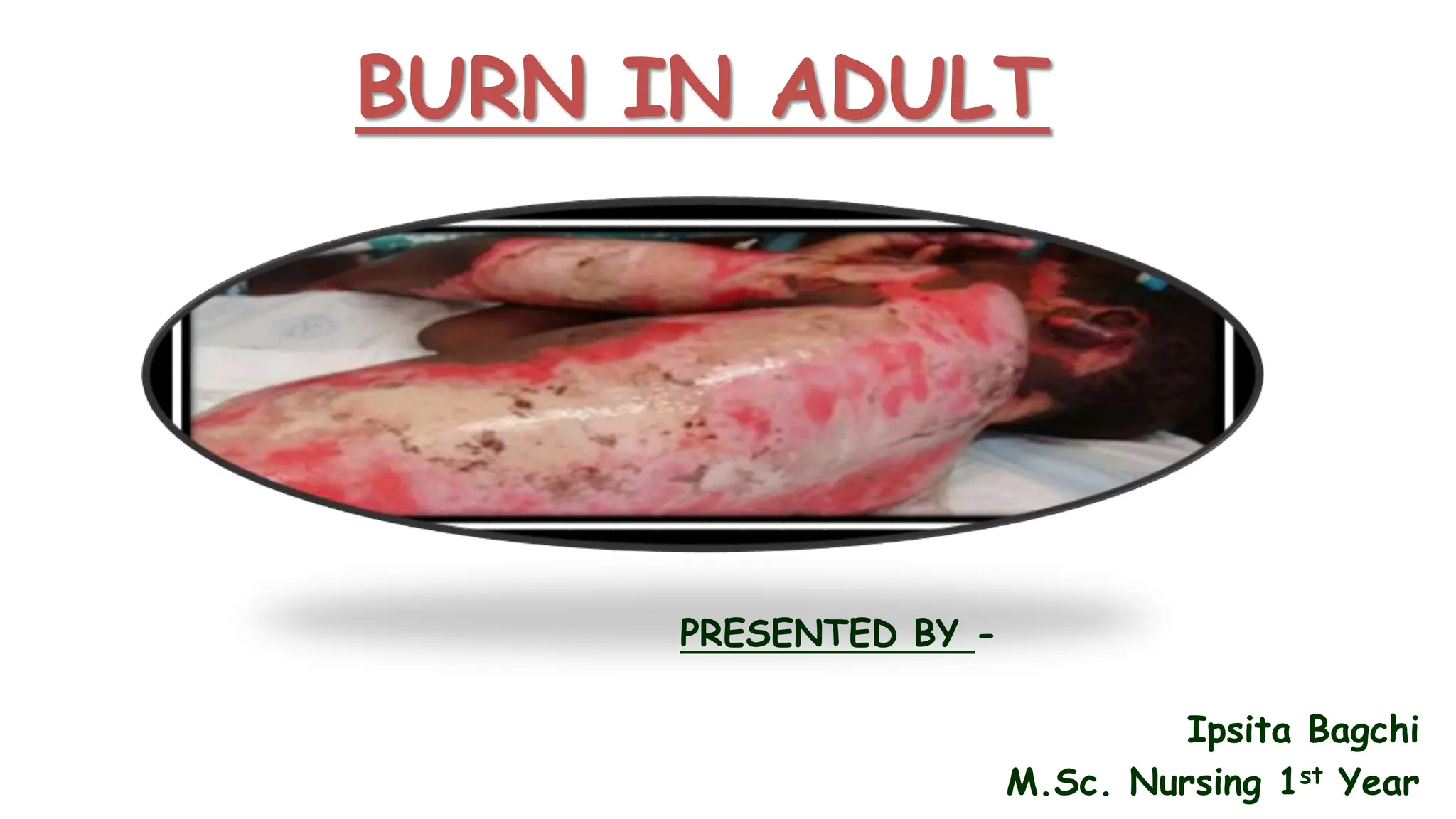
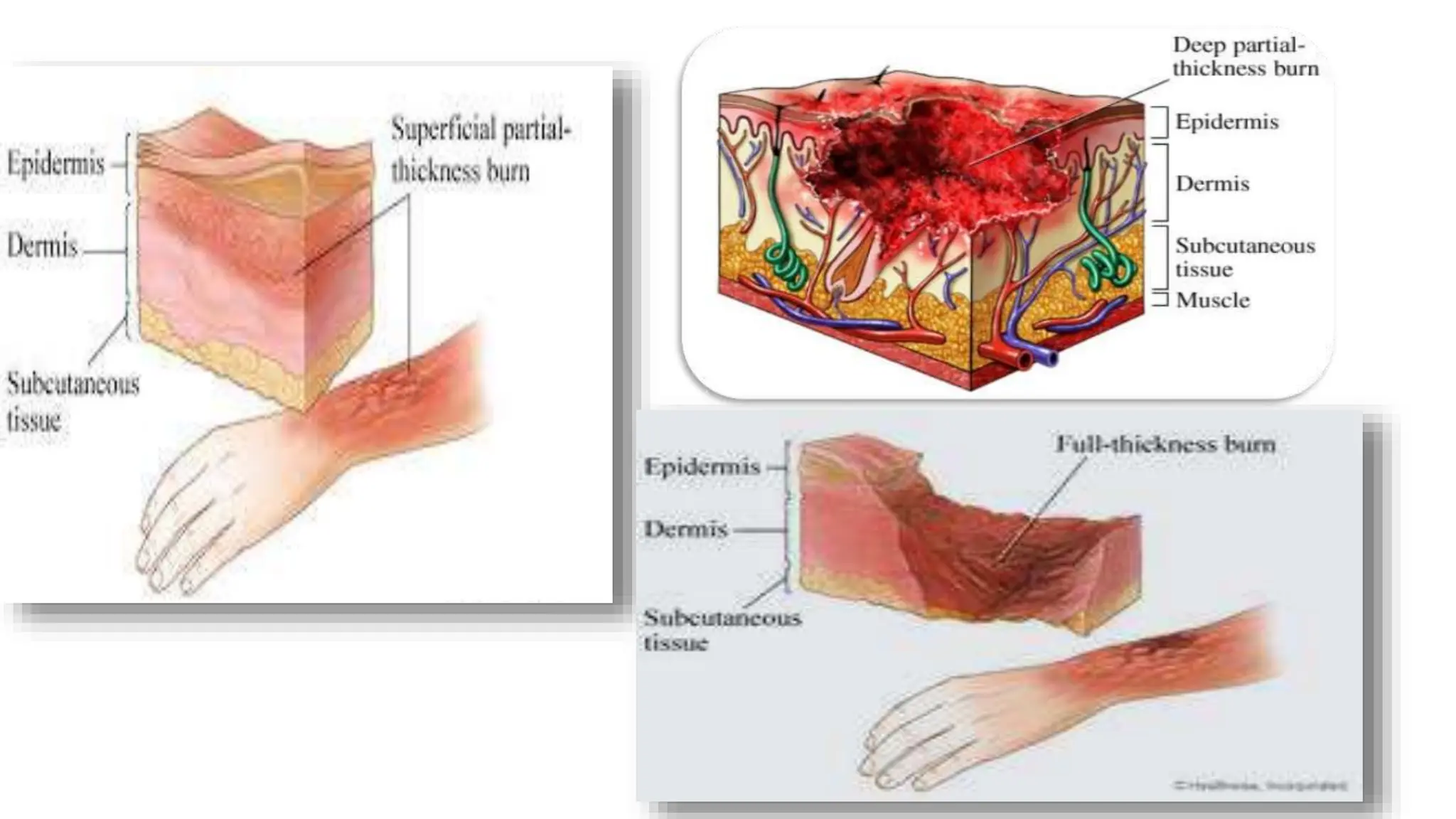
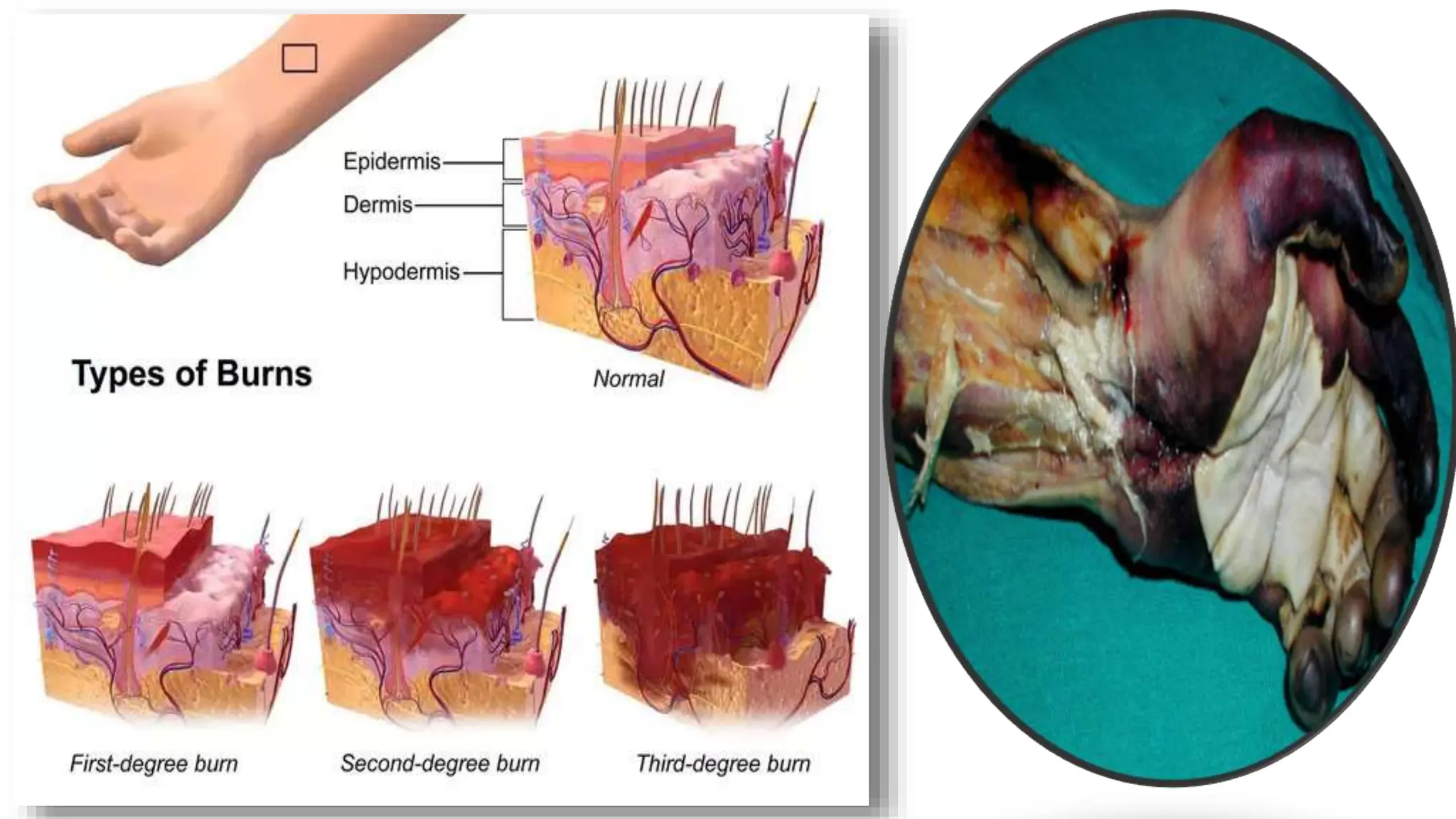
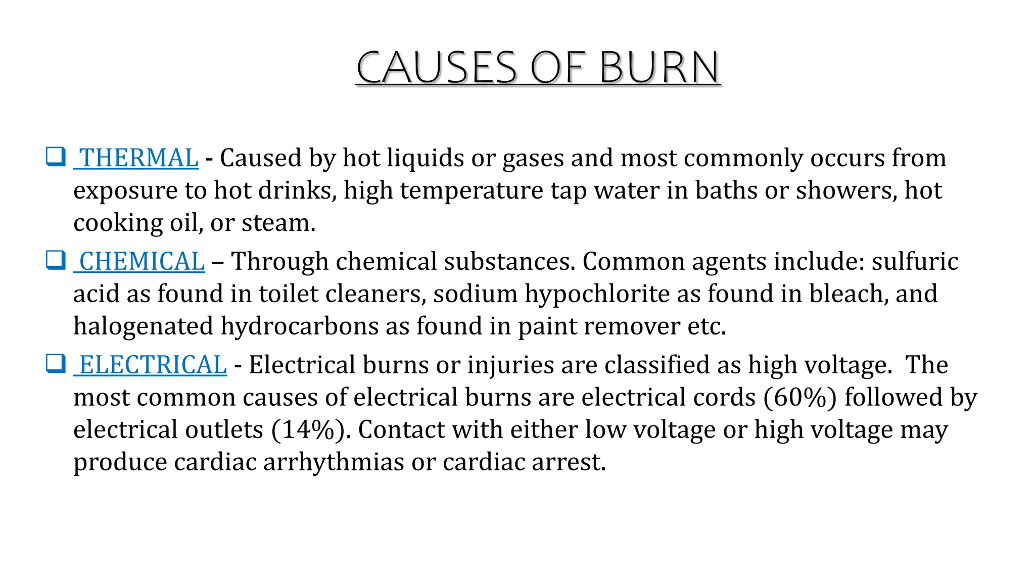
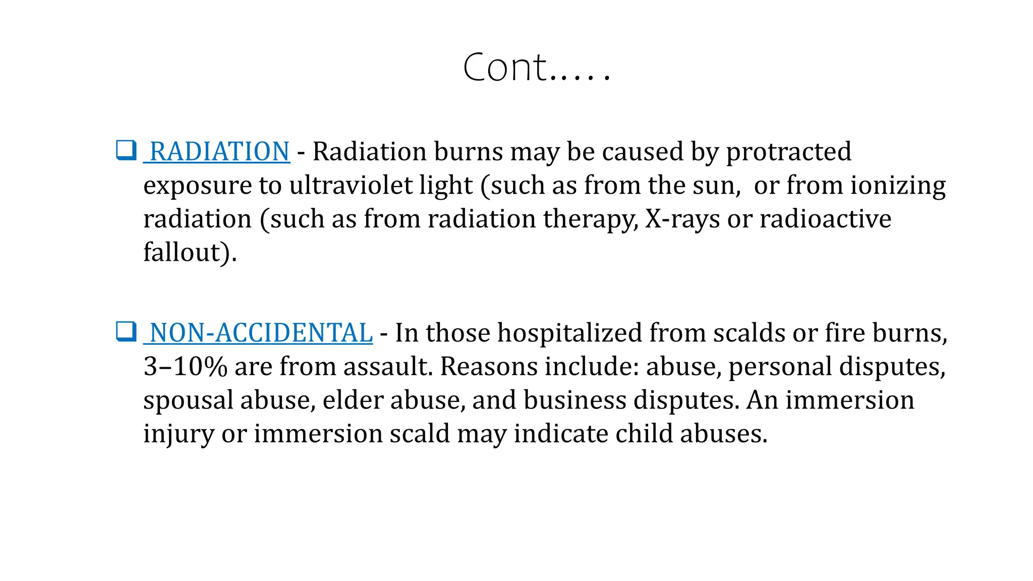
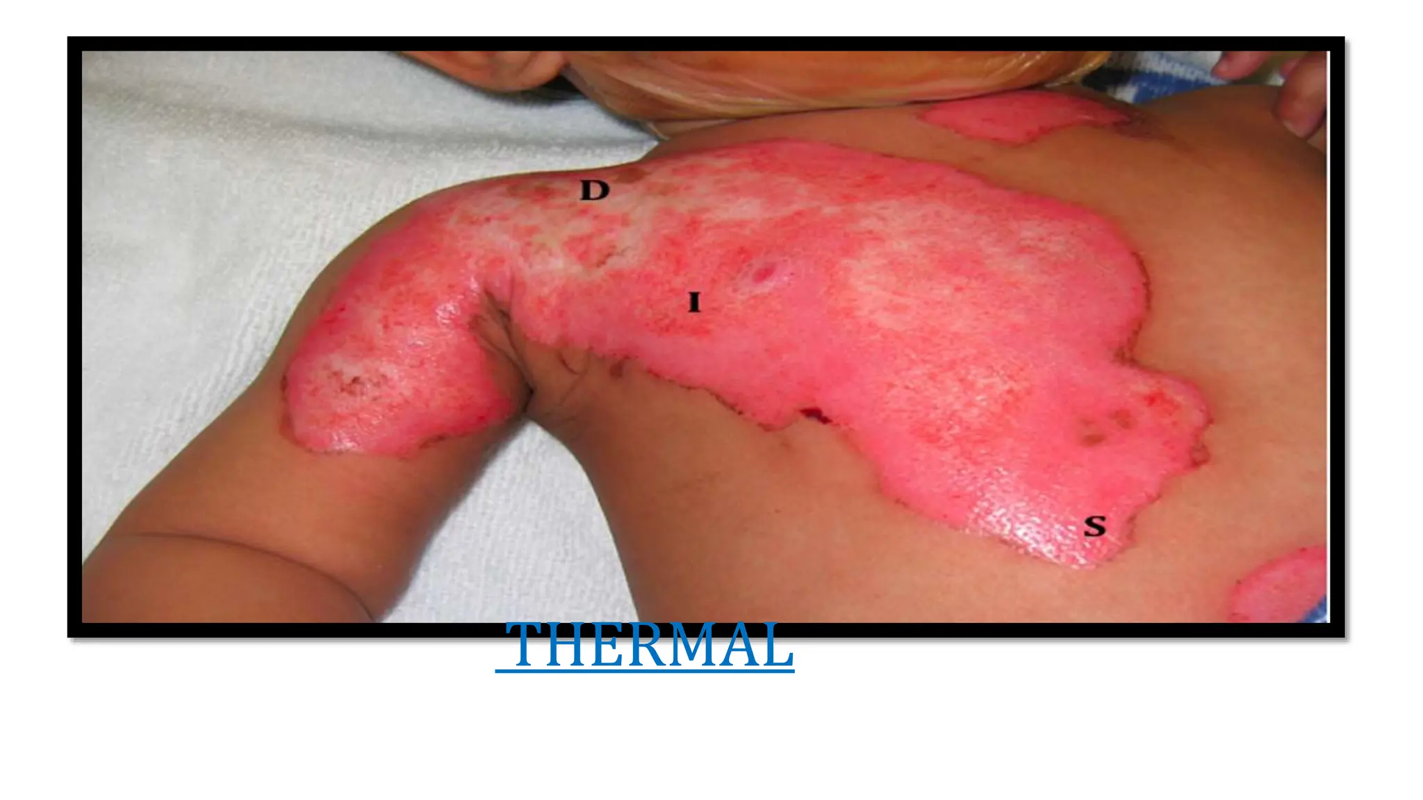
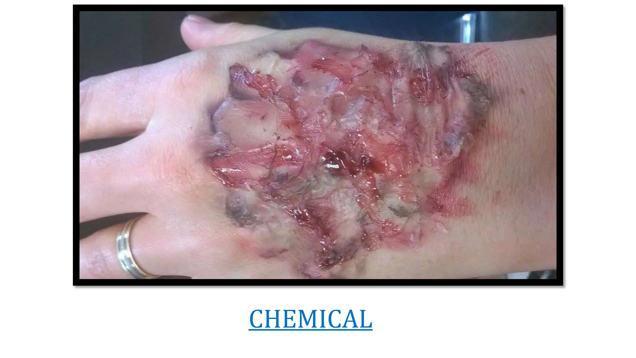
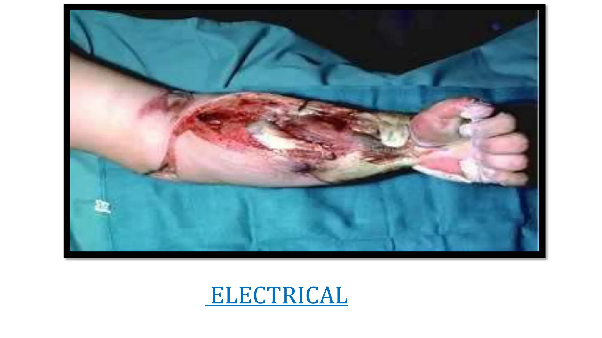
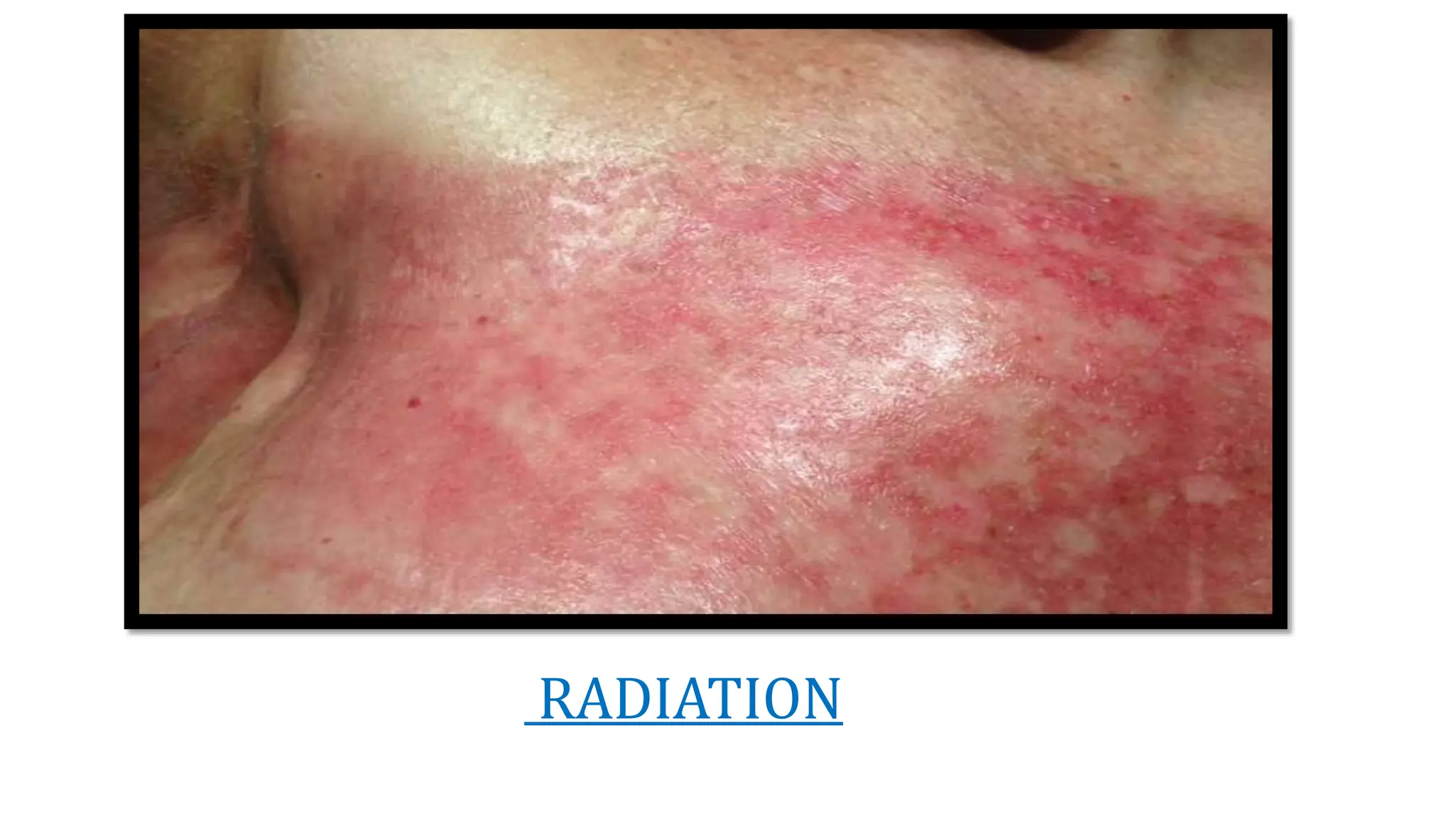
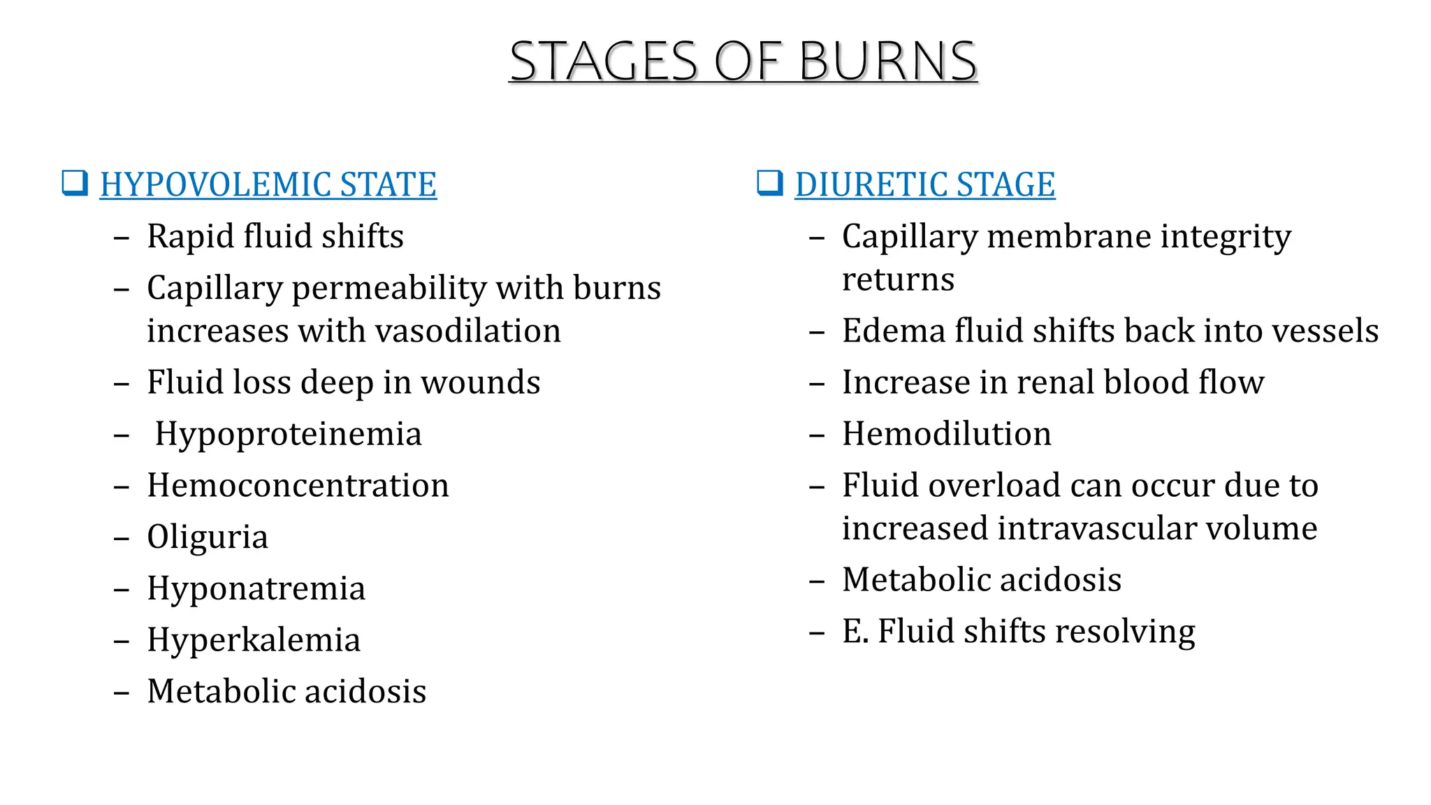
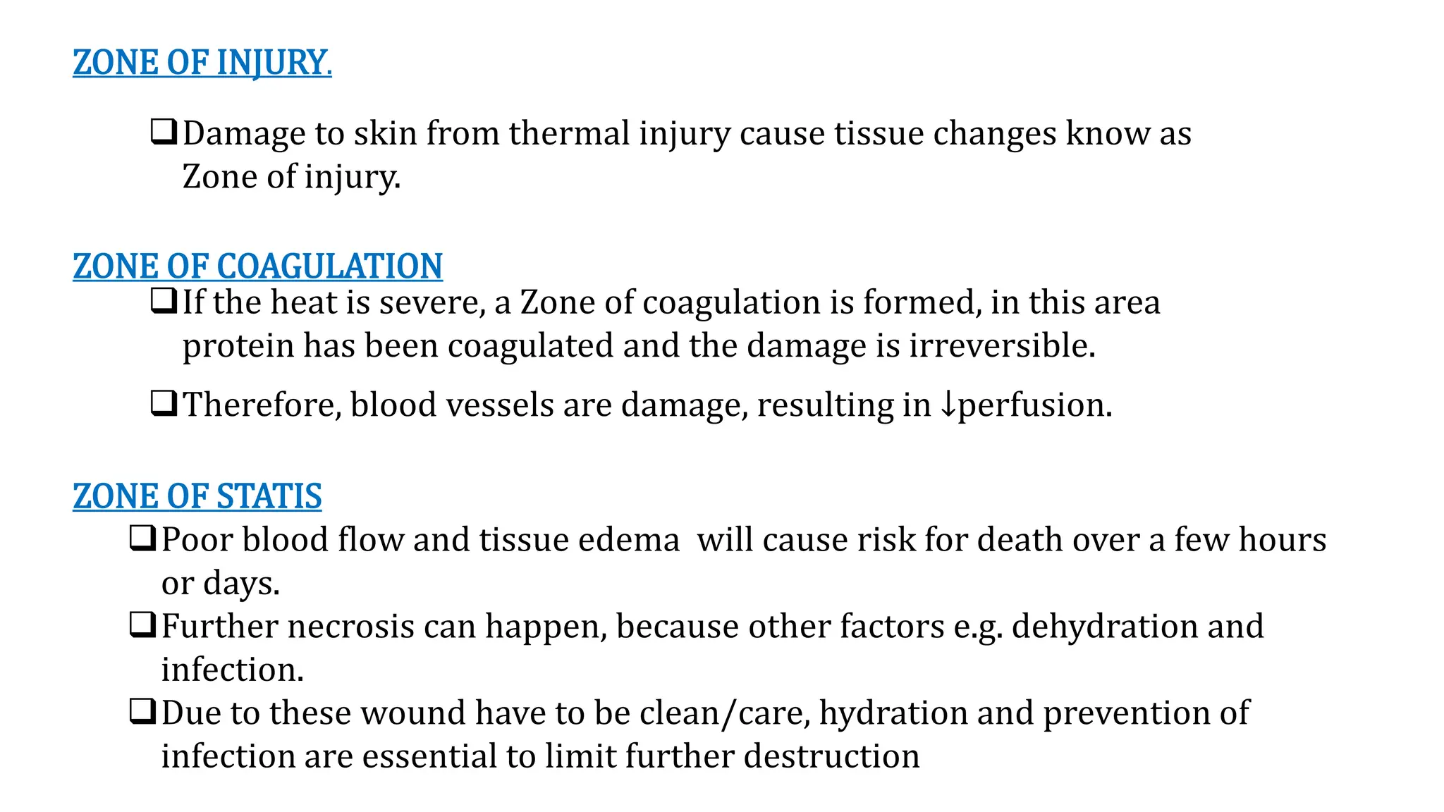
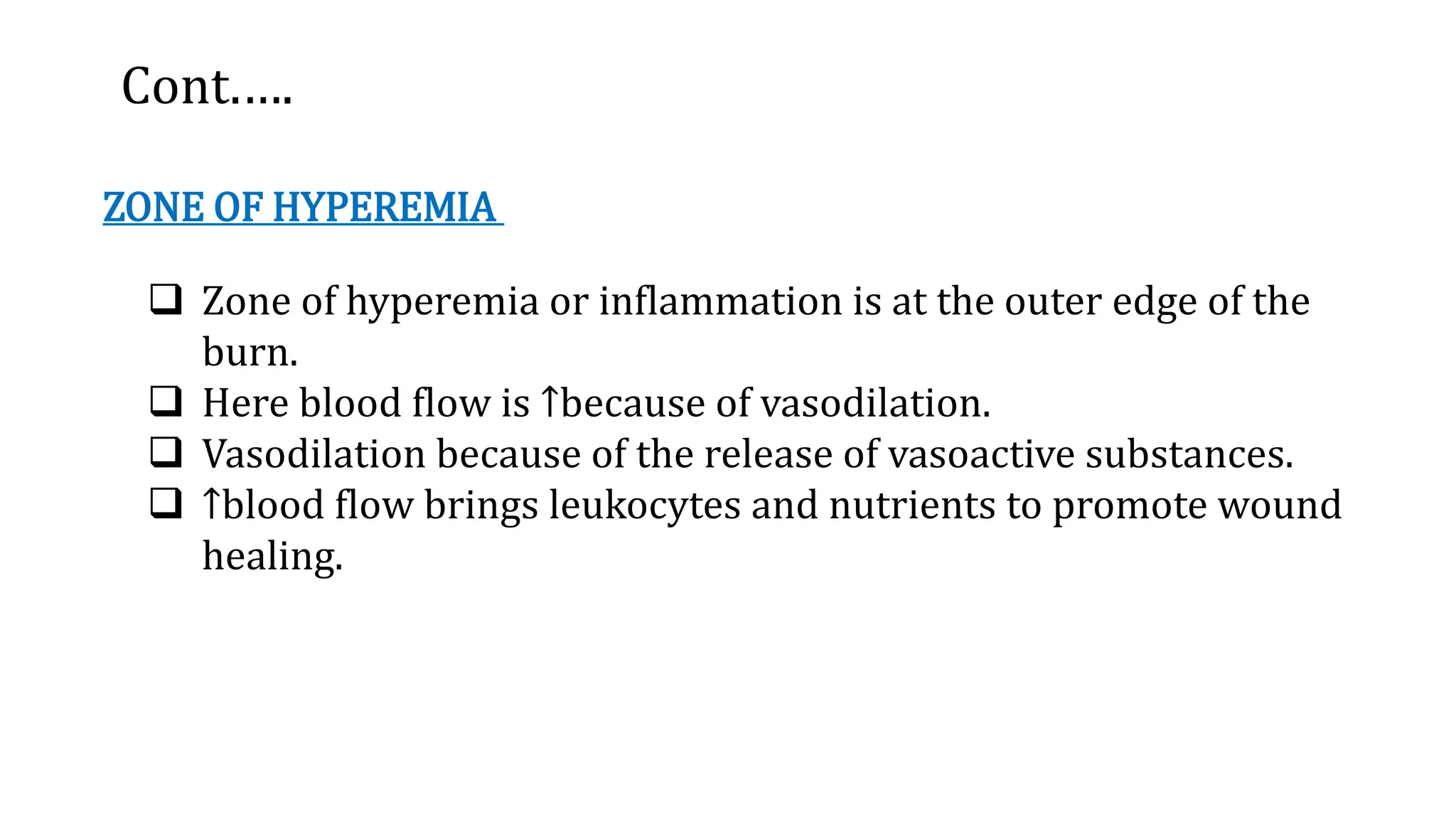
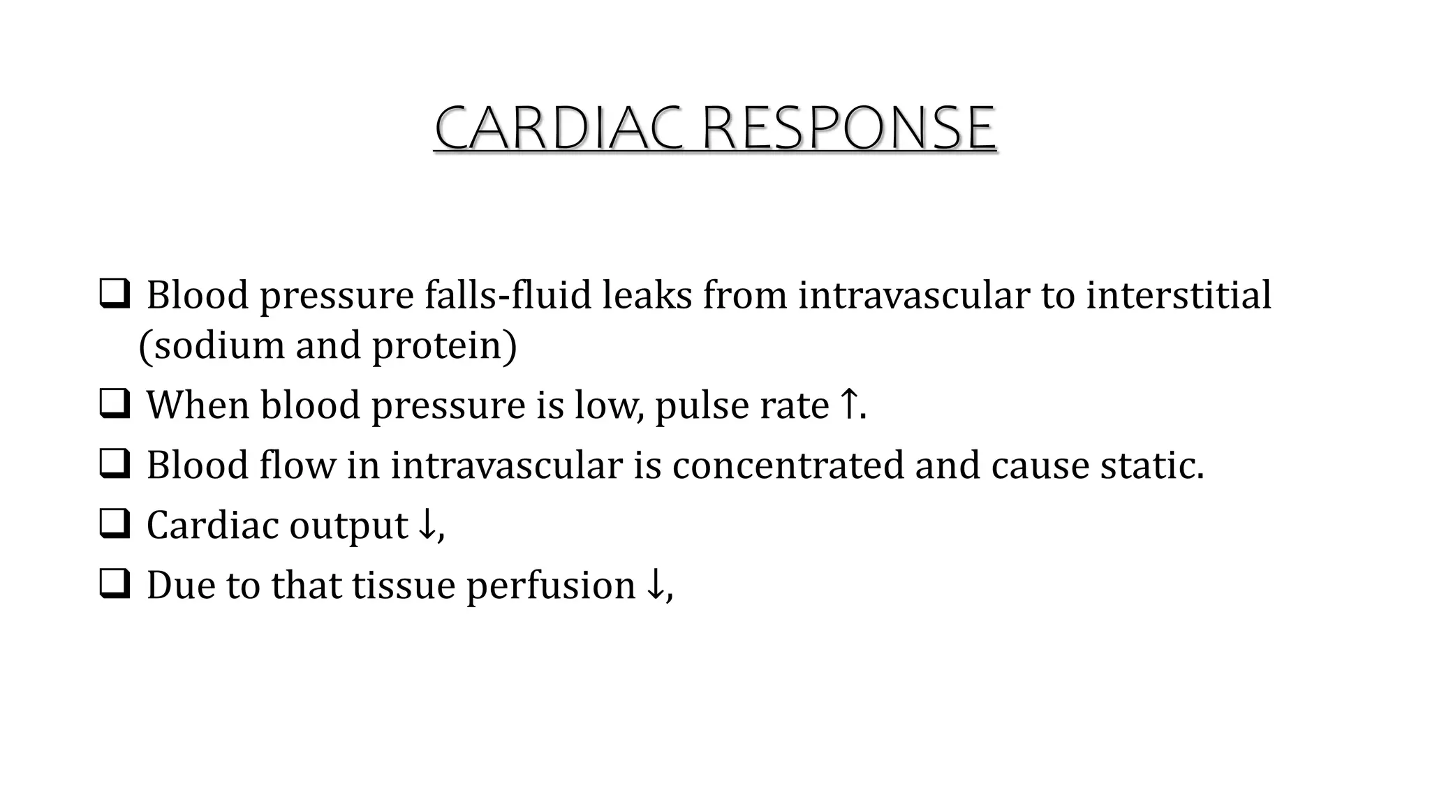
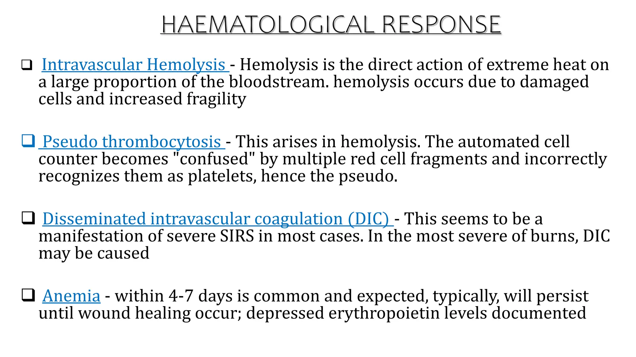
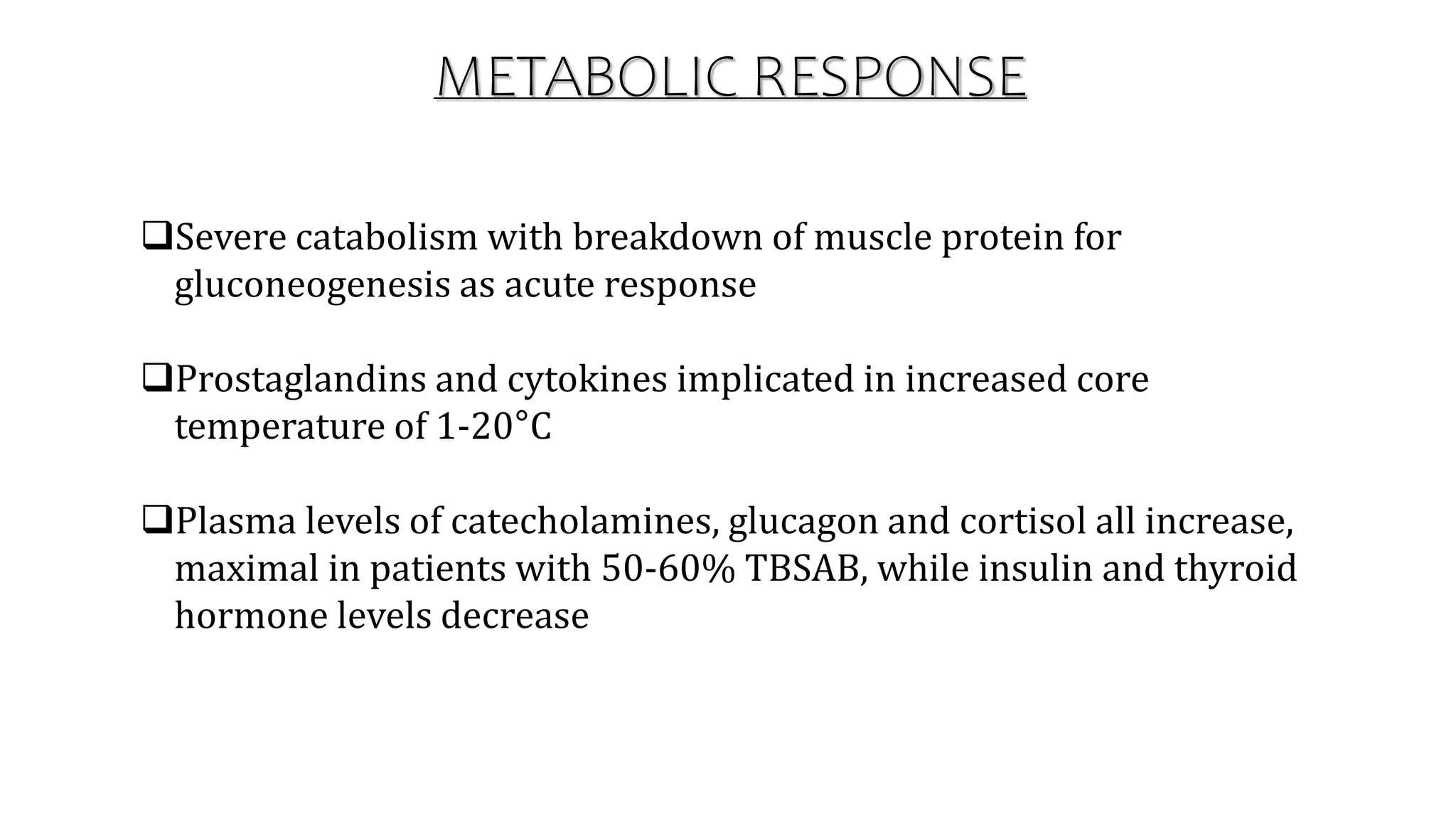
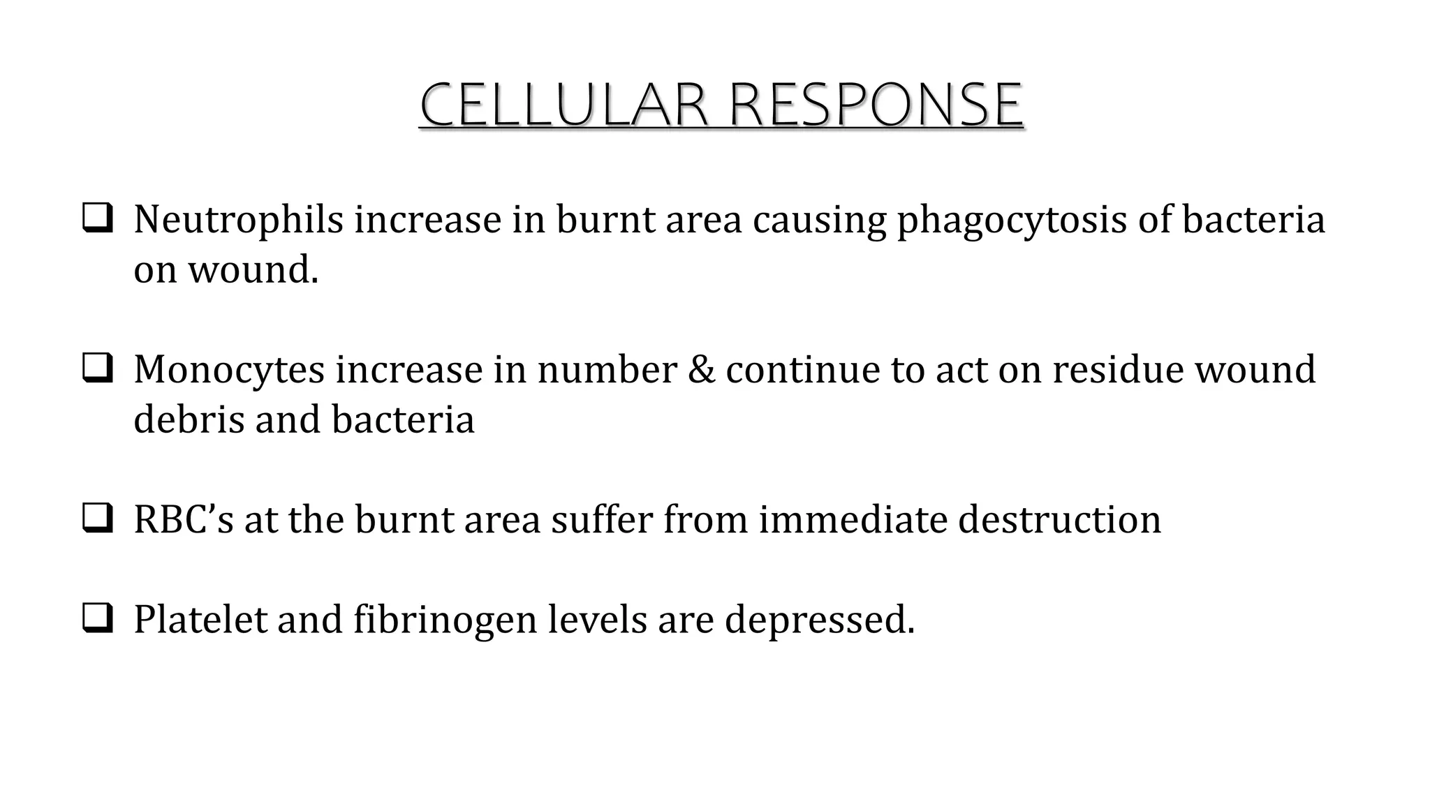
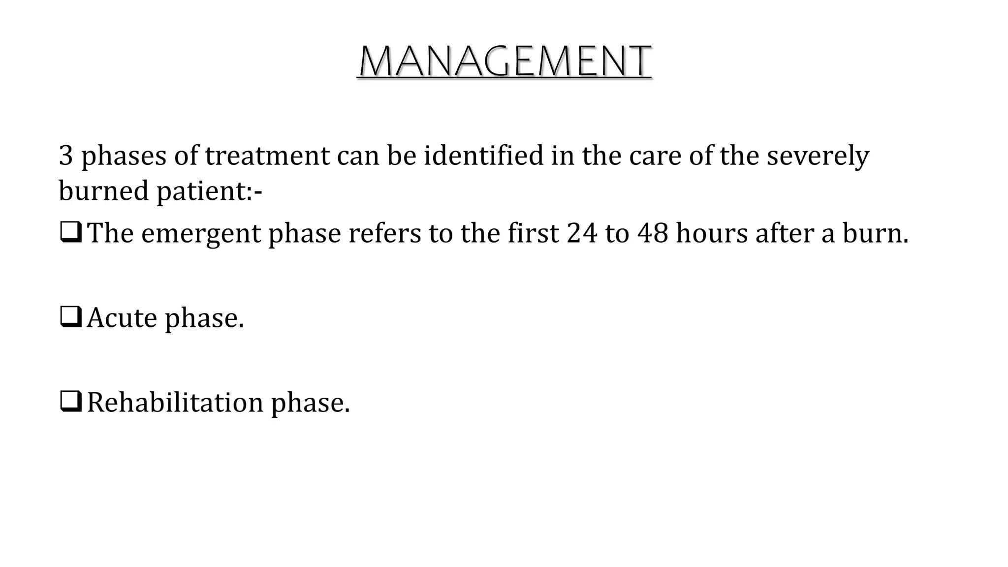
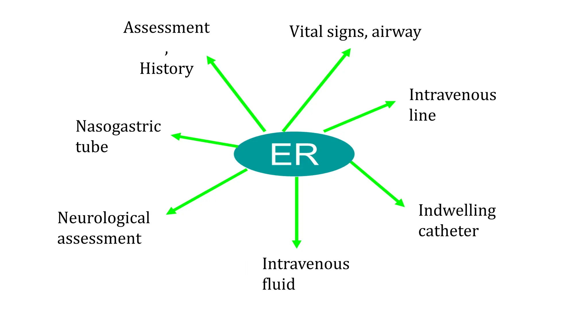
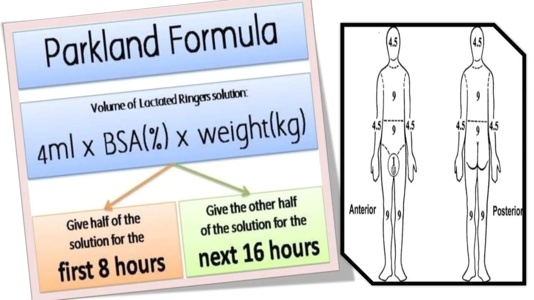
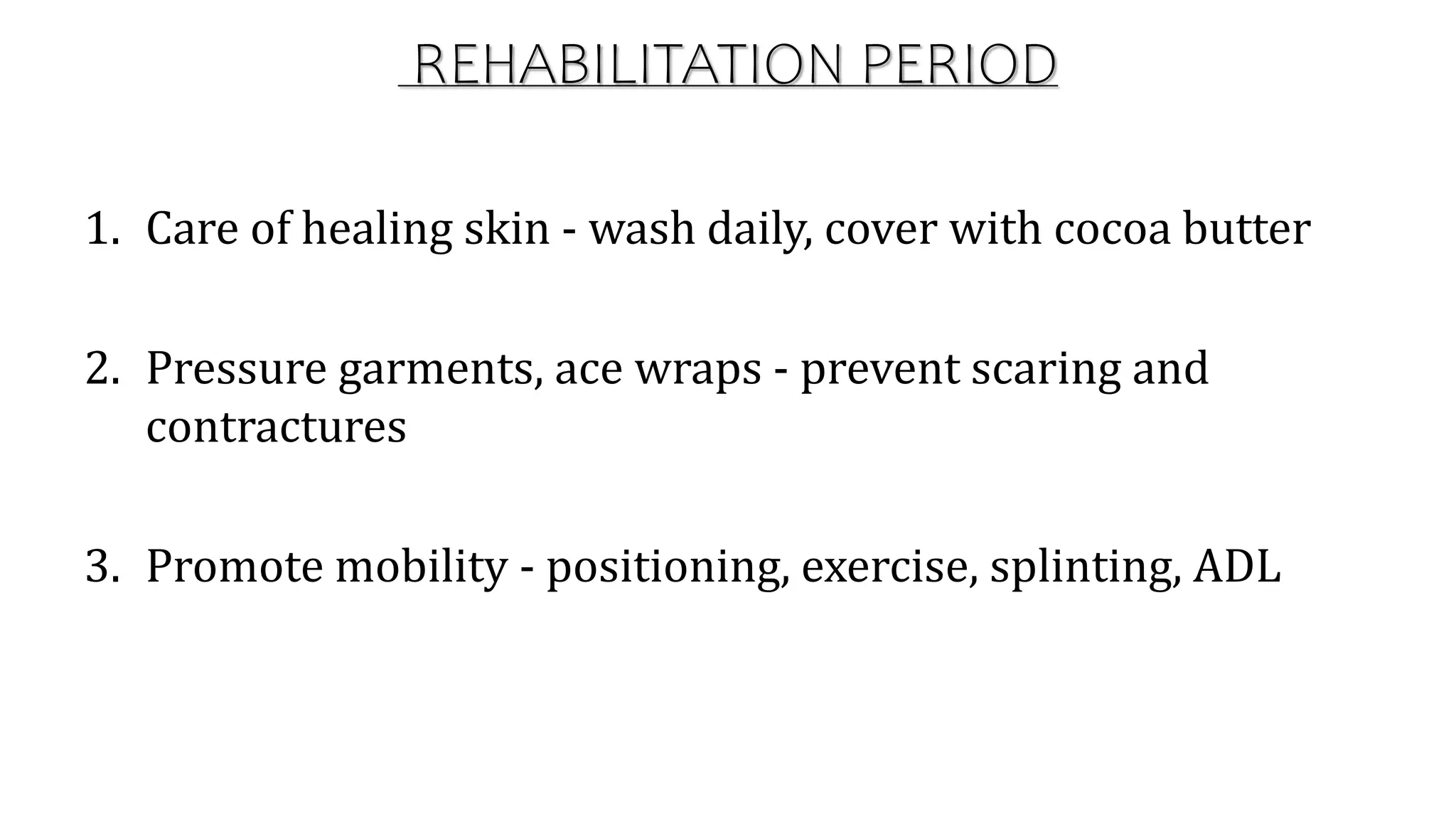
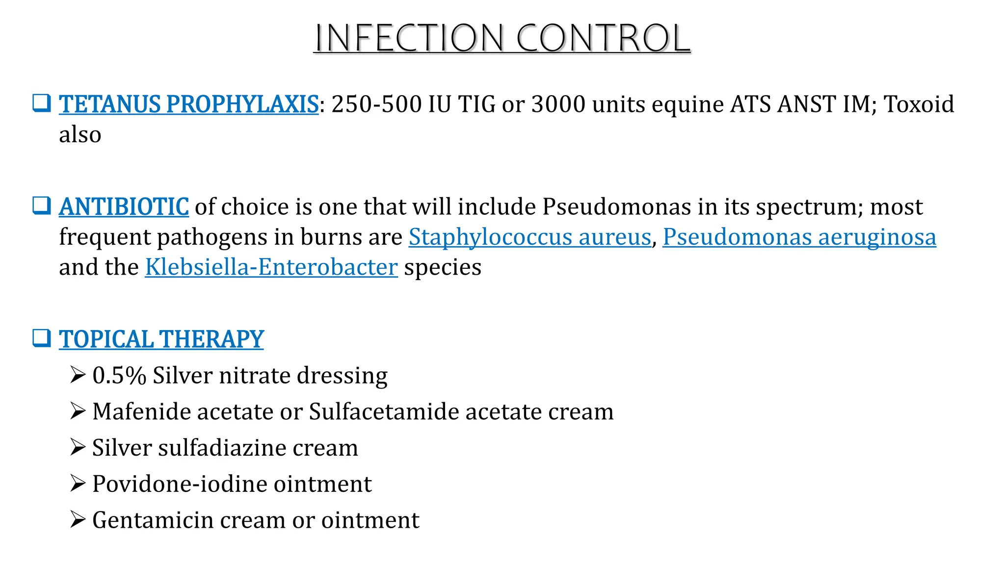
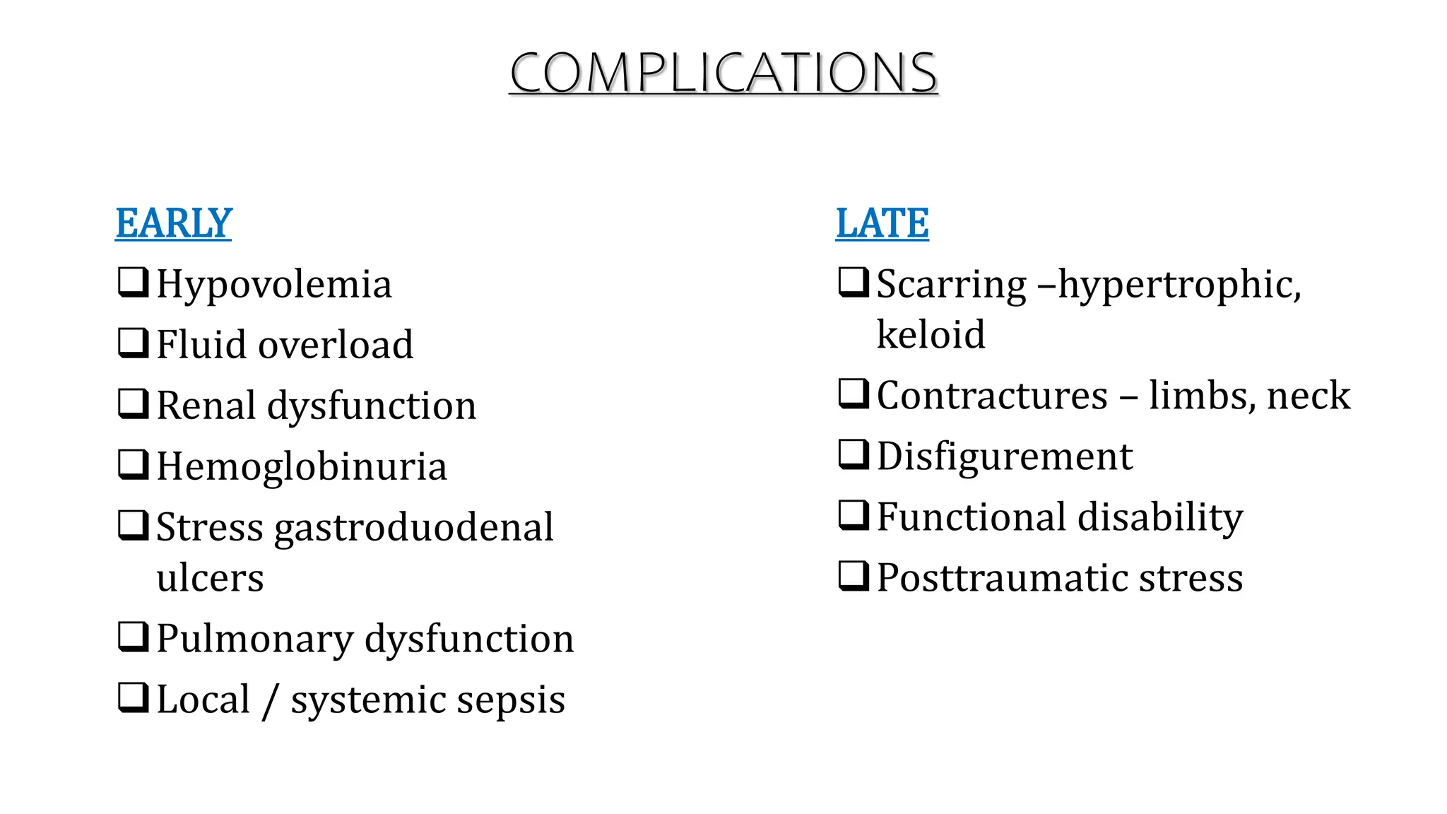
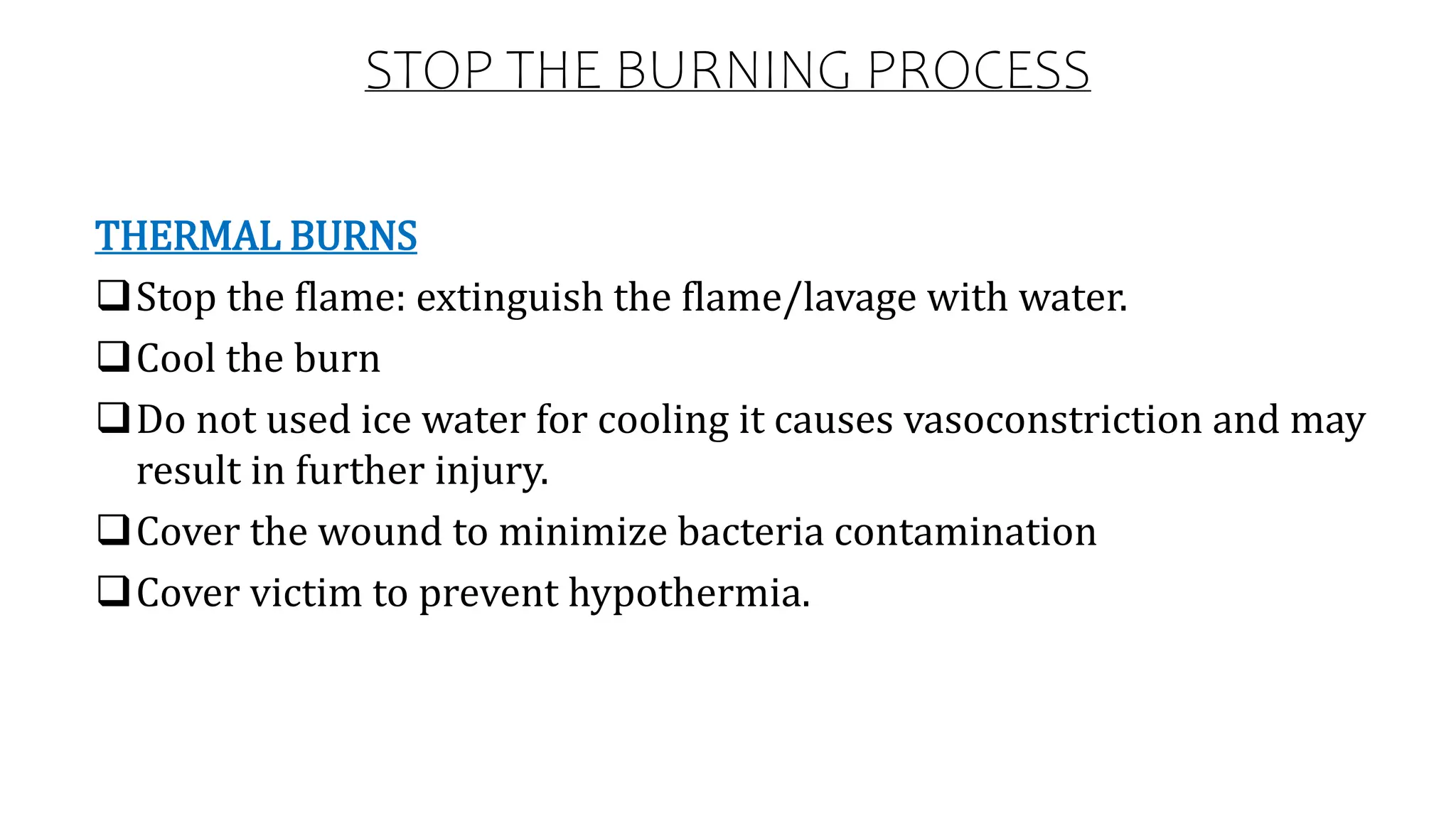
2. A burn is a type of injury to skin, or other tissues, caused by heat,
cold, electricity, chemicals, friction, or radiation. Most burns are due
to heat from hot liquids, solids, or fire. Females in many areas of the
world have a higher risk related to the more frequent use of open
cooking fires or unsafe cook stoves.
3. DEFINITION
Burn is an injury caused by thermal energy or by chemical or
physical agents having a similar effect to heating or cooling.
Burns are the tissue injury caused by the contact with heat, flame,
chemicals, electricity and radiation.
4. TYPES OF BURN
I) ACCORDING TO DEPTH OF
BURN INJURY
1. Partial thickness:
– Superficial partial
thickness
– Deep partial thickness
2. Full thickness
II) ACCORDING TO THE
EXTENT OF BURN INJURY
1. First degree burn
2. Second degree burn
3. Third degree burn
4. Fourth degree burn
5.
6.
7. CAUSES OF BURN
THERMAL - Caused by hot liquids or gases and most commonly occurs from
exposure to hot drinks, high temperature tap water in baths or showers, hot
cooking oil, or steam.
CHEMICAL – Through chemical substances. Common agents include: sulfuric
acid as found in toilet cleaners, sodium hypochlorite as found in bleach, and
halogenated hydrocarbons as found in paint remover etc.
ELECTRICAL - Electrical burns or injuries are classified as high voltage. The
most common causes of electrical burns are electrical cords (60%) followed by
electrical outlets (14%). Contact with either low voltage or high voltage may
produce cardiac arrhythmias or cardiac arrest.
8. Cont.….
RADIATION - Radiation burns may be caused by protracted
exposure to ultraviolet light (such as from the sun, or from ionizing
radiation (such as from radiation therapy, X-rays or radioactive
fallout).
NON-ACCIDENTAL - In those hospitalized from scalds or fire burns,
3–10% are from assault. Reasons include: abuse, personal disputes,
spousal abuse, elder abuse, and business disputes. An immersion
injury or immersion scald may indicate child abuses.
16. ZONE OF INJURY.
Damage to skin from thermal injury cause tissue changes know as
Zone of injury.
ZONE OF COAGULATION
If the heat is severe, a Zone of coagulation is formed, in this area
protein has been coagulated and the damage is irreversible.
Therefore, blood vessels are damage, resulting in ↓perfusion.
ZONE OF STATIS
Poor blood flow and tissue edema will cause risk for death over a few hours
or days.
Further necrosis can happen, because other factors e.g. dehydration and
infection.
Due to these wound have to be clean/care, hydration and prevention of
infection are essential to limit further destruction
17. Zone of hyperemia or inflammation is at the outer edge of the
burn.
Here blood flow is ↑because of vasodilation.
Vasodilation because of the release of vasoactive substances.
↑blood flow brings leukocytes and nutrients to promote wound
healing.
ZONE OF HYPEREMIA
Cont.….
19. CARDIAC RESPONSE
Blood pressure falls-fluid leaks from intravascular to interstitial
(sodium and protein)
When blood pressure is low, pulse rate ↑.
Blood flow in intravascular is concentrated and cause static.
Cardiac output ↓,
Due to that tissue perfusion ↓,
20. HAEMATOLOGICAL RESPONSE
Intravascular Hemolysis - Hemolysis is the direct action of extreme heat on
a large proportion of the bloodstream. hemolysis occurs due to damaged
cells and increased fragility
Pseudo thrombocytosis - This arises in hemolysis. The automated cell
counter becomes "confused" by multiple red cell fragments and incorrectly
recognizes them as platelets, hence the pseudo.
Disseminated intravascular coagulation (DIC) - This seems to be a
manifestation of severe SIRS in most cases. In the most severe of burns, DIC
may be caused
Anemia - within 4-7 days is common and expected, typically, will persist
until wound healing occur; depressed erythropoietin levels documented
21. Severe catabolism with breakdown of muscle protein for
gluconeogenesis as acute response
Prostaglandins and cytokines implicated in increased core
temperature of 1-20°C
Plasma levels of catecholamines, glucagon and cortisol all increase,
maximal in patients with 50-60% TBSAB, while insulin and thyroid
hormone levels decrease
METABOLIC RESPONSE
22. CELLULAR RESPONSE
Neutrophils increase in burnt area causing phagocytosis of bacteria
on wound.
Monocytes increase in number & continue to act on residue wound
debris and bacteria
RBC’s at the burnt area suffer from immediate destruction
Platelet and fibrinogen levels are depressed.
23. RENAL FUNCTION
Alteration in renal functions occurs due to stress response to burn injury and
hypovolemia.
There is shunting of blood away from kidneys, leading to decreased GFR
which leads to acute renal failure.
Acute tubular necrosis may occur because of inadequate fluid resuscitation
and release of large amount of hemoglobin and myoglobin from destruction of
RBC’s at the time of injury.
Sepsis
On a more exotic note, one might find their burns patient having a raise urea
due to excess protein catabolism.
24. GASTROINTESTINAL FUNCTION
Blood flow to mesenteric bed is decreased in response to
hypovolemia. This leads to acute gastric dilatation, abdominal
distention and regurgitation
Decreased blood flow to gastric mucosa occurs leading to
gastroduodenal ulceration
Paralytic ileus due to secondary to burn trauma.
Stress ulcer (stomach/duodenum) due to burn injury.
25. IMMUNOLOGIC RESPONSE
Mechanical barrier to infection is impaired because of skin
destruction
Immunoglobulin levels decreased as cytotoxic activity impaired
The reticuloendothelial system's depressed bacterial clearance is
due to decreases in opsonic function
These changes, together with a non-perfused, bacterially-colonized
overlying a wound full of proteinaceous fluid, put the patient in a
significant risk for infection
26. Majority of deaths from fire are due to smoke inhalation.
Pulmonary damage can be from direct inhalation injury or systemic
respond to the injury.
Damage to cilia and cell in the airway-inflammation.
Mucociliary transport mechanism not functioning-bronchial
congestion and infection.
Pulmonary edema, fluids escape to interstitial.
Airway obstruction.
RESPIRATORY SYSTEM
29. Cont.….
2) DEPTH OF INJURY: this is done according to the degree of burn and its sign
and symptoms.
3) SEVERITY OB BURN:
I. Minor burn
• Partial thickness burns less than 15% of TBSA.
• Full thickness burns less than 2% TBSA.
II. Moderate burn
• Partial thickness burns of 15 – 25% TBSA.
• Full thickness burn less than 10 % TBSA.
30. III. Major burns
• Partial thickness burns of 25% or greater TBSA.
• Burns involving respiratory tract injury.
• Full thickness burn of 10% or more TBSA.
• Electric burns that penetrates.
• Deep chemical burns 3rd degree burns involving face, hands
and feet.
Cont.….
31. MANAGEMENT
3 phases of treatment can be identified in the care of the severely
burned patient:-
The emergent phase refers to the first 24 to 48 hours after a burn.
Acute phase.
Rehabilitation phase.
34. ACUTE PERIOD
End of emergent period until burns heals
Focus now shifts to care of wounds and prevention of complications.
Actual range of this phase depends on degree and extent of burn
a. Assessment
b. Wound care
35. REHABILITATION PERIOD
1. Care of healing skin - wash daily, cover with cocoa butter
2. Pressure garments, ace wraps - prevent scaring and
contractures
3. Promote mobility - positioning, exercise, splinting, ADL
36. INFECTION CONTROL
TETANUS PROPHYLAXIS: 250-500 IU TIG or 3000 units equine ATS ANST IM; Toxoid
also
ANTIBIOTIC of choice is one that will include Pseudomonas in its spectrum; most
frequent pathogens in burns are Staphylococcus aureus, Pseudomonas aeruginosa
and the Klebsiella-Enterobacter species
TOPICAL THERAPY
0.5% Silver nitrate dressing
Mafenide acetate or Sulfacetamide acetate cream
Silver sulfadiazine cream
Povidone-iodine ointment
Gentamicin cream or ointment
38. STOP THE BURNING PROCESS
THERMAL BURNS
Stop the flame: extinguish the flame/lavage with water.
Cool the burn
Do not used ice water for cooling it causes vasoconstriction and may
result in further injury.
Cover the wound to minimize bacteria contamination
Cover victim to prevent hypothermia.
39. CHEMICAL BURNS
Immediately remove the clothing and a hose or shower to lavage the
involved area for a minimum 20 minutes.
ELECTRICAL BURNS
-Serious harm to victim and rescuer.
-Ensure source of electrical has been disconnected.
-Use non conductive device to remove victim.
-If victim unresponsive, assess respiration and pulse.
-Commenced CPR (cardiopulmonary resuscitation) if no pulse.
Cont.….
40. RADIATION BURN
Usually minor, involved epidermal layer of skin.
Helping the normal body mechanism to promote wound
healing
Shielding, establishing distance.
Limit time of exposure to radioactive source.
Cont.….
41. CURRENT LITERATURE
STATEMENT: Albumin administration for fluid resuscitation in burn patients: A
systematic review and meta-analysis
ABSTRACT:
This is a systematic review on albumin administration for fluid resuscitation in burn
patients.
Previous reviews suggest that albumin increases mortality in burn patients.
4 trials involving 140 patients were included in our meta-analysis.
Albumin solutions were not associated with higher mortality in burn patients.
Changes in data extraction and included trials might explain differences in outcomes.