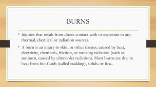
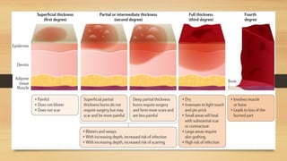
BURNS
PRESENTED BY- RISHIKA RAWAT
RN/RM
BURNS
• Injuries that result from direct contact with or exposure to any
thermal, chemical or radiation sources.
• A burn is an injury to skin, or other tissues, caused by heat,
electricity, chemicals, friction, or ionizing radiation (such as
sunburn, caused by ultraviolet radiation). Most burns are due to
heat from hot fluids (called scalding), solids, or fire..
ETIOLOGY
• Thermal burns
• Chemicals
• Electricity
• Radiations
• Inhalation injuries
.
• THERMAL BURNS
These are caused by exposure to or contact with flame, hot liquids, semi liquids
(steam), semi-solid (tar) or hot objects.
• CHEMICAL BURNS
It is caused by contact of tissue to any strong acids, alkalis or organic
compounds.
• ELECTRICAL BURNS
These are the injuries caused by heat that is generated by the electrical energy as
it passes through the body. It can result from contact with exposed or faulty
electrical wiring or high voltage power lines. People struck by lightening also
sustain electrical injury.
.
• RADIATION BURNS
These are caused by exposure to radioactive source.E.g.Nuclear- radiation
accidents. Use of ionizing radiation in industries Therapeutic radiations
Sunburns from prolonged exposure to ultraviolet rays.
• INHALATION INJURIES
It may result from exposure to asphyxiants and smoke, if the victim was
trapped in closed, smoke - filled area. It results in pulmonary pathophysiologic
changes.
.
Tissue damage due to burns is determined by:
• Extent of burn
• Depth of burn
Classification of Burns
On the basis of burn depth
• First degree injury localized to the epidermis.
• Second Degree:
I) Superficial second degree: injury to the epidermis and superficial papillary
dermis.
ii) Deep second degree: injury through the epidermis and deep up to reticular
dermis.
,
• Third degree:
full- thickness injury through the epidermis and dermis into subcutaneous fat.
• Fourth degree:
injury through the skin and subcutaneous fat into underlying muscle and bone.
Clinical Features
First degree burns:
• Reddened skin
• Pain at burn site
• Involves only epidermis
• Blanch to touch
• Have an in-tact epidermal barrier
Examples: Sun-burn, minor scald from a kitchen accident
,
Superficial second degree Burns
• Intense pain
• White to red skin
• Blisters Involves epidermis & papillary layer of dermis Spares hair follicles,
sweat glands etc.
• Erythematous & blanch to touch
• Very painful/sensitive.
• Spontaneously re-epithelialization from retained epidermal structures in 7-14
days
.
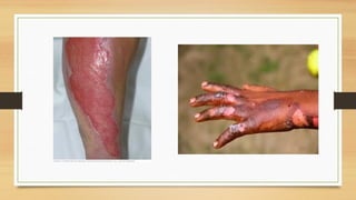
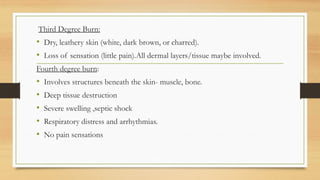
Deep second degree burns
• Injury to deeper layers of dermis, i.e., reticular dermis.
• Appears pale & mottled.
• Do not blanch to touch.
• Capillary return sluggish or absent.
• Less painful, remain painful to pinprick.
• Takes 14 to 35 days to heal by re-epithelialization from hair follicles & sweat
gland, keratinocytes often with severe scarring.
• Contractures possible.[ restriction in patients ability to move]
,
Third Degree Burn:
• Dry, leathery skin (white, dark brown, or charred).
• Loss of sensation (little pain).All dermal layers/tissue maybe involved.
Fourth degree burn:
• Involves structures beneath the skin- muscle, bone.
• Deep tissue destruction
• Severe swelling ,septic shock
• Respiratory distress and arrhythmias.
• No pain sensations
THE BURN SYNDROME
• Fluid and Electrolyte Imbalance: The burn wound becomes rapidly
edematous due to microvascular changes induced by direct thermal injury
and by release of chemical mediators of inflammation. This results in
systemic intravascular losses of water, sodium, albumin and red blood cells.
Unless intravascular volume is rapidly restored, shock develops.
• Metabolic Disturbances: This is evidenced by increased resting oxygen
consumption (hypermetabolism), an excessive nitrogen loss (catabolism), and
a pronounced weight loss (malnutrition).
.
• Bacterial Contamination of Tissues: Damaged integument creates a vast area
for surface infection and invasion of microorganisms. Burned patients with a
major thermal injury are unable to mount an adequate immunologic defense,
increasing the risks for septic shock.
• Complications from Vital Organs: All major organ systems are affected by
the burn injury. Renal insufficiency can result from hypoperfusion or from
nephron obstruction with myoglobulin and hemoglobin. Pulmonary
dysfunction may be caused from initial respiratory tract damage of from
progressive respiratory insufficiency due to pulmonary edema, adult
respiratory distress syndrome or bronchopneumonia. Gastrointestinal
complications include paralytic ileus and gastrointestinal ulcerations.
LOCAL AND SYSTEMATIC EFFECTS OF BURNS
Local Effects
• Tissue Damage: Heating of tissue results in direct cell rupture or cell
necrosis. At the periphery the cells may be viable, but injured. In addition,
collagen is denatured and damage to the peripheral microcirculation occurs.
• Inflammation: There is a marked and immediate inflammatory response. In
the areas least damaged by burning, this is manifest simply as erythema. The
precise cause of this immediate vasodilation may represent a neurovascular
response similar to Lewis's triple response.
.
• Infection: The damaged tissue represents a nidus for infection. Burn wounds
will almost inevitably be colonized by microorganisms within 24-48 hours and
this may remain as a local wound or regional infection. There may in addition be
a bacteremia or septicemia and metastatic infections may develop at other sites.
Systematic Effects of Burns
• Fluid Loss: Fluid may be lost from damaged capillaries either by visible
external loss or internally into the tissues from edema in the region of the burn.
In addition, there may be more extensive edema of the region or even of the
Ent.
• Multiple organ failure- There may be progressive failure of renal or hepatic
function or heart failure. The precise cause of these complications is uncertain
and has often been attributed to fluid loss, 'toxemia' from infection, or
uncontrol overreaction of the inflammatory response to sepsis. Multiple organ
failure may, however, occur without obvious systemic infection.
,
• Inhalation injury: These occur in those trapped in enclosed spaces. They
are particularly common in association with burns of the head and neck.
Various parts of the respiratory tract may be injured. The inhalation of hot
gases causes a thermal burn to the upper airway. This is manifest early on by
stridor, hoarse-ness, cough and respiratory obstruction.
According To The Extend of Body Surface
area involved
• The Rule of Nine
Rule of Nine is a quick way to
calculate the extents of burns. The
system assigns percentages in multiples
of nine to major body surfaces. It may
be used to estimate the total body
surface area burnt. By adding together,
the affected area the percentage of the
total body surface that is burnt can be
calculated
.
• Lund and Browder Method
Lund and Browder method, which
recognizes that the percentage of
IBSA of various anatomic parts,
especially the head and legs, and
changes with growth. By dividing the
body into very small areas and
providing an estimate of the
proportion of TBSA accounted for
by such body parts, one can obtain a
reliable estimate of the TBSA burned
.
Palm Method
• In patients with scattered burns, a
method to estimate the percentage
of burn is the palm method. The
size of the patient's palm is
approximately 1% of TBSA.
Parkland/Baxter Formula
• Lactated ringer's solution (or other balanced saline solution):
• Half to be given in first 8 hours, remaining half over next 16 hours
4 ml x kg body weight x % body surface area (BSA) burned.
Management of Burns
First Aid management for minor burns
• if the epidermis is not broken soak, it in a cool water or cold compression
• cover the burn with a sterile non adhesive bandage or clean cloth
• protect the burn from friction and pressure.
• Over the counter pain medication to help pain it may reduce swelling .
.
First Aid for Severe Burns: (Second & Third-Degree)
• Do not remove burnt clothing (unless it comes off easily), but do ensure that
the victim is not in contact with burning or smoldering materials.
• Make sure the victim is breathing. If breathing has stopped or the victim's
airway is blocked then open the airway and if necessary, begin CPR.
• Do not use a blanket or towel; a sheet is best for large burns.
• Do not apply any ointments and avoid breaking blisters.
• Elevate the burned area and protect it from pressure or friction.
• Continue to monitor the victim's vital signs (breathing, pulse, blood
pressure).
.
Do Not
• Do not apply ointment, butter, ice, medications, fluffy cotton dressing,
adhesive bandages, cream, oil spray, or any household remedy to a burn. This
can interfere with proper healing.
• Don't allow burn to become contaminated. Avoid breathing or coughing on
burned area.
• Do not disturb blisters or dead skin.
• Don't apply cold compresses and do not immerse a severe burn in cold
water. This can cause shock.
• Do not place a pillow under the victim's head if there is an airway burn and
they are lying down. This can close the airway.
Immediate Management
• Airway management
• Hyperbaric oxygen therapy [HBOT]
• Fluid management [ Ringer lactate and Normal saline]
• Wound management
• Analgesic and Sedation
Intermediate Management
• Infection prevention
• Topical antimicrobials
• Wound care
• Wound covering method
,
• A 45-year-old female patient has superficial partial thickness burns on
the posterior head and neck, front of the left arm, front and back of
the right arm, posterior trunk, front and back of the left leg, and back
of right leg. The patient weighs 91 kg. Use the Parkland Burn Formula
to calculate the total amount of Lactated Ringers that will be given
over the next 24 hours?
Answer
• 22,932 mL.
Formula: Total Amount of LR = 4 mL x BSA % x pt's weight in kg. Pt's
weight 91 kg. BSA percentage: 63%... posterior head and neck (4.5%),
front of the left arm (4.5%), front and back of the right arm (9%),
posterior trunk (18%), front and back of the left leg (18%), back of right
leg (9%) equals: 63%......4 x 63 x 91 = 22,932 mL
.
• A 30-year-old female patient has deep partial thickness burns on the
back of the right arm, posterior trunk, front of the left leg, anterior
head and neck, and perineum. The patient weighs 150 lbs. Use the
Parkland Burn Formula to calculate the total amount of Lactated
Ringers that will be given over the next 24 hours?
Answer
• 10,064 mL
Formula: Total Amount of LR = 4 mL x BSA % x pt's weight in kg. Pt's
weight 150 lbs....convert to kg....150 divided by 2.2 = 68 kg. BSA
percentage: 37%...Back of right arm (4.5%), posterior trunk (18%), front
of left leg (9%), anterior head and neck (4.5%) and perineum (1%) which
equals 37%......4 x 37 x 68 = 10,064 mL
.
• A 29-year-old male patient has superficial partial thickness burns on
the anterior right arm, posterior left leg, and anterior head and neck.
The patient weighs 78 kg. Use the Parkland Burn Formula to calculate
the total amount of Lactated Ringers that will be given over the next
24 hours?
Answer
• 5,616 ml.
Formula: Total Amount of LR = 4 mL x BSA % x pt's weight in kg. Pt's
weight 78 kg. BSA percentage: 18%...Anterior right arm (4.5%), posterior
left leg (9%), and anterior head and neck (4.5%) which equals 18%.....4 x
18 x 78 = 5,616 mL
.
• A 59-year-old male patient has full thickness burns on both of the legs
on the back, front and back of the trunk, both arms on the front and
back, and front and back of the head and neck. The patient weighs 186
lbs. Use the Parkland Burn Formula: You've already infused fluids
during the first 8 hours. Now what will you set the flow rate during the
next 16 hours (mL/hr) based on the total you calculated?
Answer
• 861 mL/hr
First calculate the total amount of fluid needed with the formula: Total
Amount of LR = 4 mL x BSA % x pt's weight in kg. The pt's weight 186
lbs...need to convert to kg: 186 divided by 2.2 = 85 kg. BSA percentage:
81%...Both of the legs on the back (18%), front and back of the trunk (36%),
both arms on the front and back (18%), front and back of the head and neck
(9%) which equals 81%.......4 x 81 x 85 = 27,540 mL. You've already infused half
of the solution during the first 8 hours...so 13,770 mL is left and it needs to be
infused over 16 hours. Hourly rate: 13,770 mL divided by 16 hours equals 861
mL/hr
.
• A 29-year-old male patient has superficial partial thickness burns on
the anterior right arm, posterior left leg, and anterior head and neck.
The patient weighs 78 kg. Use the Parkland Burn Formula to calculate
the total amount of Lactated Ringers that will be given over the next
24 hours?
Answer
• 5,616 mL.
Formula: Total Amount of LR = 4 mL x BSA % x pt's weight in kg. Pt's
weight 78 kg. BSA percentage: 18%...Anterior right arm (4.5%), posterior
left leg (9%), and anterior head and neck (4.5%) which equals 18%.....4 x
18 x 78 = 5,616 mL

Burns types and its management .pptx

  • 1.
  • 2.
    BURNS • Injuries thatresult from direct contact with or exposure to any thermal, chemical or radiation sources. • A burn is an injury to skin, or other tissues, caused by heat, electricity, chemicals, friction, or ionizing radiation (such as sunburn, caused by ultraviolet radiation). Most burns are due to heat from hot fluids (called scalding), solids, or fire..
  • 4.
    ETIOLOGY • Thermal burns •Chemicals • Electricity • Radiations • Inhalation injuries
  • 5.
    . • THERMAL BURNS Theseare caused by exposure to or contact with flame, hot liquids, semi liquids (steam), semi-solid (tar) or hot objects. • CHEMICAL BURNS It is caused by contact of tissue to any strong acids, alkalis or organic compounds. • ELECTRICAL BURNS These are the injuries caused by heat that is generated by the electrical energy as it passes through the body. It can result from contact with exposed or faulty electrical wiring or high voltage power lines. People struck by lightening also sustain electrical injury.
  • 6.
    . • RADIATION BURNS Theseare caused by exposure to radioactive source.E.g.Nuclear- radiation accidents. Use of ionizing radiation in industries Therapeutic radiations Sunburns from prolonged exposure to ultraviolet rays. • INHALATION INJURIES It may result from exposure to asphyxiants and smoke, if the victim was trapped in closed, smoke - filled area. It results in pulmonary pathophysiologic changes.
  • 7.
    . Tissue damage dueto burns is determined by: • Extent of burn • Depth of burn
  • 8.
    Classification of Burns Onthe basis of burn depth • First degree injury localized to the epidermis. • Second Degree: I) Superficial second degree: injury to the epidermis and superficial papillary dermis. ii) Deep second degree: injury through the epidermis and deep up to reticular dermis.
  • 9.
    , • Third degree: full-thickness injury through the epidermis and dermis into subcutaneous fat. • Fourth degree: injury through the skin and subcutaneous fat into underlying muscle and bone.
  • 11.
    Clinical Features First degreeburns: • Reddened skin • Pain at burn site • Involves only epidermis • Blanch to touch • Have an in-tact epidermal barrier Examples: Sun-burn, minor scald from a kitchen accident
  • 12.
    , Superficial second degreeBurns • Intense pain • White to red skin • Blisters Involves epidermis & papillary layer of dermis Spares hair follicles, sweat glands etc. • Erythematous & blanch to touch • Very painful/sensitive. • Spontaneously re-epithelialization from retained epidermal structures in 7-14 days
  • 13.
    . Deep second degreeburns • Injury to deeper layers of dermis, i.e., reticular dermis. • Appears pale & mottled. • Do not blanch to touch. • Capillary return sluggish or absent. • Less painful, remain painful to pinprick. • Takes 14 to 35 days to heal by re-epithelialization from hair follicles & sweat gland, keratinocytes often with severe scarring. • Contractures possible.[ restriction in patients ability to move]
  • 14.
    , Third Degree Burn: •Dry, leathery skin (white, dark brown, or charred). • Loss of sensation (little pain).All dermal layers/tissue maybe involved. Fourth degree burn: • Involves structures beneath the skin- muscle, bone. • Deep tissue destruction • Severe swelling ,septic shock • Respiratory distress and arrhythmias. • No pain sensations
  • 15.
    THE BURN SYNDROME •Fluid and Electrolyte Imbalance: The burn wound becomes rapidly edematous due to microvascular changes induced by direct thermal injury and by release of chemical mediators of inflammation. This results in systemic intravascular losses of water, sodium, albumin and red blood cells. Unless intravascular volume is rapidly restored, shock develops. • Metabolic Disturbances: This is evidenced by increased resting oxygen consumption (hypermetabolism), an excessive nitrogen loss (catabolism), and a pronounced weight loss (malnutrition).
  • 16.
    . • Bacterial Contaminationof Tissues: Damaged integument creates a vast area for surface infection and invasion of microorganisms. Burned patients with a major thermal injury are unable to mount an adequate immunologic defense, increasing the risks for septic shock. • Complications from Vital Organs: All major organ systems are affected by the burn injury. Renal insufficiency can result from hypoperfusion or from nephron obstruction with myoglobulin and hemoglobin. Pulmonary dysfunction may be caused from initial respiratory tract damage of from progressive respiratory insufficiency due to pulmonary edema, adult respiratory distress syndrome or bronchopneumonia. Gastrointestinal complications include paralytic ileus and gastrointestinal ulcerations.
  • 17.
    LOCAL AND SYSTEMATICEFFECTS OF BURNS Local Effects • Tissue Damage: Heating of tissue results in direct cell rupture or cell necrosis. At the periphery the cells may be viable, but injured. In addition, collagen is denatured and damage to the peripheral microcirculation occurs. • Inflammation: There is a marked and immediate inflammatory response. In the areas least damaged by burning, this is manifest simply as erythema. The precise cause of this immediate vasodilation may represent a neurovascular response similar to Lewis's triple response.
  • 18.
    . • Infection: Thedamaged tissue represents a nidus for infection. Burn wounds will almost inevitably be colonized by microorganisms within 24-48 hours and this may remain as a local wound or regional infection. There may in addition be a bacteremia or septicemia and metastatic infections may develop at other sites. Systematic Effects of Burns • Fluid Loss: Fluid may be lost from damaged capillaries either by visible external loss or internally into the tissues from edema in the region of the burn. In addition, there may be more extensive edema of the region or even of the Ent. • Multiple organ failure- There may be progressive failure of renal or hepatic function or heart failure. The precise cause of these complications is uncertain and has often been attributed to fluid loss, 'toxemia' from infection, or uncontrol overreaction of the inflammatory response to sepsis. Multiple organ failure may, however, occur without obvious systemic infection.
  • 19.
    , • Inhalation injury:These occur in those trapped in enclosed spaces. They are particularly common in association with burns of the head and neck. Various parts of the respiratory tract may be injured. The inhalation of hot gases causes a thermal burn to the upper airway. This is manifest early on by stridor, hoarse-ness, cough and respiratory obstruction.
  • 20.
    According To TheExtend of Body Surface area involved • The Rule of Nine Rule of Nine is a quick way to calculate the extents of burns. The system assigns percentages in multiples of nine to major body surfaces. It may be used to estimate the total body surface area burnt. By adding together, the affected area the percentage of the total body surface that is burnt can be calculated
  • 21.
    . • Lund andBrowder Method Lund and Browder method, which recognizes that the percentage of IBSA of various anatomic parts, especially the head and legs, and changes with growth. By dividing the body into very small areas and providing an estimate of the proportion of TBSA accounted for by such body parts, one can obtain a reliable estimate of the TBSA burned
  • 22.
    . Palm Method • Inpatients with scattered burns, a method to estimate the percentage of burn is the palm method. The size of the patient's palm is approximately 1% of TBSA.
  • 23.
    Parkland/Baxter Formula • Lactatedringer's solution (or other balanced saline solution): • Half to be given in first 8 hours, remaining half over next 16 hours 4 ml x kg body weight x % body surface area (BSA) burned.
  • 24.
    Management of Burns FirstAid management for minor burns • if the epidermis is not broken soak, it in a cool water or cold compression • cover the burn with a sterile non adhesive bandage or clean cloth • protect the burn from friction and pressure. • Over the counter pain medication to help pain it may reduce swelling .
  • 25.
    . First Aid forSevere Burns: (Second & Third-Degree) • Do not remove burnt clothing (unless it comes off easily), but do ensure that the victim is not in contact with burning or smoldering materials. • Make sure the victim is breathing. If breathing has stopped or the victim's airway is blocked then open the airway and if necessary, begin CPR. • Do not use a blanket or towel; a sheet is best for large burns. • Do not apply any ointments and avoid breaking blisters. • Elevate the burned area and protect it from pressure or friction. • Continue to monitor the victim's vital signs (breathing, pulse, blood pressure).
  • 26.
    . Do Not • Donot apply ointment, butter, ice, medications, fluffy cotton dressing, adhesive bandages, cream, oil spray, or any household remedy to a burn. This can interfere with proper healing. • Don't allow burn to become contaminated. Avoid breathing or coughing on burned area. • Do not disturb blisters or dead skin. • Don't apply cold compresses and do not immerse a severe burn in cold water. This can cause shock. • Do not place a pillow under the victim's head if there is an airway burn and they are lying down. This can close the airway.
  • 27.
    Immediate Management • Airwaymanagement • Hyperbaric oxygen therapy [HBOT] • Fluid management [ Ringer lactate and Normal saline] • Wound management • Analgesic and Sedation
  • 28.
    Intermediate Management • Infectionprevention • Topical antimicrobials • Wound care • Wound covering method
  • 29.
    , • A 45-year-oldfemale patient has superficial partial thickness burns on the posterior head and neck, front of the left arm, front and back of the right arm, posterior trunk, front and back of the left leg, and back of right leg. The patient weighs 91 kg. Use the Parkland Burn Formula to calculate the total amount of Lactated Ringers that will be given over the next 24 hours?
  • 30.
    Answer • 22,932 mL. Formula:Total Amount of LR = 4 mL x BSA % x pt's weight in kg. Pt's weight 91 kg. BSA percentage: 63%... posterior head and neck (4.5%), front of the left arm (4.5%), front and back of the right arm (9%), posterior trunk (18%), front and back of the left leg (18%), back of right leg (9%) equals: 63%......4 x 63 x 91 = 22,932 mL
  • 31.
    . • A 30-year-oldfemale patient has deep partial thickness burns on the back of the right arm, posterior trunk, front of the left leg, anterior head and neck, and perineum. The patient weighs 150 lbs. Use the Parkland Burn Formula to calculate the total amount of Lactated Ringers that will be given over the next 24 hours?
  • 32.
    Answer • 10,064 mL Formula:Total Amount of LR = 4 mL x BSA % x pt's weight in kg. Pt's weight 150 lbs....convert to kg....150 divided by 2.2 = 68 kg. BSA percentage: 37%...Back of right arm (4.5%), posterior trunk (18%), front of left leg (9%), anterior head and neck (4.5%) and perineum (1%) which equals 37%......4 x 37 x 68 = 10,064 mL
  • 33.
    . • A 29-year-oldmale patient has superficial partial thickness burns on the anterior right arm, posterior left leg, and anterior head and neck. The patient weighs 78 kg. Use the Parkland Burn Formula to calculate the total amount of Lactated Ringers that will be given over the next 24 hours?
  • 34.
    Answer • 5,616 ml. Formula:Total Amount of LR = 4 mL x BSA % x pt's weight in kg. Pt's weight 78 kg. BSA percentage: 18%...Anterior right arm (4.5%), posterior left leg (9%), and anterior head and neck (4.5%) which equals 18%.....4 x 18 x 78 = 5,616 mL
  • 35.
    . • A 59-year-oldmale patient has full thickness burns on both of the legs on the back, front and back of the trunk, both arms on the front and back, and front and back of the head and neck. The patient weighs 186 lbs. Use the Parkland Burn Formula: You've already infused fluids during the first 8 hours. Now what will you set the flow rate during the next 16 hours (mL/hr) based on the total you calculated?
  • 36.
    Answer • 861 mL/hr Firstcalculate the total amount of fluid needed with the formula: Total Amount of LR = 4 mL x BSA % x pt's weight in kg. The pt's weight 186 lbs...need to convert to kg: 186 divided by 2.2 = 85 kg. BSA percentage: 81%...Both of the legs on the back (18%), front and back of the trunk (36%), both arms on the front and back (18%), front and back of the head and neck (9%) which equals 81%.......4 x 81 x 85 = 27,540 mL. You've already infused half of the solution during the first 8 hours...so 13,770 mL is left and it needs to be infused over 16 hours. Hourly rate: 13,770 mL divided by 16 hours equals 861 mL/hr
  • 37.
    . • A 29-year-oldmale patient has superficial partial thickness burns on the anterior right arm, posterior left leg, and anterior head and neck. The patient weighs 78 kg. Use the Parkland Burn Formula to calculate the total amount of Lactated Ringers that will be given over the next 24 hours?
  • 38.
    Answer • 5,616 mL. Formula:Total Amount of LR = 4 mL x BSA % x pt's weight in kg. Pt's weight 78 kg. BSA percentage: 18%...Anterior right arm (4.5%), posterior left leg (9%), and anterior head and neck (4.5%) which equals 18%.....4 x 18 x 78 = 5,616 mL