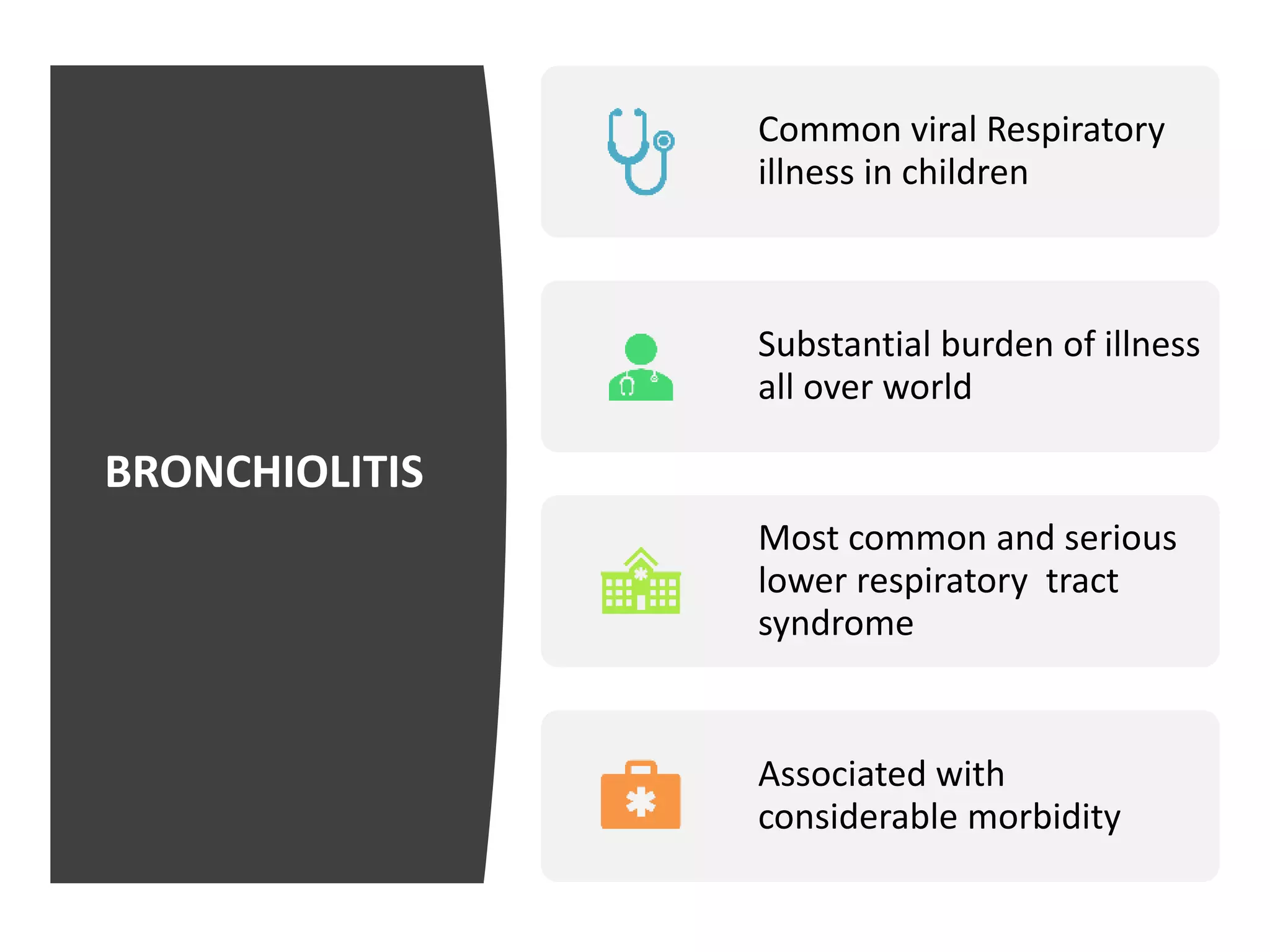
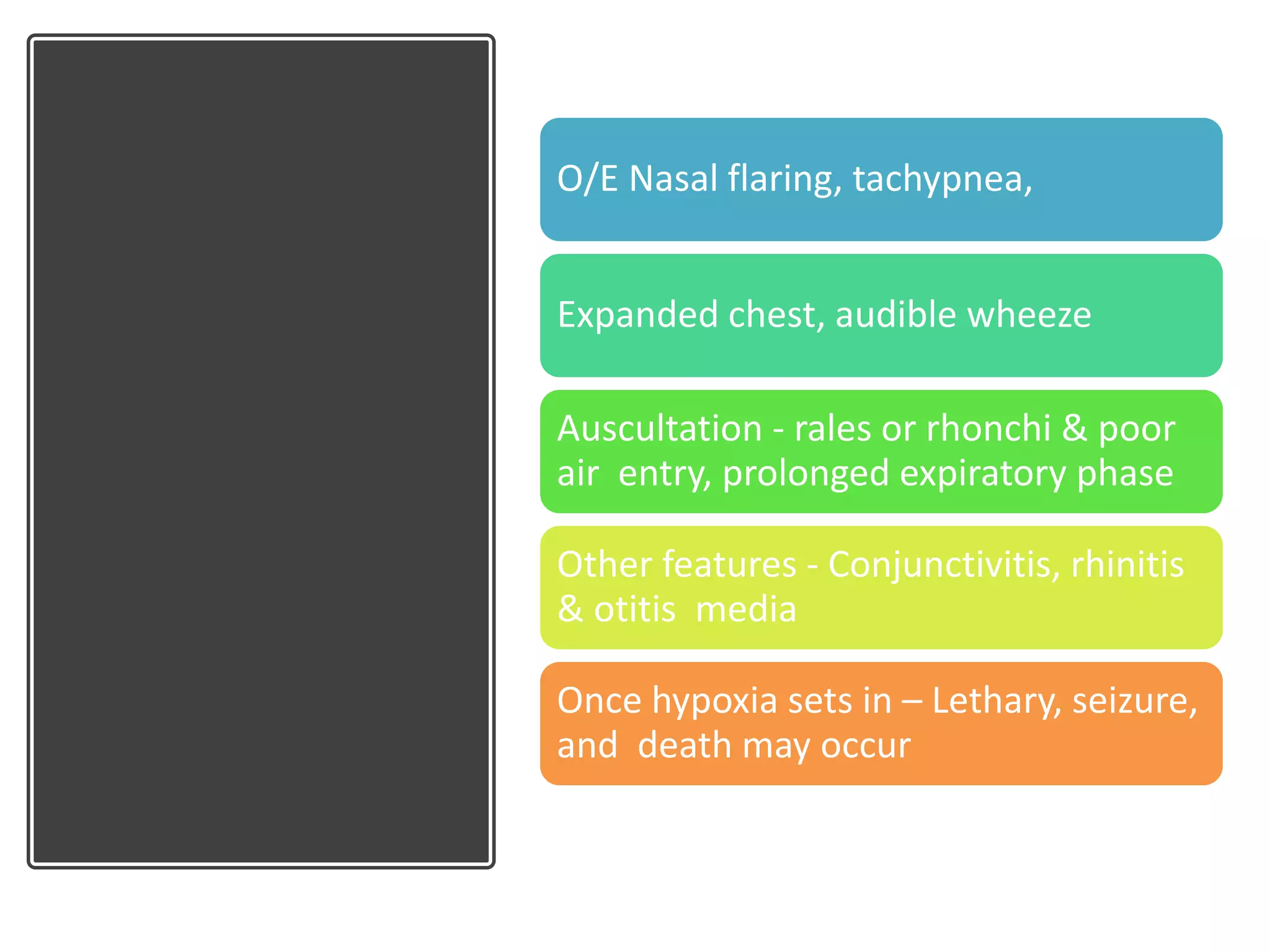
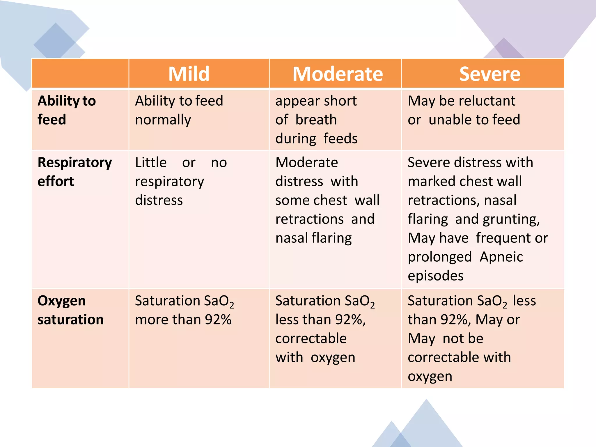
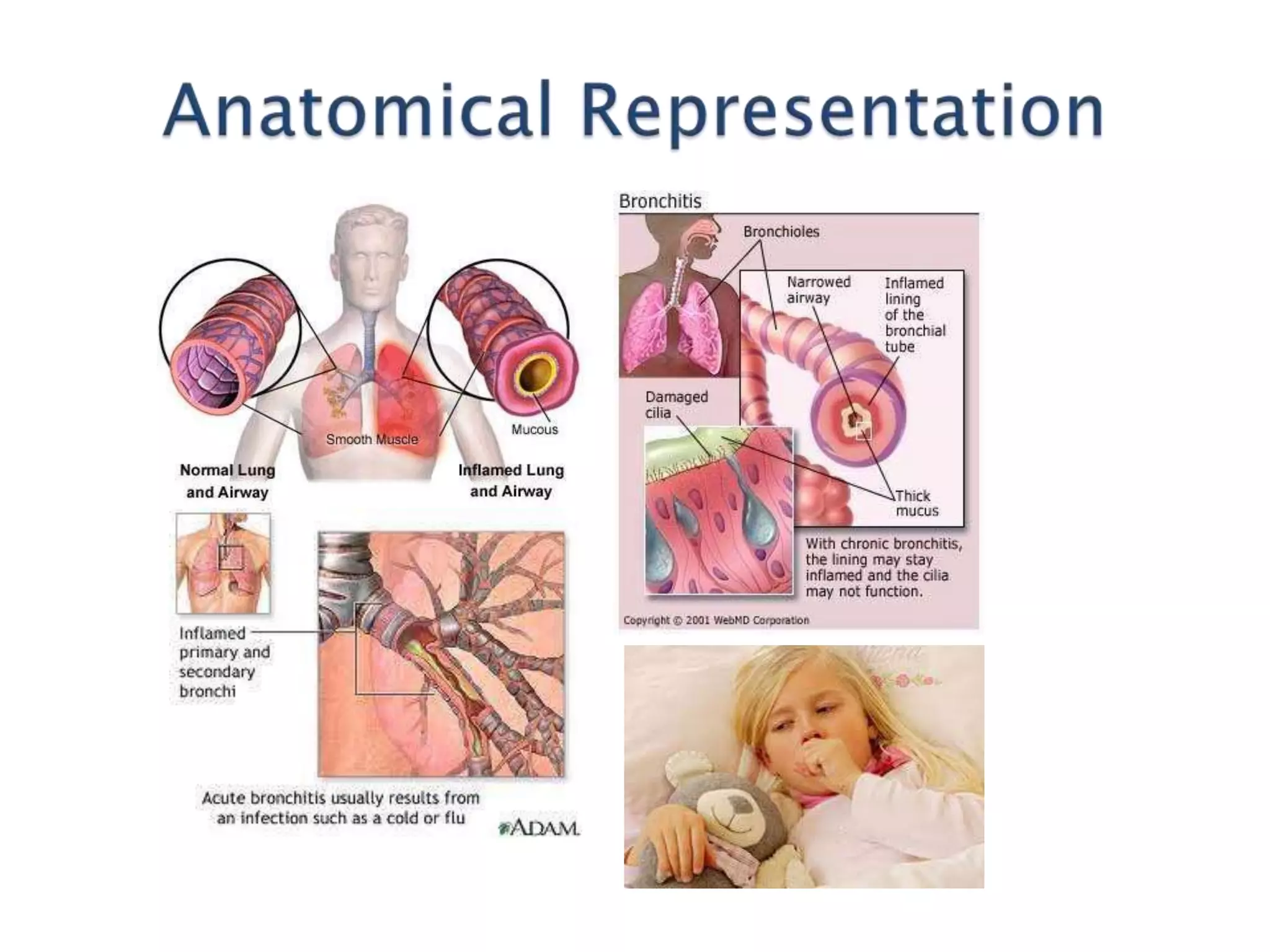
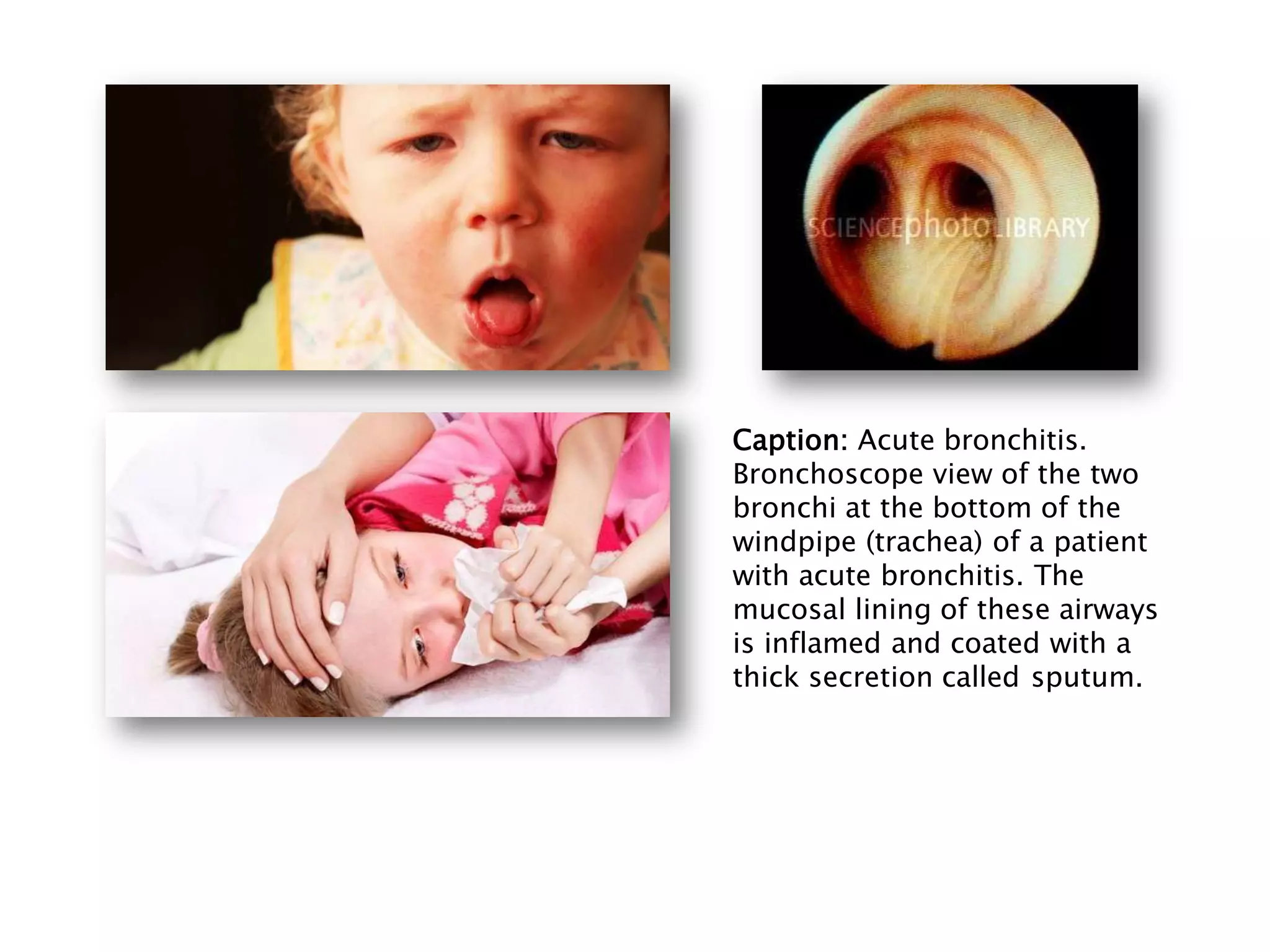
Bronchiolitis and Bronchitis
Presenter: Dr Abhishek
Moderator: Dr (Col) Wilson
Learning Objectives
• Epidemiology
• Definition
• Risk factor
• Etiology
• Pathology
• Clinical features
• Grading
• Investigations
• Differential diagnosis
• Management
• Discharge criteria
• Complications
• Prognosis
• Prevention
BRONCHIOLITIS
Common viral Respiratory
illness in children
Substantial burden of illness
all over world
Most common and serious
lower respiratory tract
syndrome
Associated with
considerable morbidity
• Generally self limiting
• Most commonly due to Respiratory Syncytial
virus Infection
DEFINITION
Clinical Syndrome
Acute onset of resp. symptoms ‐ <
2 yrs age
Initial symptoms – Upper
Respiratory Tract viral infections
Fever, coryza, progresses in 4‐6
days to
Lower Respiratory Tract
involvement ‐ Cough and wheezing
EPIDEMIOLOGY
Increased incidence
in premature
infants & children with
chronic illnesses
More common in children < 12
months
> 50% affected children ‐ 2 to 7
months of age
Infants < 6 months
are at highest
risk of clinically
significant disease
2% to 3% of
children require
hospital admission
Commonly in late autumn and
early spring
RISK FACTORS
• Infants in day care
• Exposure to passive smoke
• Crowding in the
household
• Environmental and
genetic factors do
contribute to severity of
disease
• Age less than 03 months
• Premature delivery
Congenital heart disease
Chronic lung disease
Congenital lung malformations
Tracheosophageal fistula
Immunodeficiency
Malnutrition
ETIOLOGY
M. pneumonia – though isolated not
recognized as etiological agent
Others
Influenza, Parainfluenza Adenovirus, Coronavirus, Rhinovirus
Most common – Respiratory syncytial virus
Viral
Pathology
RSV infection results in loss of
epithelia cilia and sloughing of
epithelial cells in airway
Leads to collection of
desquamated airway
epithelial cells,
polymorphoneuclear cells and
Lympocytes within the airways
Also airway mucosal oedema
Airway obstruction in acute brochiolitis –
sloughed epithelial cells, neutrophils,
lymphocytes
Complete plugging of some airway
Partial plugging of other
Leading to Localized ateclasis of some units
and over distension of other
Results in
Ventilation-
perfusion
mismatch
Causing
hypoxemia
Sloughed
epithelial cells,
Neutrophils and
lymphocytes
Airway Obstruction
Complete or partial
plugging
of airways
Atelectasis or over
distension
Ventilation and
perfusion
imbalance
Hypoxemia
CLINICAL
FEATURES
Quite variable
Initially Present
like URTI
Cough or Cold
with or without
fever
Nasal obstruction
with or without
rhinorrhea
Later disturbing
cough,
Tachypnea,
respiratory
distress
Poor feeding after
the initial onset of
symptoms
Fever ‐ higher
O/E Nasal flaring, tachypnea,
Expanded chest, audible wheeze
Auscultation ‐ rales or rhonchi & poor
air entry, prolonged expiratory phase
Other features ‐ Conjunctivitis, rhinitis
& otitis media
Once hypoxia sets in – Lethary, seizure,
and death may occur
Grading
Mild, moderate, or
severe
Based on :
• Ability to feed
• Respiratory effort
• Oxygen saturation
observed at admission
Mild Moderate Severe
Ability to
feed
Ability to feed
normally
appear short
of breath
during feeds
May be reluctant
or unable to feed
Respiratory
effort
Little or no
respiratory
distress
Moderate
distress with
some chest wall
retractions and
nasal flaring
Severe distress with
marked chest wall
retractions, nasal
flaring and grunting,
May have frequent or
prolonged Apneic
episodes
Oxygen
saturation
Saturation SaO2
more than 92%
Saturation SaO2
less than 92%,
correctable
with oxygen
Saturation SaO2 less
than 92%, May or
May not be
correctable with
oxygen
INVESTIGATIONS
Bronchiolitis is a clinical diagnosis
Nasopharyngeal aspirate (NPA) for RSV and
viral culture
Complete blood count
Electrolytes – especially if needing IV fluids
Blood culture – if temperature > 38.5°C
Arterial blood gas analysis
CXR
CHEST X‐RAY
• Need not to be done routinely
• Hyperinflation,
• Patchy infiltrates
• Typically migratory
• Attributed to Postobstructive atelectasis
DIAGNOSIS
Clinical diagnosis
Infant with short
prodromal of upper RTI
Clinical finding
• audible wheezing
• wheezing with crackles
• respiratory distress with chest
recession
DIFFERENTIAL
DIAGNOSIS
Congenital anomalies ‐ Vascular
ring, congenital heart disease
Gastroesophageal reflux
Aspiration pneumonia
Foreign body aspiration
MANAGEMENT
PRINCIPLES
Mainstay of therapy-Supportive care
Moderately ill infants ‐ require
supplementary oxygen
IVF in young infants ‐ tachypnea, partial
nasal obstruction & feeding difficulties.
Role of bronchodilators – Controversial.
• – Can have a trial with nebulised salbutamol,
Nebulised epinephrine or hpertonic saline
Viral Bronchiolitis
Mild Bronchiolitis Moderate Bronchiolitis Severe Bronchiolitis
Don’t need
investigations
HOME TREATMENT
ADMIT
Humidified oxygen to
maintain SaO2 above 95%,
IV Fluids, observer for
Deterioration
ADMIT
ICU CARE O2 to
maintain SaO2 above
95%, IV Fluids,
Cardiorespiratory
monitoring, ABG, CXR
assess need for
ventilatory
support/ICU care
IMPROVEMENT
Decrease O2 re-establish
feeding, discharge when
SaO2 above 95%
DETERIORATION
Treat as severe
bronchiolitis
OXYGEN
Drug of
Choice in
Bronchiolitis
is Oxygen
Humidified
oxygen ideal
• if SaO2 <94%, or
• in combination of clinically
significant respiratory
distress, RR > 60/min,
feeding difficulty
Supplemental
oxygen
• Maintain SaO2 above 95%
• Use nasal prongs / face mask /
hood / head box
• Hypoxemia with or without
distress, despite high O2 flow,
require ventilator support
FLUID
THERAPY
• Indications
• Moderate to severe
Bronchiolitis
or
• Severe respiratory distress
–
• Nasal flaring,
tachypnea (>60/min),
apneic episodes,
marked retractions,
tiring during feeds etc
• Normal maintenance volumes
• N/2 or N/4 dextrose saline
• Fluid volumes increased up to
20%
• if frequent or persistent
fever (>38.5°C) and/or
• markedly increased
respiratory effort
• Monitor serum electrolytes
• Sedation should be avoided
• Further compromise respiratory drive
of the child
• Antibiotic
• No role
• Aerosolized Ribavirin
• Neither prevent the need of
mechanical ventilation nor reduces
the length of hospital stay
• No role of steroids (oral or
parenteral or inhaled)
ICU
MANAGEMENT
Persistent desaturation despite oxygen
ABG evidence of respiratory failure– i.e. pO2 < 80mm Hg; pCO2 >
50mm Hg; pH < 7
Apneic episodes
associated with
desaturation
or > 15 seconds
duration
or frequent recurrent
brief episodes
Progression to severe respiratory
distress, especially in at‐risk Group
Needed in the following category :
CPAP
Continuous Positive airway
pressure
May benefit infants with
bronchiolitis by stenting
open the smaller airways
during all phases of
respiration
Prevents air trapping &
obstructive disease
Other
Nonstandard
therapies
Antirespiratory syncytial virus
preparation
• RSV-Immune globulin intravenous IGIV
• Palivizumab (RSV specific humanized
monoclonal antibody)
Heliox
• Mixture of Helium and oxygen
– 80:20%
• Flow through airways with less
turbulence and resistance
• Not recommended for routine use
DISCHARGE
Minimal respiratory distress
SaO2 > 90% in room air (Except in chronic
lung disease, heart disease, or other risk
factors)
Did not require supplemental O2 for 10 hrs.
Minimal or no chest recession
Able to take oral feeds
COMPLICATIONS
Respiratory complications ‐
most frequent
• Respiratory failure
• Apnea
• Pneumothorax
Infectious complications ‐
second most common
Cardiovascular involvement,
Electrolyte imbalance
PROGNOSIS
Generally self limiting condition
2% to 3% of children require hospitalization
Need for supplemental O2 based on SaO2 on
admission and predict length of hospital stay
Beware of rapid deterioration in high risk
group
Death is uncommon even in high risk group
Fatality rate highest – less than 03 months
age
PREVENTION
RSV is highly contagious
Spreads via inhalation or
direct transfer
Simple hand washing
Isolation of RSV infected
cases
Acute
bronchitis
 Acute bronchitis is swelling and irritation in
child's air passages.
 This irritation may cause him to cough or
have other breathing problems.
 Acute bronchitis often starts because of
another illness, such as a cold or the flu.
 The illness spreads from your child's nose
and throat to his windpipe and airways
 Acute bronchitis lasts about 2 weeks and is
usually not a serious illness.
Acute bronchitis leads to the hacking cough and phlegm production
that often follows upper respiratory tract infection. This occurs
because of the inflammatory response of the mucous membranes
within the lungs' bronchial passages. Viruses, acting alone or
together, account for most of these infections.
• Mucociliary clearance is an important primary innate defense
mechanism that protects the lungs from the harmful effects of
inhaled pollutants, allergens, and pathogens
• The mucociliary apparatus consists of 3 functional compartments:
the cilia, a protective mucus layer, and an airway surface liquid
(ASL) layer, which work together to remove inhaled particles from
the lung
Cont..
insult to the airway epithelium, such as recurrent aspiration or repeated viral infection, may contribute to
chronic bronchitis in childhood. Following damage to the airway lining, chronic infection with commonly
isolated airwayorganismsmay occur.
The most common bacterial pathogen that causes lower respiratory tractinfections in children ofall age groups
is Streptococcus pneumoniae. Nontypeable Haemophilus influenzae and Moraxella catarrhalis may be
significant pathogens in preschoolers (age < 5 y), whereas Mycoplasma pneumoniae may be significant in
school-aged children (ages 6-18 y).
Cont.. Children with tracheostomies are often colonized
with an array of flora, including alpha- hemolytic
streptococci and gamma-hemolytic streptococci.
With acute exacerbations of tracheobronchitis in
these patients, pathogenic flora may include
Pseudomonas
aeruginosa and Staphylococcus aureus (including
methicillin-resistant strains), among other
pathogens. Children predisposed to oropharyngeal
aspiration, particularly those with compromised
protective airway mechanisms, may become
infected with oral anaerobic strains of streptococci.
 Infection: Acute bronchitis is most often caused by a type of germ called a virus. It may also be
caused by other germs, such as bacteria, yeast, or a fungus.
Viral :Adenovirus, Influenza, Parainfluenza, Respiratory syncytial virus, Rhinovirus, Human
bocavirus, Coxsackievirus, Herpes simplex virus
Bacterial :S pneumoniae, M catarrhalis, H influenzae , Chlamydia
pneumoniae , Mycoplasma species
 Polluted air: Acute bronchitis can be caused when your child breathes air that has chemical
fumes, dust, or pollution.
 Cigarette smoke: If you smoke around your child, he may be at higher risk for acute bronchitis.
 Medical problems: Your child may be more likely to get bronchitis if he has other medical
problems. Examples include asthma, frequent swollen tonsils, allergies, or heart problems.
 Premature birth: Babies who are premature (born too early) may be at higher risk for
bronchitis.
• retrosternal pain during deep
breathing or coughing.
• Generally, the clinical course of
acute bronchitis is self-limited,
with complete healing and full
return to function typically seen
within 10-14 days following
symptom onset.
• constant cough. The cough
may last up to a month.
Cough may be dry, or cough
up with mucus. Mucus may
be green, yellow, white, or
• have streaks of blood in it.
Chest pain may appear when
he coughs or takes a deep
breath,fever, body aches, and
chills.
• sore throat and a runny or stuffy
nose,short of breath and wheezes
(makes a high-pitched noise) when
• breathing.
Caption: Acute bronchitis.
Bronchoscope view of the two
bronchi at the bottom of the
windpipe (trachea) of a patient
with acute bronchitis. The
mucosal lining of these airways
is inflamed and coated with a
thick secretion called sputum.
• Lungs may sound
normal.
• Crackles, rhonchi,
or large airway
wheezing, if any,
tend to be
scattered and
bilateral.
• The pharynx may
be injected.
• History of :
• Retained foreign body
• Bronchopulmonary allergy
• Immunosuppression
• Previous infections
• serum C-reactive protein screen,
• respiratory culture,
• serum cold agglutinin
• Obtain a blood or sputum culture if antibiotic therapy is under
consideration.
• test nasopharyngeal, using antigen or polymerase
chain reaction testing
• for Chlamydia species and respiratory
• syncytial, parainfluenza, and influenza viruses or viral culture.
• Gram stain, chlamydial and viral antigen assays, and
bacterial and viral cultures.
• Asthma Testing. clinical response to daily high-dose oral corticosteroids
• ,Evidence of reversible airflow obstruction revealed by pulmonary
function testing.
• Cystic Fibrosis Testing. A negative sweat test result excludecystic
fibrosis.
• Immunodeficiency . measurement of total serum immunoglobulins,
• immunoglobulin G (IgG) subclasses, and specific antibody production is
recommended.
• Chest Radiography. Chest films generally appear normal in patients with
uncomplicated bronchitis. Focal consolidation is not usually present.
• Pulmonary Function . show airflow obstruction that is reversible
with bronchodilators
• Bronchoscopy. diagnosis of chronic bronchitis is suggested if the
airways appear erythematous and friable.
 Medical therapy generally targets symptoms and includes use of
analgesics and antipyretics. Antitussives and expectorants are
often prescribed
 The prototype antitussive, codeine, has been successful in some
chronic-cough and induced-cough models, such asguaifenesin
or dextromethorphan.
 Bronchodilators ,albuterol may be worthwhile, as it may provide
significant relief of symptoms for some patients.
 Antibiotics. When bacterial etiology is suspected or as
prophylaxis to secondary infections.
 Antivirals. When viral etiology is suspected.
 Corticoids inhalative
 Analgesic and antipyreticagents
Acetaminophen (Tylenol, Aspirin-Free
Anacin, Feverall)
Ibuprofen (Ibuprin, Advil, Motrin)
 Corticosteroids, systemic

Prednisolone (Pediapred, Orapred)
Prednisone (Sterapred)
Bronchodilators
Albuterol sulfate (Proventil, Ventolin)
Metaproterenol
Theophylline (Theo-24, Uniphyl)
 Antibiotics
Erythromycin (EES, E-Mycin, Ery-Tab)
Clarithromycin (Biaxin)
Azithromycin (Zithromax)
Tetracycline (Sumycin)
Doxycycline (Vibramycin)
Amoxicillin-clavulanic acid (Augmentin)
 Antivirals
Oseltamivir (Tamiflu)
Zanamivir (Relenza)
 Corticosteroids, inhaled
Beclomethasone (Qvar)
Fluticasone (Flovent HFA, Flovent Diskus)
Budesonide inhaled (Pulmicort
Flexhaler, Pulmicort Respules)
 Referral to a pediatric pulmonologist may be
helpful for patients experiencing persistent or
recurrent symptoms and whose histories
suggest the possibility of tracheobronchial
foreign body aspiration, cystic
fibrosis, immunodeficiency, or persistent
asthma for which appropriate first-line
symptom or controller therapies have failed.
• Complications are
extremely rare and
should prompt
evaluation for anomalies
of the respiratory tract,
including immune
deficiencies.
Complications may
include the following:
• Bronchiectasis
• Bronchopneumonia
• Acute respiratory failure
Instruct older patients regarding the need for immunization against
pertussis, diphtheria, and influenza, which reduces the risk of bronchitis
due to the causative organisms.
• Instruct these patients to avoid passive environmental tobacco
smoke; to avoid air pollutants, such as wood smoke, solvents, and
cleaners; and to obtain medical attention for prolonged respiratory
infections.
• Instruct parents that children may attend school or daycare without
restrictions except during episodes of acute bronchitis with fever.
Also instruct parents that children may return to school or daycare
when signs of infection have decreased, appetite returns, and
alertness, strength, and a feeling of well-being allow.
• Acute bronchitis is almost always a self-limited process in
the otherwise healthy child.
• However, it frequently results in absenteeism from
• school and, in older patients, work.
• Chronic bronchitis is manageable with proper treatment
and avoidance of known triggers (eg, tobacco smoke).
• Proper management of any underlying disease
• process, such as asthma, cystic fibrosis,
immunodeficiency, heart
• failure, bronchiectasis, or tuberculosis, is also key.
• These patients need careful periodic monitoring to minimize
further lung damage and progression to chronic irreversible lung
disease.
thankyou..

Bronchiolitis and bronchitis in children

  • 1.
    Bronchiolitis and Bronchitis Presenter:Dr Abhishek Moderator: Dr (Col) Wilson
  • 2.
    Learning Objectives • Epidemiology •Definition • Risk factor • Etiology • Pathology • Clinical features • Grading • Investigations • Differential diagnosis • Management • Discharge criteria • Complications • Prognosis • Prevention
  • 5.
    BRONCHIOLITIS Common viral Respiratory illnessin children Substantial burden of illness all over world Most common and serious lower respiratory tract syndrome Associated with considerable morbidity
  • 6.
    • Generally selflimiting • Most commonly due to Respiratory Syncytial virus Infection
  • 7.
    DEFINITION Clinical Syndrome Acute onsetof resp. symptoms ‐ < 2 yrs age Initial symptoms – Upper Respiratory Tract viral infections Fever, coryza, progresses in 4‐6 days to Lower Respiratory Tract involvement ‐ Cough and wheezing
  • 8.
    EPIDEMIOLOGY Increased incidence in premature infants& children with chronic illnesses More common in children < 12 months > 50% affected children ‐ 2 to 7 months of age
  • 9.
    Infants < 6months are at highest risk of clinically significant disease 2% to 3% of children require hospital admission Commonly in late autumn and early spring
  • 10.
    RISK FACTORS • Infantsin day care • Exposure to passive smoke • Crowding in the household • Environmental and genetic factors do contribute to severity of disease • Age less than 03 months • Premature delivery
  • 11.
    Congenital heart disease Chroniclung disease Congenital lung malformations Tracheosophageal fistula Immunodeficiency Malnutrition
  • 12.
    ETIOLOGY M. pneumonia –though isolated not recognized as etiological agent Others Influenza, Parainfluenza Adenovirus, Coronavirus, Rhinovirus Most common – Respiratory syncytial virus Viral
  • 13.
    Pathology RSV infection resultsin loss of epithelia cilia and sloughing of epithelial cells in airway Leads to collection of desquamated airway epithelial cells, polymorphoneuclear cells and Lympocytes within the airways
  • 14.
    Also airway mucosaloedema Airway obstruction in acute brochiolitis – sloughed epithelial cells, neutrophils, lymphocytes Complete plugging of some airway Partial plugging of other Leading to Localized ateclasis of some units and over distension of other
  • 15.
    Results in Ventilation- perfusion mismatch Causing hypoxemia Sloughed epithelial cells, Neutrophilsand lymphocytes Airway Obstruction Complete or partial plugging of airways Atelectasis or over distension Ventilation and perfusion imbalance Hypoxemia
  • 17.
    CLINICAL FEATURES Quite variable Initially Present likeURTI Cough or Cold with or without fever Nasal obstruction with or without rhinorrhea Later disturbing cough, Tachypnea, respiratory distress Poor feeding after the initial onset of symptoms Fever ‐ higher
  • 18.
    O/E Nasal flaring,tachypnea, Expanded chest, audible wheeze Auscultation ‐ rales or rhonchi & poor air entry, prolonged expiratory phase Other features ‐ Conjunctivitis, rhinitis & otitis media Once hypoxia sets in – Lethary, seizure, and death may occur
  • 19.
    Grading Mild, moderate, or severe Basedon : • Ability to feed • Respiratory effort • Oxygen saturation observed at admission
  • 20.
    Mild Moderate Severe Abilityto feed Ability to feed normally appear short of breath during feeds May be reluctant or unable to feed Respiratory effort Little or no respiratory distress Moderate distress with some chest wall retractions and nasal flaring Severe distress with marked chest wall retractions, nasal flaring and grunting, May have frequent or prolonged Apneic episodes Oxygen saturation Saturation SaO2 more than 92% Saturation SaO2 less than 92%, correctable with oxygen Saturation SaO2 less than 92%, May or May not be correctable with oxygen
  • 21.
    INVESTIGATIONS Bronchiolitis is aclinical diagnosis Nasopharyngeal aspirate (NPA) for RSV and viral culture Complete blood count Electrolytes – especially if needing IV fluids Blood culture – if temperature > 38.5°C Arterial blood gas analysis CXR
  • 22.
    CHEST X‐RAY • Neednot to be done routinely • Hyperinflation, • Patchy infiltrates • Typically migratory • Attributed to Postobstructive atelectasis
  • 23.
    DIAGNOSIS Clinical diagnosis Infant withshort prodromal of upper RTI Clinical finding • audible wheezing • wheezing with crackles • respiratory distress with chest recession
  • 24.
    DIFFERENTIAL DIAGNOSIS Congenital anomalies ‐Vascular ring, congenital heart disease Gastroesophageal reflux Aspiration pneumonia Foreign body aspiration
  • 25.
    MANAGEMENT PRINCIPLES Mainstay of therapy-Supportivecare Moderately ill infants ‐ require supplementary oxygen IVF in young infants ‐ tachypnea, partial nasal obstruction & feeding difficulties. Role of bronchodilators – Controversial. • – Can have a trial with nebulised salbutamol, Nebulised epinephrine or hpertonic saline
  • 26.
    Viral Bronchiolitis Mild BronchiolitisModerate Bronchiolitis Severe Bronchiolitis Don’t need investigations HOME TREATMENT ADMIT Humidified oxygen to maintain SaO2 above 95%, IV Fluids, observer for Deterioration ADMIT ICU CARE O2 to maintain SaO2 above 95%, IV Fluids, Cardiorespiratory monitoring, ABG, CXR assess need for ventilatory support/ICU care IMPROVEMENT Decrease O2 re-establish feeding, discharge when SaO2 above 95% DETERIORATION Treat as severe bronchiolitis
  • 27.
    OXYGEN Drug of Choice in Bronchiolitis isOxygen Humidified oxygen ideal • if SaO2 <94%, or • in combination of clinically significant respiratory distress, RR > 60/min, feeding difficulty Supplemental oxygen
  • 28.
    • Maintain SaO2above 95% • Use nasal prongs / face mask / hood / head box • Hypoxemia with or without distress, despite high O2 flow, require ventilator support
  • 29.
    FLUID THERAPY • Indications • Moderateto severe Bronchiolitis or • Severe respiratory distress – • Nasal flaring, tachypnea (>60/min), apneic episodes, marked retractions, tiring during feeds etc
  • 30.
    • Normal maintenancevolumes • N/2 or N/4 dextrose saline • Fluid volumes increased up to 20% • if frequent or persistent fever (>38.5°C) and/or • markedly increased respiratory effort • Monitor serum electrolytes
  • 31.
    • Sedation shouldbe avoided • Further compromise respiratory drive of the child • Antibiotic • No role • Aerosolized Ribavirin • Neither prevent the need of mechanical ventilation nor reduces the length of hospital stay • No role of steroids (oral or parenteral or inhaled)
  • 32.
    ICU MANAGEMENT Persistent desaturation despiteoxygen ABG evidence of respiratory failure– i.e. pO2 < 80mm Hg; pCO2 > 50mm Hg; pH < 7 Apneic episodes associated with desaturation or > 15 seconds duration or frequent recurrent brief episodes Progression to severe respiratory distress, especially in at‐risk Group Needed in the following category :
  • 33.
    CPAP Continuous Positive airway pressure Maybenefit infants with bronchiolitis by stenting open the smaller airways during all phases of respiration Prevents air trapping & obstructive disease
  • 34.
    Other Nonstandard therapies Antirespiratory syncytial virus preparation •RSV-Immune globulin intravenous IGIV • Palivizumab (RSV specific humanized monoclonal antibody) Heliox • Mixture of Helium and oxygen – 80:20% • Flow through airways with less turbulence and resistance • Not recommended for routine use
  • 35.
    DISCHARGE Minimal respiratory distress SaO2> 90% in room air (Except in chronic lung disease, heart disease, or other risk factors) Did not require supplemental O2 for 10 hrs. Minimal or no chest recession Able to take oral feeds
  • 36.
    COMPLICATIONS Respiratory complications ‐ mostfrequent • Respiratory failure • Apnea • Pneumothorax Infectious complications ‐ second most common Cardiovascular involvement, Electrolyte imbalance
  • 37.
    PROGNOSIS Generally self limitingcondition 2% to 3% of children require hospitalization Need for supplemental O2 based on SaO2 on admission and predict length of hospital stay Beware of rapid deterioration in high risk group Death is uncommon even in high risk group Fatality rate highest – less than 03 months age
  • 38.
    PREVENTION RSV is highlycontagious Spreads via inhalation or direct transfer Simple hand washing Isolation of RSV infected cases
  • 39.
  • 40.
     Acute bronchitisis swelling and irritation in child's air passages.  This irritation may cause him to cough or have other breathing problems.  Acute bronchitis often starts because of another illness, such as a cold or the flu.  The illness spreads from your child's nose and throat to his windpipe and airways  Acute bronchitis lasts about 2 weeks and is usually not a serious illness.
  • 42.
    Acute bronchitis leadsto the hacking cough and phlegm production that often follows upper respiratory tract infection. This occurs because of the inflammatory response of the mucous membranes within the lungs' bronchial passages. Viruses, acting alone or together, account for most of these infections. • Mucociliary clearance is an important primary innate defense mechanism that protects the lungs from the harmful effects of inhaled pollutants, allergens, and pathogens • The mucociliary apparatus consists of 3 functional compartments: the cilia, a protective mucus layer, and an airway surface liquid (ASL) layer, which work together to remove inhaled particles from the lung
  • 43.
    Cont.. insult to theairway epithelium, such as recurrent aspiration or repeated viral infection, may contribute to chronic bronchitis in childhood. Following damage to the airway lining, chronic infection with commonly isolated airwayorganismsmay occur. The most common bacterial pathogen that causes lower respiratory tractinfections in children ofall age groups is Streptococcus pneumoniae. Nontypeable Haemophilus influenzae and Moraxella catarrhalis may be significant pathogens in preschoolers (age < 5 y), whereas Mycoplasma pneumoniae may be significant in school-aged children (ages 6-18 y).
  • 44.
    Cont.. Children withtracheostomies are often colonized with an array of flora, including alpha- hemolytic streptococci and gamma-hemolytic streptococci. With acute exacerbations of tracheobronchitis in these patients, pathogenic flora may include Pseudomonas aeruginosa and Staphylococcus aureus (including methicillin-resistant strains), among other pathogens. Children predisposed to oropharyngeal aspiration, particularly those with compromised protective airway mechanisms, may become infected with oral anaerobic strains of streptococci.
  • 46.
     Infection: Acutebronchitis is most often caused by a type of germ called a virus. It may also be caused by other germs, such as bacteria, yeast, or a fungus. Viral :Adenovirus, Influenza, Parainfluenza, Respiratory syncytial virus, Rhinovirus, Human bocavirus, Coxsackievirus, Herpes simplex virus Bacterial :S pneumoniae, M catarrhalis, H influenzae , Chlamydia pneumoniae , Mycoplasma species  Polluted air: Acute bronchitis can be caused when your child breathes air that has chemical fumes, dust, or pollution.  Cigarette smoke: If you smoke around your child, he may be at higher risk for acute bronchitis.  Medical problems: Your child may be more likely to get bronchitis if he has other medical problems. Examples include asthma, frequent swollen tonsils, allergies, or heart problems.  Premature birth: Babies who are premature (born too early) may be at higher risk for bronchitis.
  • 47.
    • retrosternal painduring deep breathing or coughing. • Generally, the clinical course of acute bronchitis is self-limited, with complete healing and full return to function typically seen within 10-14 days following symptom onset. • constant cough. The cough may last up to a month. Cough may be dry, or cough up with mucus. Mucus may be green, yellow, white, or • have streaks of blood in it. Chest pain may appear when he coughs or takes a deep breath,fever, body aches, and chills. • sore throat and a runny or stuffy nose,short of breath and wheezes (makes a high-pitched noise) when • breathing.
  • 48.
    Caption: Acute bronchitis. Bronchoscopeview of the two bronchi at the bottom of the windpipe (trachea) of a patient with acute bronchitis. The mucosal lining of these airways is inflamed and coated with a thick secretion called sputum.
  • 49.
    • Lungs maysound normal. • Crackles, rhonchi, or large airway wheezing, if any, tend to be scattered and bilateral. • The pharynx may be injected.
  • 50.
    • History of: • Retained foreign body • Bronchopulmonary allergy • Immunosuppression • Previous infections
  • 51.
    • serum C-reactiveprotein screen, • respiratory culture, • serum cold agglutinin • Obtain a blood or sputum culture if antibiotic therapy is under consideration. • test nasopharyngeal, using antigen or polymerase chain reaction testing • for Chlamydia species and respiratory • syncytial, parainfluenza, and influenza viruses or viral culture. • Gram stain, chlamydial and viral antigen assays, and bacterial and viral cultures.
  • 52.
    • Asthma Testing.clinical response to daily high-dose oral corticosteroids • ,Evidence of reversible airflow obstruction revealed by pulmonary function testing. • Cystic Fibrosis Testing. A negative sweat test result excludecystic fibrosis. • Immunodeficiency . measurement of total serum immunoglobulins, • immunoglobulin G (IgG) subclasses, and specific antibody production is recommended. • Chest Radiography. Chest films generally appear normal in patients with uncomplicated bronchitis. Focal consolidation is not usually present. • Pulmonary Function . show airflow obstruction that is reversible with bronchodilators • Bronchoscopy. diagnosis of chronic bronchitis is suggested if the airways appear erythematous and friable.
  • 53.
     Medical therapygenerally targets symptoms and includes use of analgesics and antipyretics. Antitussives and expectorants are often prescribed  The prototype antitussive, codeine, has been successful in some chronic-cough and induced-cough models, such asguaifenesin or dextromethorphan.  Bronchodilators ,albuterol may be worthwhile, as it may provide significant relief of symptoms for some patients.  Antibiotics. When bacterial etiology is suspected or as prophylaxis to secondary infections.  Antivirals. When viral etiology is suspected.  Corticoids inhalative
  • 54.
     Analgesic andantipyreticagents Acetaminophen (Tylenol, Aspirin-Free Anacin, Feverall) Ibuprofen (Ibuprin, Advil, Motrin)  Corticosteroids, systemic  Prednisolone (Pediapred, Orapred) Prednisone (Sterapred) Bronchodilators Albuterol sulfate (Proventil, Ventolin) Metaproterenol Theophylline (Theo-24, Uniphyl)  Antibiotics Erythromycin (EES, E-Mycin, Ery-Tab) Clarithromycin (Biaxin) Azithromycin (Zithromax) Tetracycline (Sumycin) Doxycycline (Vibramycin) Amoxicillin-clavulanic acid (Augmentin)  Antivirals Oseltamivir (Tamiflu) Zanamivir (Relenza)  Corticosteroids, inhaled Beclomethasone (Qvar) Fluticasone (Flovent HFA, Flovent Diskus) Budesonide inhaled (Pulmicort Flexhaler, Pulmicort Respules)
  • 55.
     Referral toa pediatric pulmonologist may be helpful for patients experiencing persistent or recurrent symptoms and whose histories suggest the possibility of tracheobronchial foreign body aspiration, cystic fibrosis, immunodeficiency, or persistent asthma for which appropriate first-line symptom or controller therapies have failed.
  • 56.
    • Complications are extremelyrare and should prompt evaluation for anomalies of the respiratory tract, including immune deficiencies. Complications may include the following: • Bronchiectasis • Bronchopneumonia • Acute respiratory failure
  • 57.
    Instruct older patientsregarding the need for immunization against pertussis, diphtheria, and influenza, which reduces the risk of bronchitis due to the causative organisms. • Instruct these patients to avoid passive environmental tobacco smoke; to avoid air pollutants, such as wood smoke, solvents, and cleaners; and to obtain medical attention for prolonged respiratory infections. • Instruct parents that children may attend school or daycare without restrictions except during episodes of acute bronchitis with fever. Also instruct parents that children may return to school or daycare when signs of infection have decreased, appetite returns, and alertness, strength, and a feeling of well-being allow.
  • 58.
    • Acute bronchitisis almost always a self-limited process in the otherwise healthy child. • However, it frequently results in absenteeism from • school and, in older patients, work. • Chronic bronchitis is manageable with proper treatment and avoidance of known triggers (eg, tobacco smoke). • Proper management of any underlying disease • process, such as asthma, cystic fibrosis, immunodeficiency, heart • failure, bronchiectasis, or tuberculosis, is also key. • These patients need careful periodic monitoring to minimize further lung damage and progression to chronic irreversible lung disease.
  • 59.