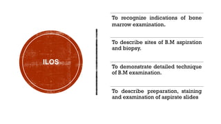
To recognize indications of bone marrow examination.
To describe sites of B.M aspiration and biopsy.
To demonstrate detailed technique of B.M examination.
To describe preparation, staining and examination of aspirate slides
Bone marrow aspirationand
biopsy
Amira Shehata, M.D
.
Clinical Pathology Department
Faculty of Medicine-Menoufia University
2.
ILOS
To recognize indicationsof bone
marrow examination.
To describe sites of B.M aspiration
and biopsy.
To demonstrate detailed technique
of B.M examination.
To describe preparation, staining
and examination of aspirate slides
3.
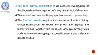
The bonemarrow examination is an essential investigation for
the diagnosis and management of many hematological disorders.
The aspirate and trephine biopsy specimens are complementary.
The final interpretation requires the integration of patient history,
clinical examination, PB counts and smear, B.M aspirate and
biopsy findings, together with the results of supplementary tests
such as immunophenotyping, cytogenetic analysis and molecular
genetic studies.
4.
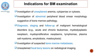
Indications for BMexamination
Investigation of unexplained anemia, cytopenias or cytoses.
Investigation of abnormal peripheral blood smear morphology
suggestive of bone marrow pathology.
Diagnosis, staging and follow-up of malignant hematological
disorders (e.g., acute and chronic leukemias, myelodysplastic
neoplasm, myeloproliferative neoplasms, lymphomas, plasma
cell myeloma, amyloidosis, mastocytosis)
Investigation of suspected bone marrow metastases.
Unexplained focal bony lesions on radiological imaging.
5.
Continued....
Unexplained organomegalyor presence of mass lesions inaccessible
for biopsy.
Microbiological culture for investigations of pyrexia of unknown origin
or specific infections, e.g., military tuberculosis, leishmaniasis, malaria.
Evaluation of iron stores.
Investigation of lipid/glycogen storage disorders.
Confirmation of normal bone marrow if bone marrow is being aspirated
for allogeneic transplantation.
Follow-up and monitoring of therapy.
6.
BM examination
Preliminaryprocedures:
It is important that the operator is aware of the clinical indications for
the marrow examination and the specimens required to be taken.
The operator should also be aware of the need for adequate
analgesia, and of safety issues with regard to thrombocytopenia or
coagulopathic risks ???.
Special considerations of the site for BM examination and positioning
of the patient should be given to immobile patients, obese patients,
pediatric patients, patients with lytic bone lesions, or who have had
prior radiotherapy???.
7.
Before theBM examination, the following should be performed:
The procedure should be explained in detail to the patient.
The past clinical history of the patient should be obtained, any
allergies and co-morbidities documented, and any pre-
medications explained.
Informed consent should be obtained from the patient
(discussion of the risks and benefits of the procedure).
A blood count and smear should be obtained if these have not
been collected in the previous 2 days.
8.
Anatomic site
The posterior superior iliac spine is the preferred anatomic site for
BM aspiration and trephine biopsy.
The anterior iliac spine can be used if the patient is immobile.
The medial surface of the tibia can also be used in infants.
A sternal aspirate may be appropriate in certain circumstances, e.g.
if the patient is immobile, has received radiotherapy to the pelvis or
other sites have yielded a ‘dry tap’ or if a trephine biopsy is not
required.
It should be noted that sternal aspiration should not be attempted in
patients with suspected plasma cell myeloma and in children
why???.
9.
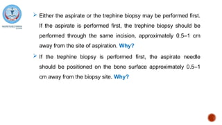
Either theaspirate or the trephine biopsy may be performed first.
If the aspirate is performed first, the trephine biopsy should be
performed through the same incision, approximately 0.5–1 cm
away from the site of aspiration. Why?
If the trephine biopsy is performed first, the aspirate needle
should be positioned on the bone surface approximately 0.5–1
cm away from the biopsy site. Why?
10.
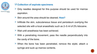
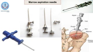
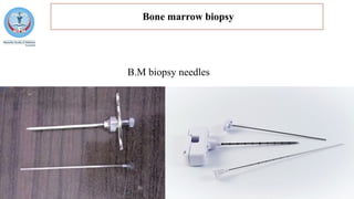
Collection ofaspirate specimens
Only needles designed for this purpose should be used for marrow
aspiration.
Skin around the area should be cleaned; How?
Infiltrate the skin, subcutaneous tissue and periosteum overlying the
selected site with a local anaesthetic such as 2–5 ml of 2% lidocaine.
Wait until anesthesia has been achieved.
With a penetrating movement, pass the needle perpendicularly into
the cavity of the bone.
When the bone has been penetrated, remove the stylet, attach a
syringe and suck up marrow contents.
It isrecommended that the aspirate should be drawn with a 10-
or 20-ml plastic syringe, to provide adequate negative pressure,
attached to the aspiration needle.
To preserve morphology, the syringe should not contain
anticoagulant.
Approximately 0.5 ml of the first draw of the aspirate should be
collected to make BM smears. Why?
13.
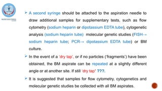
A secondsyringe should be attached to the aspiration needle to
draw additional samples for supplementary tests, such as flow
cytometry (sodium heparin or dipotassium EDTA tube), cytogenetic
analysis (sodium heparin tube) molecular genetic studies (FISH→
sodium heparin tube; PCR→ dipotassium EDTA tube) or BM
culture.
In the event of a ‘dry tap’, or if no particles (‘fragments’) have been
obtained, the BM aspirate can be repeated at a slightly different
angle or at another site. If still ‘dry tap’ ???.
It is suggested that samples for flow cytometry, cytogenetics and
molecular genetic studies be collected with all BM aspirates.
14.
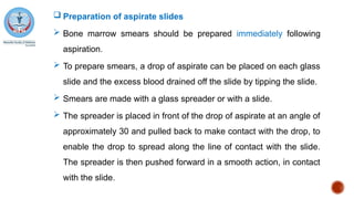
Preparation ofaspirate slides
Bone marrow smears should be prepared immediately following
aspiration.
To prepare smears, a drop of aspirate can be placed on each glass
slide and the excess blood drained off the slide by tipping the slide.
Smears are made with a glass spreader or with a slide.
The spreader is placed in front of the drop of aspirate at an angle of
approximately 30 and pulled back to make contact with the drop, to
enable the drop to spread along the line of contact with the slide.
The spreader is then pushed forward in a smooth action, in contact
with the slide.
15.
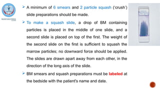
A minimumof 6 smears and 2 particle squash (‘crush’)
slide preparations should be made.
To make a squash slide, a drop of BM containing
particles is placed in the middle of one slide, and a
second slide is placed on top of the first. The weight of
the second slide on the first is sufficient to squash the
marrow particles; no downward force should be applied.
The slides are drawn apart away from each other, in the
direction of the long axis of the slide.
BM smears and squash preparations must be labeled at
the bedside with the patient's name and date.
16.
Staining ofBM aspirate slides
Two air-dried smears and one squash slide should be fixed with
absolute methanol as soon as they are thoroughly dry for
subsequent staining by a Romanowsky method or Perls stain for
iron.
All BM smears should be cover-slipped using a mounting
medium.
Additional slides may be used for cytochemistry (e.g.
myeloperoxidase) or archived as unfixed, unstained smears, as
required.
17.
Microscopy
TheBM smear should first be viewed under low power
magnification to determine:
the number and cellularity of particles.
the number and morphology of megakaryocytes, and to
scan for clumps of abnormal cells and for abnormal cells of low
incidence.
Areas of well-spread marrow cells in the cellular trails of the BM
smear behind the particles are selected for assessment at higher
magnification for morphological assessment of cells, including
cytological detail, parasites or cell inclusions.
18.
BM smearsare particularly useful for cellular detail and
differential cell counts.
The squash preparation is useful for the assessment of cellularity,
megakaryocyte numbers, focal disease (e.g. lymphoma, plasma
cell myeloma, mast cells, metastatic carcinoma, storage
histiocytes, granulomas), and the detection of abnormal cells of
low incidence.
In the absence of particles, megakaryocytes or other hemopoietic
precursors, the sample should be reported as a ‘blood tap’.
19.
In theabsence of particles, but in the presence of
megakaryocytes or other precursor cells, the sample should be
reported as a dilute BM sample and a qualitative evaluation can
be performed.
In the presence of particles with absent or very reduced
cellularity, only a qualitative description should be provided.
The patient’s blood count and a PB smear stained with a
Romanowsky stain should always be reviewed in conjunction with
the aspirate slides.
20.
The nucleateddifferential count (NDC)
A marrow NDC should be performed to assess hemopoietic activity,
to compare the proportions of the different cell lineages and to
quantify abnormal cells, if present.
The NDC should be performed in the cellular trails of the BM smear
behind the particles.
BM cells should be counted in an area where the cells are well
dispersed with good cytological detail.
The NDC should comprise blast cells, promyelocytes, myelocytes,
metamyelocytes, band forms, segmented neutrophils, eosinophils,
basophils, mast cells, promonocytes and monocytes, lymphocytes,
plasma cells and erythroblasts.
21.
At least500 cells should be counted in at least 2 smears when a
precise percentage of an abnormal cell type is required for
diagnosis.
To reduce imprecision from sampling error, the total number of
cells counted in the NDC should be increased, by counting in
another smear, or counted by a second observer, if the abnormal
cell count is very close to a critical threshold for disease
stratification or to a low threshold (e.g. 5%) or when the
appearance suggests a patchy involvement of the BM with
abnormal cells.
22.
The myeloid:erythroid(M:E) ratio should be calculated by
expressing the ratio of all granulocytes and monocytes and their
precursors (i.e. myeloblasts, promyelocytes, myelocytes,
metamyelocytes, band forms, segmented neutrophils,
eosinophils, basophils, promonocytes and monocytes) to
erythroblasts (at all stages of differentiation).
Flow cytometric differential counts should not be used as
surrogates for the NDC obtained from the smear.
Flow cytometry and morphology are complementary methods
that give different and valuable information, but the degree of
correlation varies greatly.
23.
Storage iron
A Prussian Blue stain should be performed on a methanol-fixed
marrow smear for the evaluation of storage iron and sideroblasts.
A BM smear with increased iron stores should be included as a
positive control.
An iron stain should be performed on all initial BM aspirates, but
may not be necessary on follow-up BM aspirates, e.g. for
leukemia.
The presence or absence of iron stores should be evaluated by
examining BM macrophages in several particles in the BM smear.
24.
Iron storesin smears may be graded subjectively as absent,
reduced, normal, increased, or markedly increased.
The total number of sideroblasts (normal, reduced or increased)
should be reported and the frequency and location (cytoplasmic
or perinuclear) of siderotic granules should be noted.
Ring sideroblasts are defined by the presence of five or more
siderotic granules encircling one third or more of the nucleus in
an iron-stained smear.
At least 100 erythroblasts should be evaluated for the percentage
of ring sideroblasts, if present.
25.
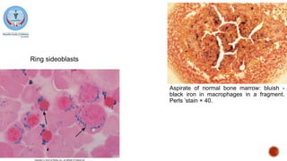
Aspirate of normalbone marrow: bluish -
black iron in macrophages in a fragment.
Perls ’stain × 40.
Ring sideoblasts
26.
The aspiratereport
The aspirate report should include the results of the blood count
and a PB smear comment.
The adequacy of the aspirate should be mentioned in the report. If
the aspiration was a ‘dry’ or hemodilute tap should be stated.
The cellularity of BM particles should be evaluated by assessing
several particles in smears or squash preparations.
Cellularity is better assessed in squash preparations than smears.
It should be noted that overall BM cellularity is generally better
assessed in trephine biopsy sections than the aspirate.
27.
In makinga subjective assessment of the cellularity of films prepared from
aspirates, the cellularity of fragments is of more importance than the
cellularity of trails, although occasionally the presence of quite cellular trails
– despite hypocellular fragments – suggests that the marrow cellularity is
adequate.
An average fragment cellularity between 25% and 75% is usually taken to
indicate normality, except at the extremes of age.
The cellularity of sections of fragments is expressed in terms of hemopoietic
tissue as a percentage of the total of hemopoietic and adipose tissue.
Cellularity can be described as reduced, normal, increased or
markedly increased.
28.
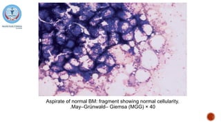
Aspirate of normalBM: fragment showing normal cellularity.
May–Grünwald– Giemsa (MGG) × 40
.
29.
The numbers(decreased, normal or increased), whether
maturation is normal or abnormal and the morphology of the
erythroid and myeloid lineages should be commented upon and
described if abnormal.
The numbers of blast cells should be reported.
The numbers of lymphocytes and plasma cells and whether their
morphology is normal or abnormal should also be noted.
Megakaryocyte numbers and morphology should be
documented; their numbers are best assessed in the trephine
biopsy specimen.
30.
When macrophagenumbers are increased, this should be
documented and abnormalities of morphology (e.g. hemo- or
erythro-phagocytosis, presence of inclusions such as micro-
organisms, crystals, or vacuoles) recorded.
Any abnormal cells or metastatic tumour cell aggregates should
be described and the presence of significant numbers of
smudged cells should also be documented.
The results of the iron stain and other cytochemical investigations
should be reported.
31.
Relevant flowcytometric findings, if available, should be
summarized in the aspirate report.
The aspirate report should not be delayed whilst awaiting results
of supplementary investigations and results that are pending
should be noted in the initial report.
When the results of molecular genetic studies are known and
they impact upon diagnosis, they should be commented upon in
a Supplementary Report, to be issued when the results of these
or other supplementary investigations are available.
32.
A modifiedreport may also be required if the final conclusion and
diagnosis is altered as a consequence of these additional test
results.
The conclusion of the BM aspirate report should document the
diagnosis or differential diagnosis.
Major findings may be summarized and other investigations to be
undertaken mentioned.
The findings should be compared with previous BM reports if the
aspirate was performed for disease monitoring.
If the aspirate was performed to confirm a clinical diagnosis and
the result was negative, this should be stated.
33.
Turnaround Time
The processing TAT for the BM aspirate, or the time from
collection of the aspirate to the time when slides are available for
microscopy should be about two to six working hours.
If results are required urgently by the requesting clinician, the
Reporting TAT, or the time from when the slides are available for
microscopy to the time when a verbal or written report is issued,
should be about three working hours for a verbal report or up to
24 h for a written report.
In less urgent cases, a written report on the aspirate alone should
be available in 48 h.


![PERI-PROSTHETIC FRACTURE NAIL-PLATE CONSTRUCT [NPC].pptx](https://cdn.slidesharecdn.com/ss_thumbnails/drarunkumardrmohamedashrafperiprostheticfrasturenail-plateconstructnpc-260209164459-7e9d15a1-thumbnail.jpg?width=640&height=640&fit=bounds)
![ONFH[AVN HIP] -TRIPLE REGIME -A NOVAL SURGICAL CONCEPT .pptx](https://cdn.slidesharecdn.com/ss_thumbnails/onfhavnhip2026koaconcalicutdrgokuldevdrmashraf-260210064517-213ec005-thumbnail.jpg?width=640&height=640&fit=bounds)

